
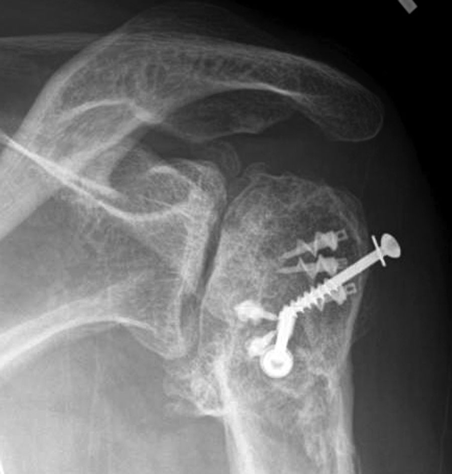
As we've emphasized before (see here), two plain x-rays are necessary and sufficient to make most diagnoses of shoulder arthritis.
Here is an anteroposterior (AP) and an axillary view typical of shoulders with post traumatic arthritis.
The upper view, the AP shows collapse of the humeral head, osteophyte formation, sclerosis, screws and suture anchors.

Follow on twitter: https://twitter.com/shoulderarth
Follow on facebook: click on this link
Follow on facebook: https://www.facebook.com/frederick.matsen
Follow on LinkedIn: https://www.linkedin.com/in/rick-matsen-88b1a8133/
Here are some videos that are of shoulder interest
Shoulder arthritis - what you need to know (see this link).
How to x-ray the shoulder (see this link).
The ream and run procedure (see this link).
The total shoulder arthroplasty (see this link).
The cuff tear arthropathy arthroplasty (see this link).
The reverse total shoulder arthroplasty (see this link).
The smooth and move procedure for irreparable rotator cuff tears (see this link).
Shoulder rehabilitation exercises (see this link)