
These authors present a current concepts review on 'glenoid dysplasia'. Their bullet points are reproduced here:
➤ Subtle forms of glenoid dysplasia may be more common than previously thought and likely predispose some patients to symptomatic posterior shoulder instability. Severe glenoid dysplasia is a rare condition with characteristic radiographic findings involving the posteroinferior aspect of the glenoid that often remains asymptomatic.
➤ Instability symptoms related to glenoid dysplasia may develop over time with increased activities or trauma. Physical therapy focusing on rotator cuff strengthening and proprioceptive control should be the initial management.
➤ Magnetic resonance imaging and computed tomographic arthrograms are useful for detecting subtle glenoid dysplasia by revealing the presence of an abnormally thickened or hypertrophic posterior part of the labrum, increased capsular volume, glenoid retroversion, and posteroinferior glenoid deficiency.
➤ Open and arthroscopic labral repair and capsulorrhaphy procedures have been described for symptomatic posterior shoulder instability. Glenoid retroversion of >10° may be a risk factor for failure following soft-tissue-only procedures for symptomatic glenoid dysplasia.
➤ Osseous procedures are categorized as either glenoid reorientation (osteotomy) or glenoid augmentation (bone graft), and no predictable results have been demonstrated for any surgical strategy. Glenoid osteotomies have been described for increased retroversion, with successful results, although others have noted substantial complications and poor outcomes.
➤ In severe glenoid dysplasia, the combination of bone deficiency and retroversion makes glenoid osteotomy extremely challenging. Bone grafts placed in a lateralized position to create a blocking effect may increase the risk of the development of arthritis, while newer techniques that place the graft in a congruent position may decrease this risk.
Comment: This article does a nice job of summarizing what is known about the evaluation and management of glenoid dysplasia. As the authors point out, "a variety of terms and definitions have been used to describe abnormal glenoid morphology, including dysplasia, hypoplasia, glenoid cleft, and retroversion, which contribute to confusion regarding diagnosis and management." Dysplasia (from the Greek δυσ- dys-, "bad" or "difficult" and πλάσις plasis, "formation"). As such glenoid dysplasia should be used to refer to abnormal glenoid development.
This got a bit confused when a type C glenoid shape was defined (see this link) as " a glenoid retroversion of more than 25 degrees, regardless of erosion; retroversion was primarily of dysplastic origin and explained the early event of osteoarthritis. In primary GHOA, this classification of the glenoid can discriminate retroversion between posterior erosion and dysplasia." It is important to distinguish glenoid 'bad formation' from glenoid retroversion that is acquired from osteoarthritis or capsulorrhaphy arthropathy. Likewise in cases of glenoid dysplasia it is important to distinguish symptoms of painful stiffness without arthritis or instability from dysplasia + instability or dysplasia + arthritis.


The result can be failure of these soft tissues leading to posterior instability and / or bone on bone arthritis.

In our hands, the best approach seems to be to use stretching and strengthening external rotation to avoid an internal rotation contracture and to optimize posterior stability.
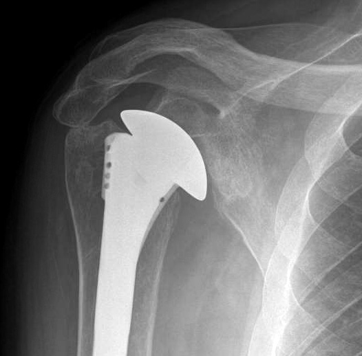
Should non-opeartive management fail, consideration can be given to an arthroplasty. In our hands the safest and most successful is a well balanced hemiarthroplasty.


===
Use the "Search" box to the right to find other topics of interest to you.
You may be interested in some of our most visited web pages including:shoulder arthritis, total shoulder, ream and run, reverse total shoulder, CTA arthroplasty, and rotator cuff surgery as well as the 'ream and run essentials'
You may be interested in some of our most visited web pages including:shoulder arthritis, total shoulder, ream and run, reverse total shoulder, CTA arthroplasty, and rotator cuff surgery as well as the 'ream and run essentials'