Showing posts with label glenoid inclination. Show all posts

Tuesday, March 5, 2024

What happens when glenoid version and inclination are "corrected"?


Posteriorly augmented glenoid components are increasingly being advocated for use in anatomic total shoulder arthroplasty. As pointed out in Augmented anatomic glenoid components - are they necessary? their value in terms of improving clinical patient outcomes is yet to be demonstrated. In addition to their increased cost, their use may be associated with increased loosening moments and difficulty in fitting the component to the glenoid bone.


Furthermore, the use of stepped, wedged, or half wedged glenoid components assumes that the two concavities in an eroded glenoid lie with one anterior and the other posterior. 


However it has become evident that the pathologic concavity need not be confined to the posterior direction and is not infrequently in the posterior superior aspect of the glenoid.


As a result, it may not be well fit by a posteriorly augmented glenoid component.

The authors of Three-dimensional analysis of biplanar glenoid deformities: What are they and can they be virtually reconstructed with anatomic total shoulder arthroplasty implants? discuss the three dimensional deformity in the arthritic glenoid and its implications for the use of augmented components. They defined "biplanar" glenoid deformities as those with a combined increase in both superior inclination and retroversion and suggest that these deformities are associated with difficulties in glenoid implantation and inferior clinical outcomes.

They analyzed 268 patients with glenohumeral osteoarthritis indicated for total shoulder arthroplasty.  Glenoids with superior inclination ≥10˚ and retroversion ≥20˚ were considered to have biplanar deformity. 49% of these shoulders had type B2 pathoanatomy. The direction of the deformity was directly posterior in 57% and posterior superior in 24%.

Their parameters for acceptable glenoid reconstruction included glenoid polyethylene implant position with  90% seating and less than 20% cancellous bone exposure, no central peg perforation, no more than a single peripheral peg perforation, and maintenance of prosthetic joint line lateral to pathologic joint line. 

Accepting less correction increased the rate of satisfactory reconstruction and decreased the planning system's suggested use of augmented glenoid components:

(1) The shoulders were first virtually planned for anatomic TSA attempting correction to neutral inclination and version.  Virtual aTSA planning indicated that 41% of the shoulders could not be reconstructed to neutral inclination and version using any implant. Of those that could, the system suggested that 94% have augmented implants. 

(2) The shoulders were then virtually planned for anatomic TSA accepting correction to 5˚ superior inclination and 10˚ retroversion. Virtual aTSA planning indicated that 10% could not be reconstructed with any implant.  Of those that could, the system suggested that 58% have augmented implants.

Final implant insertion commonly involved removal of substantial amounts of bone,  unseating in the posterosuperior quadrant, cancellous exposure in the anteroinferior quadrant, and vault perforation.


As the authors point out despite classical recommendations for 80% as the threshold for adequate glenoid implant seating, recent evidence suggests that even with 84% seating, the risk of glenoid loosening increases by up to 28% compared to full backside support. Seating values below 90% are associated with increases in bone stress and critical cement volumes. Furthermore, the importance of subchondral bone preservation is well-established, but specific thresholds for the amount of cancellous bone exposure are not defined. 

Comment: This is an important study. It makes us ask, is it more important to "correct" the glenoid pathoanatomy or to preserve glenoid bone stock and "accept" increased glenoid version and inclination? (see Glenoid version: acceptors and correctors). How should a surgeon optimize version, inclination, cancellous bone exposure, and component seating? Many of the publications attempting to address these questions are based on finite element analysis, simulation modeling, in vitro studies using Sawbones, theory, or studies using outdated components (e.g. those with keels or non-ingrowth pegs).  These studies can create a view of a "correct time zero implant position" or "acceptable resurfacing parameters" that may or may not relate to the outcome of the patients we treat. 

This study points out that correcting a biplane deformity to neutral version and inclination frequently frequently results in peg perforation, exposure of subchondral cancellous bone and inadequate backside support of the implant.

It may be that the most robust approach to glenoid component placement is to conservatively ream the glenoid face to a single concavity, accepting glenoid version and inclination, preserving maximal bone quality and quantity, and completely seating a standard round backed glenoid component that matches the reamed concavity. 

You can support cutting edge shoulder research and education that are leading to better care for patients with shoulder problems, click on this link.

Follow on twitter: https://twitter.com/RickMatsen or https://twitter.com/shoulderarth
Follow on facebook: click on this link
Follow on facebook: https://www.facebook.com/frederick.matsen
Follow on LinkedIn: https://www.linkedin.com/in/rick-matsen-88b1a8133/

Here are some videos that are of shoulder interest
Shoulder arthritis - what you need to know (see this link).
How to x-ray the shoulder (see this link).
The ream and run procedure (see this link).
The total shoulder arthroplasty (see this link).
The cuff tear arthropathy arthroplasty (see this link).
The reverse total shoulder arthroplasty (see this link).
The smooth and move procedure for irreparable rotator cuff tears (see this link).
Shoulder rehabilitation exercises (see this link).


Tuesday, March 7, 2023

Reverse total shoulder: the importance of inferior angulation of the base plate and how to achieve it.

Inferior inclination of the reverse total shoulder baseplate reduces the risk of component loosening, scapular notching, and limited range of motion.


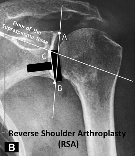
The authors of The reverse shoulder arthroplasty angle: a new measurement of glenoid inclination for reverse shoulder arthroplasty, point out that the key to achieving inferior inclination is to recognize that (1) the baseplate should be placed on the inferior aspect of the bony glenoid and 
(2) the preoperative inclination of the inferior glenoid (C-D) is different than the inclination of the entire glenoid (A-B).
The supraspinatus fossa line is a useful reference line to measure glenoid inclination because the sclerotic line of the supraspinatus fossa line is visible on both plain radiographs and CT scans. 

They defined the preoperative reverse shoulder arthroplasty angle as the angle between a line perpendicular to a line drawn along the floor of the supraspinous fossa and the plane of the inferior glenoid (A-S) (below left). 

The postoperative reverse shoulder arthroplasty angle is the angle between a line perpendicular to a line drawn along the floor of the supraspinous fossa and the plane of the baseplate (below right). 

They suggest that the ideal inclination of the baseplate is perpendicular to the line drawn along the floor of the supraspinous fossa, i.e. the postoperative reverse shoulder arthroplasty angle is zero.


 In 47 shoulders with rotator cuff tear arthropathy the preoperartive reverse shoulder arthroplasty angles measured 25±8 degrees on plain radiographs, 20±6 on reformatted 2D CT scans, and 21±5 on 3D reconstructions: thus, each method gave comparable values.

The Favard classification describes the common patterns of glenoid erosion seen with rotator cuff tear arthropathy: no erosion (E0), varying degrees of concentric central erosion (E1), and eccentric superior erosion (E2 and E3). The blue wedges indicate the amount of correction of inclination necessary to place the baseplate perpendicular to a line drawn along the floor of the supraspinous fossa. 

Correction of the inclination can be accomplished by reaming the inferior glenoid, adding bone graft beneath the upper part of the baseplate, or using a superiorly augmented baseplate. 

Comment: The preoperative reverse shoulder arthroplasty angle is useful for determining the amount of correction needed while the postoperative reverse shoulder arthroplasty angle is useful for determining whether the desired correction was achieved.

You can support cutting edge shoulder research that is leading to better care for patients with shoulder problems, click on this link.

Follow on twitter: https://twitter.com/shoulderarth
Follow on facebook: click on this link
Follow on facebook: https://www.facebook.com/frederick.matsen
Follow on LinkedIn: https://www.linkedin.com/in/rick-matsen-88b1a8133/

Here are some videos that are of shoulder interest
Shoulder arthritis - what you need to know (see this link).
How to x-ray the shoulder (see this link).
The ream and run procedure (see this link).
The total shoulder arthroplasty (see this link).
The cuff tear arthropathy arthroplasty (see this link).
The reverse total shoulder arthroplasty (see this link).
The smooth and move procedure for irreparable rotator cuff tears (see this link).
Shoulder rehabilitation exercises (see this link).

Monday, February 8, 2016

What is the value of patient-specific instrument guidance in shoulder arthroplasty?

Patient-specific instrument guidance of glenoid component implantation reduces inclination variability in total and reverse shoulder arthroplasty.

The authors present 36  shoulder arthroplasties (12 TSAs, 24 RSAs) of which 18 procedures (6 TSAs, 12 RSAs) were executed using preoperative 3D planning and patient-specific (PSI) guides to position the central guide pin for glenoid component implantation. In 9 cases, the glenoid anatomy was severely distorted through wear or previous surgery. 

The inclination of the glenoid component was measured using the angle between the glenoid baseplate and the floor of the supraspinatus fossa (angle β) on postoperative radiographs.

For TSA, the average angle β was 74 ± 9 in the PSI group and 86 ± 12 in the non-PSI group. 
For RSA, the average angle β was 83 ± 7 in the PSI group and 90 ± 17 in the non-PSI group. 

The x-rays below show the subtle difference between a reverse glenoid inserted without PSI (left) and with PSI (right).


Extreme values of glenoid component inclination were more likely to occur in the non-PSI group than in the PSI group (P < .001 for TSA; P = .02 for RSA).

Comment: A few days ago we posted on an article stating that accurate measurement of glenoid inclination could not be made without 3D reconstruction of CT scans (link).

Here we have a 'case controlled' study, comparing glenoids placed with and without patient-specific instrument (PSI) guidance. However, the authors do not explain how patients were assigned to the two groups or whether the pathoanatomy was comparable. The clinical outcomes of the two groups are not compared. The increment in cost, preparation time and operative time for the PSI method is not provided. As a result, the value (benefit/cost) cannot be determined and we cannot be sure that the patient whose x-rays are shown on the right above will fare better than the one whose x-rays are shown on the left.

As the authors point out, there are many variables other than inclination (AP and superior/inferior orientation of the starting point, anterior and posterior inclination, bone quantity and quality, glenoid component geometry) to be considered in the selection and positioning of a glenoid component. They also point out that 
"(1) the manufacturing the PSI guide depends on an accurately defined surgical plan on a correct 3D model of the patient’s scapula. Segmenting an accurate 3D model  of the scapula, however, can be difficult because of cartilage loss, severe bone deformity, calcified labrum, and difficulty in segmenting glenoid bone from humeral bone.
(2) fitting the PSI guide on the glenoid and finding its stable and correct position depend on a meticulous exposure  of the anterosuperior glenoid rim with sufficient removal of the soft tissues.and 
(3)  the accuracy of the system is also dependent on the surgeon’s ability to ream to the proper depth in line with the central guide pin."

A final set of questions emerge in regard to such complex technologies: 
is it the recommendation that such a device should be used on all shoulder arthroplasties?
is the technology more appropriate for high volume or for less experienced surgeons?
what is the learning curve?
where does the money come from to cover the incremental costs?
if we learn to depend on such a system, what happens if it is not available?

We will have to ask and answer these questions for each new technology that is brought forward.

Friday, February 5, 2016

Preoperative imaging of the arthritic shoulder: the glenoid inclination angle


These authors explore methods for measuring the glenoid inclination angle ( β-angle), formed by the intersection of a line on the floor of the supraspinatus fossa and glenoid fossa line.

In 51 shoulders they compared measurements on (1) an anteroposterior (AP) x-ray view of the shoulder, (2) unformatted 2-dimensional (2D) computed tomography (CT) scan, and (3) reformatted 2D CT scan in the scapular plane to their 'gold standard': glenoid inclination angle calculated by the 3D software.

The mean differences from the gold standard was 
1° (SD), 0.5°) for the β-angle measured on reformatted CT scans
3° (SD, 0.7°) for the  β-angle measured on AP plain radiographs and
10° (SD, 0.9°) for the β-angle measured on unformatted 2D CT scans 

The incremental costs and time for the CT scan with reformatting are not presented, so that the value (benefit/cost) of the two degrees of increased accuracy in comparison to plain films cannot be determined.

Comment: While there is no question that measurements of pre and postoperative glenoid orientation are of interest, the paper's statement that "An accurate measurement method for preoperative glenoid inclination is essential for intraoperative decision making to maximize implant longevity and function" has yet to be documented.
We find that the necessary and sufficient information for the planning and conduct of shoulder arthroplasty can be gained from the economical standardized AP view in the plane of the scapula


and the standardized axillary view taken with the arm elevated 90 degrees in the plane of the scapula (position of function).

===

Check out the new Shoulder Arthritis Book - click here.


You may be interested in some of our most visited web pages including:shoulder arthritis, total shoulder, ream and runreverse total shoulderCTA arthroplasty, and rotator cuff surgery as well as the 'ream and run essentials'