Showing posts with label stemless humeral components. Show all posts

Saturday, November 29, 2025

Five stemless humeral arthroplasty components - design differences and regional humeral bone density

Stemless humeral arthroplasty depends on fixation of the humeral component to the proximal humeral metaphysis. The article referenced below maps the typical bone density of this region from the article Spatial mapping of humeral head bone density


Stemless components consist of (a) the humeral head and (b) a fixation device that connects the humeral head to the bone of the humeral metaphysis - we'll call that the "nucleus".

The first stemless humeral head component was the TESS (Total Evolutive Shoulder System), developed by Zimmer Biomet and introduced around 2004-2005. The TESS used a nucleus consisting of central peg with metaphyseal fins for fixation. 


The TESS had a relatively short clinical lifespan - it was discontinued after several years due to concerns about loosening and suboptimal outcomes. The device struggled with achieving reliable fixation, particularly in patients with poor bone quality.

In spite of this initial failure, many shoulder arthroplasty surgeons are currently successfully using newer stemless humeral components because of their ability to position the articular surface where the surgeon desires, rather than having it positioned as dictated by a humeral stem. 

For many patients the cancellous bone of the humeral metaphysis is sufficient for secure fixation of the implant. Thus, as long as the head cut is made properly and the bone is of good quality, things usually work out well.

The challenge is that in older patients or patients with diminished quality of the cancellous bone in the humeral metaphysis, the fixation may not be adequate.  

Some surgeons advocate preoperative CT imaging to examine local bone density. However, because the quality of fixation depends not only on bone quality but also on the design of the fixation device - the nucleus - that links the humeral head component to the humeral bone it seem more practical to insert the trial nucleus to assess fixation quality; if it's insufficient the surgeon can switch to a stemmed component.

Below I've endeavored to present five stemless shoulder arthroplasty systems arranged in chronological order of U.S. FDA clearance, examining their design philosophy, and mechanical advantages for osteoporotic bone quality. 

N.B. Please note that I have no conflict of interest with any of the companies selling these implants. Note also that I used Claude.ai to help in the analysis and preparation of this post. As such there may be errors and I'd welcome any evidence-based corrections/comments. Finally, this analysis does not present clinical outcome, radiographic followup data, or complexity of revision among these options - such a comparison would require a very large number of patients and careful control of bone density and surgical technique.

1. Simpliciti (Wright Medical/Stryker)

FDA CLEARANCE: March 10, 2015

Design Geometry

Three central fins (~10mm length) arranged in a star pattern with central contact through a nucleus. While the nucleus provides broad cancellous contact with minimal risk cortical perforation, the central fin placement relies on cancellous bone which is weaker in patients with diminished bone desitity. 

[Churchill RS et al. J Bone Joint Surg Am. 2016;98(7):552-560 (prospective two-year multicenter FDA IDE study, n=149). Churchill RS. J Shoulder Elbow Surg. 2014;23(9):1409-1414 (stemless shoulder arthroplasty current status review). First stemless system cleared in the United States, with clinical use in France beginning in 2010.]

2. Comprehensive Nano (Zimmer Biomet)

FDA CLEARANCE: April 2019


Design Geometry

Single central peg (15-20mm length, 8-10mm diameter) with tapered profile plus modular metaphyseal shells. Load concentrated in cancellous bone without peripheral bone engagement.

[Razmjou H et al. J Shoulder Elbow Surg. 2012;21(10):1-9 (early TESS/stemless design study). IDE study began with first U.S. implant in October 2013 at University of Virginia. Built on clinical heritage of Biomet T.E.S.S. stemless shoulder available in Europe since 2004. Comprehensive Nano IDE clinical trial data on file, Zimmer Biomet.]

3. Eclipse (Arthrex)

FDA CLEARANCE: August 1, 2019 (510(k) K183194)


Design Geometry

Fenestrated central screw (6.5mm diameter, 35mm length) penetrates into cancellous bone.  A collar-bearing baseplate is compressed against cancellous bone and the cortical rim.  The degree of compression depends on the purchase achieved by the screw in the central cancellous bone which can be weak.

Clinical Evidence

[Habermeyer P et al. J Shoulder Elbow Surg. 2015;24(9):1463-1472 (midterm results). Habermeyer P et al. JSES Open Access. 2019;3(4):234 (9-13 year results). Gallacher S et al. J Shoulder Elbow Surg. 2018;27(12):2191-2197 (independent 2-year study). Uschok S et al. J Shoulder Elbow Surg. 2017;26(2):225-232 (Eclipse vs Univers II RCT). Over 20,000 successful implantations in Europe and Canada prior to U.S. FDA clearance. Longest clinical track record of any stemless system (2005-present)].

4. CS EDGE (DJO/Enovis)

FDA CLEARANCE/LAUNCH: September 30, 2020

Design Geometry

Three peripheral serrated fins arranged in tripod configuration (~15mm length, 2-3mm thickness) plus a short central peg. Fins engage peripheral bone (greater tuberosity, calcar region). Insertion needs to avoid fin perforation of cortex.

Clinical Evidence

Product launch announcement September 30, 2020 (DJO/Enovis).  No published peer-reviewed clinical studies available to date.


5. OsseoFit (Zimmer Biomet)

FDA CLEARANCE: December 13, 2024

Design Geometry

Asymmetric 3-4 peripheral fins with variable lengths (12-18mm) scaled to engage the asymmetrical peripheral bone in humeal head. Posterior-inferior fin longest, medial fin engages calcar, superior fins engage greater tuberosity. Requires precise surgical technique for correct rotational alignment

Clinical Evidence

FDA clearance announcement December 13, 2024 (Zimmer Biomet). No published peer-reviewed clinical studies available to date.


Fundamental Principle: Humeral head bone architecture exhibits a shell-and-core structure with peripheral bone density significantly exceeding central metaphyseal bone quality, especially in elderly patients and those with osteoporosis. Peripheral fixation designs theoretically exploit the strongest available bone, while central fixation systems depend on the bone that may be compromised in typical arthroplasty candidates.


It's all about fixation


Acorn Woodpecker
Madera Canyon, AZ
2020


Follow on twitter/X: https://x.com/RickMatsen
Follow on facebook: https://www.facebook.com/shoulder.arthritis
Follow on LinkedIn: https://www.linkedin.com/in/rick-matsen-88b1a8133/

Here are some videos that are of shoulder interest
Shoulder arthritis - what you need to know (see this link).
How to x-ray the shoulder (see this link).
The ream and run procedure (see this link)
The total shoulder arthroplasty (see this link)
The cuff tear arthropathy arthroplasty (see this link).
The reverse total shoulder arthroplasty (see this link).
The smooth and move procedure for irreparable rotator cuff tears (see this link)
Shoulder rehabilitation exercises (see this link).


Saturday, February 22, 2025

Pre-operative planning for anatomic shoulder arthroplasty and ream and run hemiarthroplasty – what can be accomplished using plain x-rays?

Dwight D. Eisenhower, our 34th President, said “the plan is nothing, planning is everything”, meaning that while specific plans may change, the act of planning itself is crucial because it prepares us to anticipate, adapt and respond effectively.

Thus, while there are important qualities of each patient’s arthritic shoulder that are only be revealed once the osteophytes have been removed and the contracted soft tissues are released, we can anticipate some elements of the surgery with consideration of the patient’s pre surgical anatomy as seen on plain radiographs and the preoperative physical examination. 

Michael Hachadorian, one of our current fellows, put together this blog post addressing this topic. He pointed to 4 major considerations that can help guide us in the planning of the shoulder arthroplasty

(1) Is the patient a better candidate for an anatomic total shoulder or for a ream and run procedure?

(2) Is the patient stiff (i.e. <100º of presurgical forward elevation and ≤0º of external rotation)?

(3) Does the patient exhibit posterior decentering on the preoperative axillary “truth” view?

(4) Is "overstuffing" only a humeral problem, or is it a potential issue with the global lateralization that can accompany the addition of a polyethylene glenoid component? (see Seven ways to overstuff an anatomic arthroplasty)

Here he presents five cases as illustrations of an approach to preoperative planning for shoulder arthroplasty that does not utilize CT scans or 3D planning software.

Case 1, The preoperatively centered, stiff arthritic shoulder

Here is a 44 year old male with severe OA who desired a ream and run. His preoperative examination reveals a stiff left shoulder with FE 80º, ER to 0º, ERA to 5º and IRA to 5º.


His plain radiographs showed many loose bodies and osteophytes. His axillary “truth” view shows a humeral head centered on an A2 type glenoid, suggesting that posterior instability is unlikely to be an issue with this shoulder.  

In this case the most important thing is to restore mobility to this shoulder, rather than trying to restore the "premorbid" anatomy. 

The PACS (Picture Archiving and Communication System) is available to most shoulder surgeon. It has tools for “trying” different prosthetic head sizes as shown below.

50 mm diameter of curvature.

54 mm diameter of curvature

The  54 diameter of curvature intersects the 3 points of the “perfect circle” and reveals the medialization resulting from arthritic humeral head bone loss. However, given the considerable presurgical stiffness that is likely to recur after surgery despite vigorous soft tissue relases, one can anticipate that the 50 mm diameter of curvature might be a better choice if confirmed by intraoperative testing.

Using angle tool at 135º relative to the humeral shaft, the PACS tools can then be used to explore the location of the head cut for this size head component, including its relation to the cuff insertion superiorly and to the osteophytes inferiorly. 

Based on this head cut, the thickness of the humeral head implant can be estimated measuring the distance from the medial aspect of the circle to the cut line. This example shows a 20 mm thickness head component.


However, given that this patient has considerable stiffness, what might it look like if we used the same head cut then downsized the head height to 50x18, deliberately “understuffing “? We can change the measurement tool to 18 by dragging the medial marker of this tool slightly lateral until the measurement reads “18.0 mm”. Then we can match the circle by dragging the entire circle laterally. Note that our perfect circle is no longer “perfect” in that it now extends slightly lateral to the GT.


Intraoperatively, both of these configurations were trialed and it was found that the range of motion was better with the 50 18 head and the stability was adequate. Here is the postoperative film with the  50 18 in place.

With the subscapularis closed his forward flexion was 165 degrees and his external rotation 30 degrees.


Case 2, The preoperatively decentered, stiff arthritic shoulder

This patient is an active 64 M who desired a ream and run procedure. His preoperative exam showed 90º forward elevation, external rotation at the side of 10º, external rotation in abduction to 30º, internal rotation in abduction to 0, and cross body adduction of 30 cm to contralateral acromioclavicular joint. 

His preoperative x-rays are shown below. His axillary “truth” view demonstrates essentially complete posterior decentering on a posteriorly eroded glenoid.


So here we have the challenging combination of stiffness and posterior instability. How can preoperative planning guide our efforts?

A 54 size head seems to fit well, but there was concern about posterior instability with an "anatomic" humeral reconstruction given his preoperative decentering. Note that while in the previous example, we elected to “undersize” the humeral head, we anticipate that the same strategy in this case could increase concern about postoperative instability.


On the “truth” view about 75% of the humeral head would lie posterior to the perpendicular bisector of the glenoid face.


We marked out the head height (18 mm) and the desired cut angle 135º. This head position would leave a 1 mm berm. We wanted to avoid a "too high" head because a head that is high with the arm at the side is posterior with the arm flexed forward at 90º


However, at surgery after osteophyte resection and anterior soft tissue releases,  the planned 54 18 concentric head component was (as anticipated) posteriorly unstable.

As a result, a 54 18  anteriorly eccentric humeral head was placed on a short stem (selected to assure implant stability in the humerus). 

The postoperative AP view shows the head in the planned position. 

The reconstruction did not substantially alter the global lateralization in comparison to the preoperative position.


The postoperative “truth” view shows the anteriorly eccentric humeral head centered on the glenoid that was reamed conservatively without glenoid version "correction". 


While some might argue that "correcting' glenoid version would have stabilized the shoulder against posterior instability, that approach seems less certain that what was performed here. While some are concerned about the effect of the anterior offset head on the subscapularis repair, the use of the 4 mm eccentric head has not been associated with tendon failure (see Total shoulder arthroplasty with an anterior-offset humeral head in patients with a B2 glenoid). As shown in this case, the amount of anterior prominence of the humeral head component is small.

This reconstruction provided a stable shoulder with postoperative flexion to 150º and external rotation to 30º.


Case 3 Total shoulder with overstuffing resulting from the addition of the glenoid polyethylene.

While focus on the reconstruction of the humeral head is reasonable for ream and run surgery, the effect of the addition of a 4 mm thick polyethylene glenoid component in total shoulder arthroplasty needs to be accounted for. In these cases, an anatomically reconstructed humeral head may lead to excessive global lateralization and resulting stiffness.

Here are the preoperative radiographs from a patient who had longstanding shoulder OA and was notably stiff on preoperative examination (FE 80, ER to 10, ERA 20 and IRA 10). 


The “truth” view indicated some posterior decentering. This patient had elected to undergo total shoulder arthroplasty.


In this case, 50 x 20 appeared to be like a reasonable option to re-create humeral anatomy. 


Post-operative radiographs demonstrated re-creation of humeral anatomy.   


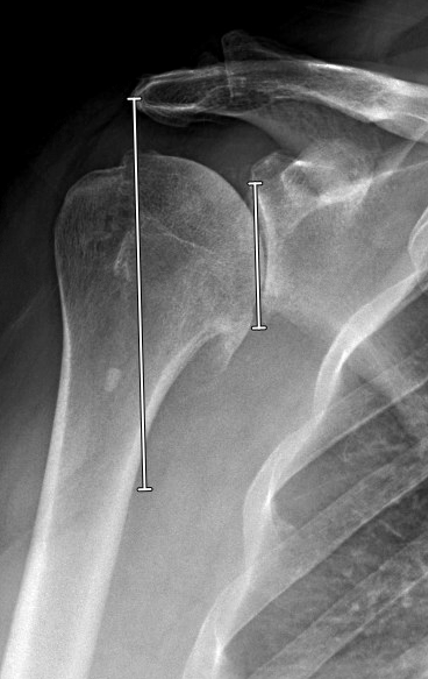
However by drawing a “plum line” down from the lateral aspect of the acromion parallel to the glenoid face we see that the center of rotation of the humeral head is now lateral to the acromion whereas it was medial to the acromion preoperatively. Despite re-creation of humeral anatomy, the humerus now has more global lateralization. This patient had stiffness postoperatively which required a manipulation under anesthesia.


 Case 4. Total shoulder with undersizing of the humeral head to accommodate the stuffing effect of the glenoid component.

Here are preoperative radiographs from a patient who had longstanding shoulder OA and was notably stiff on preoperative examination (FE 90, ER to neutral, ERA 10 and IRA 10). The “truth” view did not indicate posterior instability. 


A head size of 54 would nicely reapproximate the patient’s humeral anatomy, 


however there was concern that the 4 mm glenoid component could lead to a stiff shoulder by increasing the global lateralization.


Downsizing to a size 50 x 18 head would avoid excessive global lateralization once the thickness of the glenoid component is factored in. 


The revised plan is shown below 

And here is the postoperative x-ray


While the humeral component may appear to be undersized, the reconstruction did not increase the global lateralizing, avoiding tightening of the shoulder. 


The humeral head remained concentric on the postoperative “truth” view despite under sizing the humeral head component.


Case 5: Total shoulder in a posteriorly decentered humeral head without stiffness. 


An anatomic head cut was templated using a 50 20 humeral head.


During surgery, it was determined that an anterior eccentric humeral head was needed given persistent posterior instability with all head sizes. After trialing the 50 20 anterior eccentric head offered the desired stability. 

The postoperative "truth" view showed the anteriorly eccentric humeral head centered on the glenoid. Note the minimal anterior prominence of the eccentric head.

Comment: First of all, thanks to Mike for the heavy lifting in putting this together. Second, thanks to all our past shoulder fellows and colleagues such as Armodios (Armand) Hatzidakis and Surena Namdari for their active role in continuing to shape our thoughts about shoulder arthroplasty.

You can support cutting edge shoulder research that is leading to better care for patients with shoulder problems, click on this link

Follow on twitter/X: https://x.com/RickMatsen
Follow on facebook: https://www.facebook.com/shoulder.arthritis
Follow on LinkedIn: https://www.linkedin.com/in/rick-matsen-88b1a8133/

Here are some videos that are of shoulder interest
Shoulder arthritis - what you need to know (see this link).
How to x-ray the shoulder (see this link).
The ream and run procedure (see this link).
The total shoulder arthroplasty (see this link).
The cuff tear arthropathy arthroplasty (see this link).
The reverse total shoulder arthroplasty (see this link).
The smooth and move procedure for irreparable rotator cuff tears (see this link)
Shoulder rehabilitation exercises (see this link).