Wednesday, August 31, 2011

Rotator Cuff 6 - Clinical examination for a rotator cuff tear

Today, we'll consider how the physical examination of a shoulder can help discern if a rotator cuff problem may be present


Inspection can reveal atrophy, as well as incisions and scars indicating previous surgery and penetration. The physical examination may reveal subacromial roughness from hypertrophic bursa or from the superior edges of torn tendon rubbing against the coracoacromial arch that can be felt by a hand placed over the acromion as the shoulder is rotated.

Palpation can reveal gaps in the cuff tendon as shown in the figures below.


The range of motion examination can reveal restrictions due to contracture surrounding the area of injury or scarring in the humeroscapular motion interface. Limited range of motion is particularly common in the presence of partial thickness tears of the rotator cuff. The most common partial thickness tear is that of the supraspinatus tendon. In this situation it is characteristic to have loss of the motions that places this tendon under tension – internal rotation with the arm at the side


 internal rotation of the arm in 90 degrees of abduction


 and cross body movement

While, in the past, pain on these maneuvers has been attributed to ‘impingement,’ it is now recognized as being due to the pull on the partially torn tendon attachment which is analogous to the pain experienced on stretching the origin of the extensor carpal radialis brevis in tennis elbow.


Cuff strength is conveniently examined using manual tests of isometric torque. Isometric testing removes potential interference from pain on motion, from crepitance, or from stiffness. These tests examine the integrity of the supraspinatus

 the infraspinatus

and the subscapularis


Pain or weakness on these maneuvers constitutes an abnormal tendon sign for the specified tendon-muscle unit. These tests are relatively specific to each muscle, but are not specific to the cause of weakness; for example, a suprascapular nerve lesion or a cuff tear may each produce abnormal supraspinatus and infraspinatus tendon signs. 

Using these simple tests, the examiner can assess crepitance, loss of range of motion and pain or weakness on examination of specific tendons.


--
Here are some videos that are of shoulder interest
Shoulder arthritis - what you need to know (see this link).
How to x-ray the shoulder (see this link).
The ream and run procedure (see this link).
The total shoulder arthroplasty (see this link).
The cuff tear arthropathy arthroplasty (see this link).
The reverse total shoulder arthroplasty (see this link).
The smooth and move procedure for irreparable rotator cuff tears (see this link).
Shoulder rehabilitation exercises (see this link).

Monday, August 29, 2011

Rotator Cuff 5 - Can the rotator cuff tear be repaired?


Yesterday's post covered some of the key factors bearing on the repairability of a rotator cuff tear.


Below are listed findings that have been shown to be encouraging or discouraging about the prospect of the shoulder having a durably reparable cuff tear. It is of interest that many of these factors can be determined without advanced testing.

ENCOURAGING                                          DISCOURAGING

History
    Age less than 55                                             Age over 65
    Acute traumatic onset                                     Insidious, atraumatic onset
    No relation to work                                        Attribution of tear to work
    Short duration of weakness                            Weakness over 6 weeks
    No history of smoking                                    Many smoking pack-years
    No steroid injections                                       Repeated steroid injections
    No major medications                                     Steroids/antimetabolites
    No concurrent disease                                     Inflammatory joint disease
    No infections                                                   History of previous infection
    No previous shoulder surgery                          Previous cuff surgery
    Benign surgical history                                    History of failed tissue repairs

Physical Examination
   Good nutrition                                                  Poor nutrition/obesity
   Mild weakness                                                  Severe weakness
   No spinatus atrophy                                          Severe spinatus atrophy
   Stable shoulder                                                 Anterior superior instability
   Intact acromion                                                 Previous acromioplasty
   No stiffness                                                       Stiffness

Radiographs
   Normal radiographs                                          Upwards head displacement
                                                                              Cuff tear arthropathy

MRI or Ultrasound                                            
  Good tendon quality                                         Thin tendon
  One tendon tear                                                 Multiple tendon involvement
  Small gap to close                                             Severe retraction


--
Here are some videos that are of shoulder interest
Shoulder arthritis - what you need to know (see this link).
How to x-ray the shoulder (see this link).
The ream and run procedure (see this link).
The total shoulder arthroplasty (see this link).
The cuff tear arthropathy arthroplasty (see this link).
The reverse total shoulder arthroplasty (see this link).
The smooth and move procedure for irreparable rotator cuff tears (see this link).
Shoulder rehabilitation exercises (see this link).

Saturday, August 27, 2011

Rotator Cuff 4 - Mechanisms of Tear, Factors Affecting Repair, "Impingement"

Initiation of Cuff Failure


Cuff fiber failure commonly results from the sudden application of eccentric loads, for example when the muscle attempts to resist a downward force on the arm
while the cuff seems to be better able to tolerate concentric loads, for example in a controlled lift away from the side. 


An anatomic factor predisposing to deep surface failure of the cuff insertion is internal abutment, where the corner of the glenoid contacts the deep aspect of the cuff at its tuberosity insertion 
This is most likely to be a problem for throwers who have stretched out their anterior capsule, allowing increased external rotation.


It is apparent that the most common form of cuff fiber failure, that which occurs on the articular surface of the cuff tendon, cannot be attributed to scuffing of the bursal surface by the acromion: so-called subacromial ‘impingement.’ In fact, the cuff insertion is well under the acromion at relatively small angles of elevation where it is protected from contact with the coracoacromial arch. Current evidence indicates that the most rotator cuff tears arise from tension overload and age-related attrition, rather than 'impingement'.

Readers might be interested in a recent review of the literature regarding the diagnosis of "impingement syndrome".


Factors Compromising Tendon Healing


Deep surface rotator cuff fiber failure exposes the defect to joint fluid. This joint fluid prevents the formation of a fibrin clot and, thus, healing is contravened.

Furthermore, tension at the edge of the cuff tear compromises the circulation to the margin of the tendon

For these reasons, left to their own devices, cuff defects tend to progress rather than healing. An optimal cuff repair surgery will bring healthy tendon into contact with vascularized bone and exclude joint fluid from the repair site. Subsequent posts will review the principles of surgical repair in some detail.


Factors Affecting Reparability

In considering the potential for surgically restoring a durable tendon insertion to bone, the surgeon needs to consider the quality of the tissue to be used in the repair. The ability of the cuff tendon tissue to withstand tensile loads is compromised by age, disuse, steroid injections, smoking, and poor general health. These predisposing factors can dispose the cuff tendons to fail with minimal force – essentially an atraumatic fiber failure. Cuff fibers that fail atraumatically may be so constitutionally weak that they cannot hold up even if repaired back to the bone. Thus, in chronic atraumatic cuff tears there is reason to consider a non-operative approach to improving shoulder function by rehabilitating the muscle–tendon units that remain intact.


Acute, traumatic cuff detachments that result from major force application are likely to be repairable

If acute traumatic cuff tears are not repaired promptly, the muscle may undergo intramuscular contracture, atrophy, and fatty degeneration and the tendon may become progressively reabsorbed. These degenerative changes compromise the opportunity for surgical repair. Thus, as with any other tendon avulsion from bone, time is of importance in the repair of acute tears of the rotator cuff. 


Loss of the rotator cuff subjects the superior glenoid to increased loads that can contribute to its erosion 


Progressive upward displacement of the humeral head produces secondary changes in the coracoacromial arch 

Once the humeral head has ascended so that its equator is above the residual cuff, contraction of the cuff muscles lock the humeral head in the superiorly displaced position

Chronic upwards displacement of the humeral head from cuff deficiency and superior glenoid erosion can result in cuff tear arthropathy,

--


Use the "Search the Blog" box to the right to find other topics of interest to you.

You may be interested in some of our most visited web pages including: shoulder arthritis, total shoulder, ream and runreverse total shoulderCTA arthroplasty, and rotator cuff surgery.


Friday, August 26, 2011

Rotator Cuff 3 - Patterns of tearing

In partial thickness cuff tears loss of some of the tendon fibers reduces the ability of the attached muscle fibers to deliver force to the bone.  This disruption typically starts on the deep surface of the cuff.

, but may also occur within the tendon
or on the bursal side.  
Partial tears may be accompanied by avulsion of a piece of the greater tuberosity
 A thinned tendon contains fewer fibers than one of normal thickness; thus, a muscle with a thinned tendon has lost the optimal attachment of its contractile elements and increasing load is placed on the remaining fibers


Partial tendon tears can also prevent effective use of the muscle by producing pain on muscle contraction.  In this respect a partial thickness cuff tear resembles a tennis elbow, a condition in which the extensor carpi radialis brevis is partially torn from the latter epicondyle.  Contraction of the muscle produces pain at the tendon attachment to bone.  This condition is also similar to partial thickness tears at the insertion of the Achilles and patellar tendons.  While these conditions are often thought to be inflammatory (“tendonitis”), the problem is actually mechanical. When a tendon is partially torn, there is disproportionate load on the fibers at the edge of the tear – like when a zipper is pulled open, or like a nylon stocking developing a run in it, or like a piece of paper that is partially torn – the load is on the connection adjacent to the tear.  This force concentration is sometimes referred to as the ‘notch’ phenomenon – a mechanism by which partial tears can propagate progressively

This progressive tendon tearing is often progressive, each step in the progression may be interpreted as episodes of ‘tendonitis’ until it is recognized that the problem is a cuff tear.


--

Here are some videos that are of shoulder interest
Shoulder arthritis - what you need to know (see this link).
How to x-ray the shoulder (see this link).
The ream and run procedure (see this link).
The total shoulder arthroplasty (see this link).
The cuff tear arthropathy arthroplasty (see this link).
The reverse total shoulder arthroplasty (see this link).
The smooth and move procedure for irreparable rotator cuff tears (see this link).
Shoulder rehabilitation exercises (see this link).

Rotator Cuff 2 - Clinical Considerations Relating to Shoulder Strength

Neurogenic and Muscular Causes

The most devastating injury to a muscle is the loss of its nerve supply. Losses of the innervation of the deltoid, supraspinatus, and infraspinatus can be congenital (Erb’s Palsy), inflammatory (brachial neuritis), degenerative (cervical radiculopathy), traumatic (penetrating or stretch), compressive (a ganglion in the spinoglenoid notch), or iatrogenic (dissection or screw placement). These conditions along with myopathies, such as facioscapular muscular dystrophy, must be considered when the shoulder is weak.

The day this paragraph was written, we saw a patient sent in for a cuff repair. He had been in a water-skiing accident and landed on the water in a way that pressed his head to the left and his right shoulder down. At the moment of impact he experienced a shock down his right arm and noted severe weakness of his right shoulder. His physician sent him for an MRI that showed a partial tear of his supraspinatus. On exam in our office he had, six weeks after injury, atrophy and weakness of his spinati, deltoid, and biceps – a classic Erb’s type injury.

Electromyography, nerve conduction velocities, and muscle biopsies may be needed to sort out these diagnoses.

Loss of the Humeral Fulcrum

If the humeral head does not remain centered during muscle contraction, the forces intended to cause rotation will cause translation. As an example, the shoulder with posterior instability: when the arm is elevated forward, the humeral head slips out the back, losing its fulcrum. In this situation, valuable muscle excursion is consumed by displacement of the head and the strength of elevation is lost. The effects of loss of centering are also seen with the cuff and superior labral deficient shoulder in which contraction of the deltoid produces ascension of the humeral head rather than abduction


However, if the glenoid concavity is intact, the humeral head may remain stabilized in the glenoid in spite of superior cuff deficiency. This is why many shoulders with cuff tears remain functional.


Alternatively, if the coracoacromial arch remains intact, the humeral head of the cuff deficient shoulder may be secondarily stabilized in a new position, superiorly displaced against the arch.


Thus in the presence of cuff deficiency, it is vitally important to protect and maintain the coracoacromial arch. Without it, the humeral head will escape anterosuperiorly when the deltoid contracts – a complication that can result from acromioplasty and resection of the coracoacromial ligament.


This phenomenon is nicely demonstrated in this short video clip of a patient before surgery.


The treatment of anterosuperior instability may require a reverse total shoulder.

--
Here are some videos that are of shoulder interest
Shoulder arthritis - what you need to know (see this link).
How to x-ray the shoulder (see this link).
The ream and run procedure (see this link).
The total shoulder arthroplasty (see this link).
The cuff tear arthropathy arthroplasty (see this link).
The reverse total shoulder arthroplasty (see this link).
The smooth and move procedure for irreparable rotator cuff tears (see this link).
Shoulder rehabilitation exercises (see this link).

Thursday, August 25, 2011

Rotator Cuff 1 - Overview

The rotator cuff has two main functions in the shoulder: (1) it stabilizes the humeral head in the glenoid socket and (2) it works with the deltoid and other shoulder muscles to help provide the strength necessary for the shoulder to do its work.


In this video, one can see how the supraspinatus tendon, the most commonly injured tendon of the rotator cuff, normally passes smoothly below the coracoacromial arch as the arm is lifted from the side in this cadaver demonstration.


For a short cut to an overview of the cuff and the spectrum of rotator cuff disease, see
this page and the gallery on its right.

You may also like to see our recent
review of rotator cuff failure from the New England Journal of Medicine.

---


We'll take a step back and consider some of the principles of shoulder strength in depth.


When a muscle contracts, it approximates its effective origin and insertion with a force limited by its physiological cross sectional area. Muscles that have large cross sections, like the deltoid, can provide a larger maximal force than a muscle with a small cross section like the subclavius. The contractile elements within a muscle are connected to tendon fibers that connect to the bony origin and insertion. The tendon insertion is structured so that there is a smooth mechanical transition from flexible tendon to stiff bone.


This smooth transition enables the insertion to manage the repeated bending loads to which it is subject.


When the tissues providing this smooth transition degenerate they become stiff and weak so that the tendon insertion becomes increasingly vulnerable to failure.


The strength of a muscle is noted in terms of the torque it can generate. We recall that torque results when a force is exerted at a distance from a fixed center of rotation. The magnitude of the torque is the product of the length of the line connecting the center of rotation to the effective attachment of the muscle (the lever arm) and the magnitude of the force perpendicular to this line.


The effective points of attachment depend on the position of the arm and are not necessarily the anatomic insertions .


The weight of a dumbbell that can be held with the arm out to the side is determined by the sum of the products of the forces and their respective lever arms divided by the lever arm of the weight to be lifted.


Throughout this discussion, the lever arms have been described as the distance between the point of action of the muscle force and the center of rotation. In order for torques to be realized, the humerus must rotate around a stable center. This is why we have placed such emphasis on the mechanisms for centering of the head in the section on Stability. Without this precise centering, the effectiveness of the muscle contractions would be lost.


Muscles are also characterized by their excursion – the change in length over which they can provide force. In order to be effective throughout a range of motion, the centimeters of excursion of a muscle must match the product of the muscle’s lever arm in centimeters and the range of motion in degrees divided by the number of degrees in a radian.


So, while longer lever arms result in more torque per unit muscle force, they also require greater muscle excursion.


Muscles provide the maximal amount of force when operating close to the middle of their excursion with a drop off in maximal force as the muscle length approaches maximal extension or maximal contraction.


Muscles that have been chronically detached, as in long standing cuff tears, tend to lose their excursion. Even if they are reattached, the length over which they can exert an effective force is often diminished.

A special feature of the shoulder is that the powerful thoracoscapular muscles can position the entire glenohumeral joint along with the deltoid and the rotator cuff through a range of approximately 40 degrees of adduction/abduction


and 40 degrees of protraction/retraction


This ’portability’ of the glenohumeral joint enables the scapulohumeral muscles to carry out most shoulder functions in the mid-range of their excursion where they are the strongest. It is of note that the humeroscapular position is essentially the same for the knockout punch, the bench press, the point of racquet contact with the ball in the tennis serve, and the moment of release for the baseball pitch, even though the scapulothoracic positions are quite different.

One of the relatively unexplored facets of active shoulder strength is the requirement for muscular balance. In the knee, the muscles generate torques about a relatively fixed axis: that of flexion-extension. If the quadriceps pull is a bit off-center, the knee still extends. In the shoulder, no such fixed axis exists. In a specified position, each muscle creates a unique set of rotational moments. Imagine a rope attached to a sphere. The motion resulting from pulling on the rope depends on the orientation of the sphere as well as the direction of pull on the rope. If some of the resulting motion were undesired, it would need to be cancelled out by attaching another rope and pulling on it to resist the unwanted motion. So, for example, the anterior deltoid exerts moments in forward elevation, internal rotation, and cross-body movement.


If elevation without cross body movement is desired, the posterior deltoid must negate the cross body moment of the anterior deltoid.


Similarly, if elevation without rotation is desired, the cross-body and internal rotation moments of this muscle must be resisted by other muscles (such as the posterior deltoid and infraspinatus). These balancing activities take place at an additional energy cost. However, if the infraspinatus function is lacking, it is difficult to flex the arm without internal rotation.

--

Here are some videos that are of shoulder interest
Shoulder arthritis - what you need to know (see this link).
How to x-ray the shoulder (see this link).
The ream and run procedure (see this link).
The total shoulder arthroplasty (see this link).
The cuff tear arthropathy arthroplasty (see this link).
The reverse total shoulder arthroplasty (see this link).
The smooth and move procedure for irreparable rotator cuff tears (see this link).
Shoulder rehabilitation exercises (see this link).


How to stretch and how not to stretch

Some patients have had difficulty maintaining their range of motion after shoulder arthroplasty.
Sometimes the issue is that they are not correctly monitoring their range of elevation.
Ideally the range should be monitored weekly by a consistent physical therapist for the first 6 to 12 weeks after surgery. The therapist can report to the surgeon if there is any question about the onset of stiffness or the loss of range of motion.
Alternatively, the patient may use photos taken from the side to compare to those shown in this link to make sure they are attaining at least 150 degrees of elevation (the angle between the body and the arm).

Some very motivated patients still need coaching on the best stretching technique. We like to avoid a lot of quick stretches because they result in temporary 'elastic' lengthening of the tissue that returns immediately to the pre stretching length as illustrated by stretching the bungee in the video below.


Other well motivated patients stretch their shoulder too roughly, so that tissue is torn giving rise to pain and additional scar tissue. This is illustrated by pulling on a stick of cold chewing gum in the video below.


Ideally, stretches should be comfortable and held for a long time (one or two minutes for each repetition) so that 'plastic' or permanent lengthening of the tissues is achieved. This is shown in the video below as I stretch some bubble wrap (note that I had to take out some of the stretching time so the video would fit). Note also that the amount of lengthening achieved was definite, but not large. But if a bit of permanent lengthening is achieved each time, the shoulder motion will increase. To prevent 'backsliding' the stretches should be done frequently through the day.


--

===

To see the topics covered in this Blog, click here

Use the "Search" box to the right to find other topics of interest to you.

You may be interested in some of our most visited web pages including:shoulder arthritis, total shoulder, ream and runreverse total shoulderCTA arthroplasty, and rotator cuff surgery as well as the 'ream and run essentials'


To see other similar posts, click on the label of interest below.


Reverse Total Shoulder for Combined Shoulder Arthritis and Massive Rotator Cuff Tear and for Failed Conventional Total Shoulder Replacement

The reverse total shoulder replacement can restore comfort and function to the arthritic shoulder combined with a massive rotator cuff tear and for failed conventional total shoulder replacement.  This combination of conditions can result in major loss of stability and active motion of the shoul­der. The reverse total shoulder provides stability of the shoulder joint so that the deltoid muscle can power the shoulder through a useful range of motion. In this procedure the arthritic ball is replaced by a socket fixed to the arm bone (humerus) by a stem that fits within it. A metal ball is fixed to the bone of the arthritic socket with screws. Success requires technical excellence of the surgery and a commitment to the rehabilitation program until the desired range of motion can be achieved comfortably. The figure below left shows the humeral stem, cup, and white polyethylene cup as well as the glenosphere (ball) and screws used for fixation into the scapula (shoulder blade). The figure below right is an x-ray of this prosthesis in place.

First, a review of some basics.

What Are The Key Parts Of The Normal Shoulder Joint?

The ball (humeral head) fits in the socket (glenoid) and is held there by the rotator cuff 


What Is Shoulder Arthritis?
Shoulder arthritis is a condition in which de­generation, injury, inflammation or previous surgery destroys the normally smooth carti­lage on the ball (humeral head-below left) and socket (glenoid-below right).


How Is Shoulder Arthritis Diagnosed?
Carefully standardized X-rays reveal the loss of the space between the hu­meral head and glenoid that is normally occupied by cartilage, leaving bone on bone contact.

What Is A Conventional Total Shoulder?

In a conventional total shoulder, the arthritic surface of the ball is replaced with a metal ball with a stem that is press fit in the inside of the arm bone (humerus-below left) and the socket (glenoid) is resurfaced with a high density polyethylene component (below right).
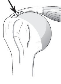
When Will A Conventional Shoulder Not Work?
When the rotator cuff is sufficiently torn that it no longer provides the necessary stability for the joint, the humeral head slides upwards. This results in slackening of the deltoid (below left) no longer able to raise the hand to carryout nor­mal activities. A conventional shoulder cannot restore the necessary stability in this situation (below right).
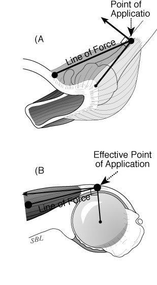
What Is A Reverse Total Shoulder?
In a reverse total shoulder the ball is located on the shoulder blade (glenoid) and the socket is located on the arm bone (humerus), exactly the opposite of the situation in a conventional total shoulder. This configuration provides sta­bility because the muscles around the shoul­der compress the ball and socket together. 
How Are The Parts Of A Reverse Total Shoulder Hooked To The Bones?
The ball (glenosphere) is screwed to the bone of the shoulder blade. The cup (humeral sock­et) is fixed to a stem that is cemented down the inside of the arm bone (humerus).

What Is The Incision Like?
After a general or regional anesthetic, this procedure is performed through an incision between the deltoid and the pectoralis major muscles on the front of the shoulder. It includes release of adhesions and con­tractures and removal of bone spurs that may block range of motion. Our team of surgeons, anesthesi­ologists, and surgical assistants usually perform this procedure in less than two hours.
Who Should Consider A Reverse Total Shoulder?


Surgery for shoulder arthritis and rotator cuff deficiency should only be considered when the condition of the shoulder is limiting the quality of the patient’s life and after a trial of physical therapy and mild analgesics to determine if non-operative management is helpful.  If severe disability persists, patients may consider the reverse total shoulder – no other surgical proce­dure has the ability to restore the stability needed in the absence of a functioning rotator cuff. The ideal patient is healthy, active, motivated and committed to complying with the rehabilita­tion program.

Who Should Probably Not Consider A Reverse Total Shoulder?
This procedure is less likely to be successful in individuals with depression or obesity. Pa­tients who use narcotic medication or who use tobacco may have increased difficult recover­ing from this procedure.

What Are The Keys To Success Of A Reverse Total Shoulder?
Success requires technical excellence of the surgery and a commitment by the patient to fol­low the rehabilitation program prescribed by the surgeon.

How Does A Patient Prepare For A Reverse Total Shoulder?
As for all elective surgical procedures, the patient should be in the best possible physical and mental health at the time of the procedure. Any heart, lung, kidney, bladder, tooth, or gum problems should be managed before surgery. Any infection may be a reason to delay the operation. Any skin problem (acne, scratches, rashes, blisters, burns, etc) on the shoulder or arm should be resolved before surgery. The shoulder surgeon needs to be aware of all health issues, including allergies as well as the non-prescription and prescription medications being taken. For instance, aspirin and anti-inflammatory medication may affect the way the blood clots. Some of these may need to be modified or stopped around the time of surgery.

What Happens After Surgery?
The reverse total shoulder is a major surgical procedure that involves cutting of skin, tendons and bone. The pain from this surgery is managed by the anesthetic and by pain medications. Immediately after surgery, strong medications (such as morphine or Demerol) are often given by injection. Within a day or so, oral pain medications (such as hydrocodone or Tylenol with codeine) are usually sufficient. The shoulder rehabilitation program is started on the day of surgery. The patient is encouraged to be up and out of bed soon after surgery and to pro­gressively reduce their use of pain medications. Hospital discharge usually takes place on the second or third day after surgery. The arm is kept in a sling for six weeks after the proce­dure to allow for healing, but the patient can use the hand for eating.  Driving is not recom­mended during this time. Thus the patient needs to be prepared to have less arm function for the six weeks after surgery than immediately before surgery. For this reason, patients usually require some assistance with self-care, activities of daily living, shopping and driving.  Man­agement of these limitations requires advance planning to accomplish the activities of daily living during the period of recovery.

What About Rehabilitation?
After the six weeks in a sling, progressive use of the shoulder for usual daily activities is en­couraged. Formal physical therapy is often not needed.

When Can Ordinary Daily Activities Be Resumed?
The reverse total shoulder is not a procedure that is designed for heavy use or sports.  It is designed to help the patient regard the gentle activities of daily living.  Every precaution should be taken to avoid falls on the operated shoulder.

What Problems Can Complicate A Reverse Total Shoulder And How Can They Be Avoided?
Like all surgeries, the reverse total shoulder can be complicated by infection, nerve or blood vessel injury, fracture, instability, component loosening, and anesthetic complications. Fur­thermore, this is a technically exacting procedure and requires an experienced surgeon to optimize the bony, prosthetic and soft tissue anatomy after the procedure.  The procedure can fail if the reconstruction is too tight, too loose, improperly aligned, insecurely fixed or if unwanted bone-to-bone contact occurs. 

Conclusion

Summary reverse total shoulder replacement for the arthritic shoulder combined with a massive rotator cuff tear. 
The reverse total shoulder is a a technically challenging surgical procedure that can restore comfort and function to shoulders with arthritis and mas­sive defects in the rotator cuff or in failed conventional total shoulder replacement. In the hands of an experienced surgeon, the reverse total shoulder can be an effective method for treating shoulders arthritis and severe rotator cuff deficiency. Pre-planning and persistent rehabilitation efforts will help assure the best possible result for the patient.


--

Use the "Search the Blog" box to the right to find other topics of interest to you.

You may be interested in some of our most visited web pages including: shoulder arthritis, total shoulder, ream and runreverse total shoulderCTA arthroplasty, and rotator cuff surgery.