
These authors point out that glenohumeral arthritic pathoanatomy is often evaluated using advanced imaging, which is more expensive and less practical than plain radiographs.
They compared the assessment of the Walch glenoid type using plain x-rays to that using magnetic resonance imaging in 50 patients assessed for shoulder arthroplasty by 5 raters. The inter-rater agreement for x-ray images and MRIs was “moderate” (κ = 0.42 and κ = 0.47, respectively) for the 5-category Walch classification (A1, A2, B1, B2, C) and “moderate” (κ = 0.54 and κ = 0.59, respectively) for the 3-category Walch classification (A, B, C). The agreement between x-ray images and consensus MRI was much lower: “fair-to-moderate” (κ = 0.21-0.51) for the 5-category and “moderate” (κ = 0.36-0.60) for the 3-category Walch classification.
Based on these results the authors concluded that x-ray images are inferior to advanced imaging when assessing glenoid wear.
Comment: The results of this paper do not appear to support its conclusions. The inter-rater agreement for both x-rays and MRI's were essentially identical.
There are some other important issues in this paper.
First, the axillary views were not taken in a standardized manner (see our recommended technique here).
Second, the example shown in the paper shows the identical glenoid type for both the axillary view and the MRI (even though the authors conclude that the x-ray image is classified as type B2, but magnetic resonance imaging reveals a type C glenoid). No new information was gained from spending the money on the MRI.


Finally, it has not been shown that shoulder arthroplasties performed after expensive imaging (MRI or CT) have superior outcomes to those performed with the less expensive standardized plain radiographs.
More on this topic here:
These authors observe that the Walch classification provides a useful frame of reference when assessing subluxation and glenoid morphology in primary glenohumeral osteoarthritis.


They compared the use of computed tomography (CT) and axillary radiographs to determine arthritic glenoid pathoanatomy (Walch type) in 75 consecutive shoulders with primary glenohumeral osteoarthritis.
The average intraobserver agreement for radiographs was 0.66.
The average intraobserver agreement for CT scans was 0.60.
Pairwise comparisons between observers showed higher agreement for radiographs than for CT scans (0.48 vs. 0.39).
The average agreement for observations on radiographs and CT scans was 0.42 (moderate; 0.40, 0.37, and 0.50).
In their study, the B2 glenoid was found in 40% of the cases.

In their study intraobserver agreement using the Walch classification based on axillary radiographs was substantial and compared favorably with agreement based on CT scans.
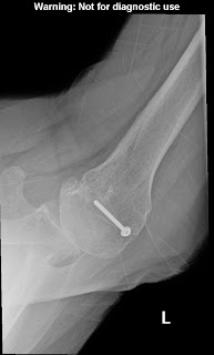
Comment: The purpose of imaging of the shoulder is to help establish the diagnosis, to determine the severity of the pathoanatomy, to help in surgical planning, and to enable the surgeon to illustrate the condition of the shoulder for the patient. Unless a specific research protocol is in place, we resist the temptation to ‘over-image’ , i.e. obtaining scans or reconstructions that are not necessary for the care of the patient such as that shown below.

The observation that CT scans may offer a few degrees of increased precision in the measurement of glenoid version does not convince us that this precision improves the quality of the surgery or the clinical outcome. Almost always standardized plain films are sufficient to garner the needed information and, as is shown below, information can be gathered from properly taken plain films that cannot be gathered on CT scans . In that proper radiographic technique (like surgical technique) is necessary to achieve the desired outcome, we take time to assure that our x-ray technologists know what we are seeking in the images.
The first key view is the anteroposterior in the plane of the scapula taken so that the x-ray beam passes through the glenohumeral joint. This view shows the superior-inferior position of the humeral head relative to the glenoid, the presence of osteophytes on the humeral head and glenoid, joint space narrowing, the degree of medial displacement of the humerus in relation to the lateral acromial line, the quality of the humeral and glenoid bone, the presence of loose bodies, and the presence of humeral head collapse or deformity.
The second key view is the axillary view taken with the arm in the functional position of elevation in the plane of the scapula and oriented so that both the spinoglenoid notch and the scapular neck are visible. This view shows a different perspective of the humeral anatomy, the amount of glenoid bone, the shape of the glenoid, its version in relation to the plane of the scapula and the relationship of the humeral head to the glenoid fossa. We have named the axillary view taken in with the arm elevated in the plane of the scapula the ‘truth’ view. This is because it demonstrates the glenohumeral relationships in the functional position of elevation; this is in contrast to CT scans, which have the disadvantage of being taken with the arm in the adducted position

Unfortunately, many of the ‘axillary views’ sent to us on patients for consultation are taken without standardization, making it impossible to determine the important features of the glenohumeral joint as shown below.

When taken properly, the standardized anteroposterior and axillary views indicate the thickness of the cartilage space between the humerus and the glenoid, the relative positions of the humeral head and the glenoid, the presence of osteophytes, the degree of osteopenia, and the extent of bony deformity and erosion.


Since arthritis usually involves the central aspect of the humeral head,


joint space narrowing is most evident on the truth view as opposed to images made with the arm at the side. Of even greater importance is the ability of the axillary ‘truth’ view to show posterior subluxation or ‘functional decentering’ that is not evident in images taken with the arm at the side.


The degree of posterior subluxation can be measured as (a) the position of the center of the humeral head in relation to the plane of the scapula, (b) the position of the center of the humeral head in relation to the glenoid face or (c) the point of contact of the humeral articular surface on the glenoid articular surface. We prefer the latter because it is this point of contact that reflects the degree of centering of the net humeral joint reaction force on the glenoid. It is the malcentering of this joint reaction force that leads to posterior instability, posterior glenoid wear and to rocking horse loosening of prosthetic glenoid components. The standardized axillary view also enables the surgeon to see the shape of the glenoid surface. Three main types have been described: concentric wear (type A)


eccentric posterior wear (type B),

and dysplastic (type C)


In actual practice, there are so many intermediate types of glenoid pathoanatomy that rigorous separation into a few distinct classes is difficult.


An important aspect of glenoid pathology is the amount of the glenoid that is involved in the pathologic concavity, known as the ‘neoglenoid. Finally, the standardized axillary view enables the measurement of the degree of glenoid retroversion in relation to the body of the scapula. Thus, on the standardized axillary view, the surgeon can usually determine the major important characteristics of glenohumeral arthritic pathoanatomy: the amount of joint space narrowing, the degree of retroversion, the degree of posterior subluxation with the arm in a functional position, the glenoid shape, the percentage of the glenoid involved in the pathologic concavity and the angle of retroversion.


Because of their low cost and freedom from metal artifacts, standardized axillary views provide a practical and reliable way to document the postoperative anatomy sequentially over time and to compare it to what was present before surgery.
A third view, the templating view, is obtained when humeral arthroplasty is being considered. This view is an anteroposterior (AP) view of the humerus taken with the arm in 30 degrees of external rotation relative to the x-ray beam with a magnification marker added. This view places the humeral neck in maximal profile and allows a comparison of proximal humeral anatomy with that of various humeral prostheses. In templating, it is important to recognize that the humeral canal is not cylindrical – the medial-lateral dimension is usually wider than the anteroposterior dimension so that the AP view may overestimate the size of the stem that will fit the diaphysis. This view is also useful for determining whether sufficient osteoporosis is present to merit special consideration at the time of arthroplasty

Advanced imaging may be useful in the unusual case where the anatomy is distorted by prior injury or surgery, when there is concern about the amount of bone available for reconstruction, or when the standardized plain films cannot be obtained. In the great majority of cases, however, the extra cost and radiation of the CT scan can be avoided through the use of these standardized plain films. In that we can learn what we need to know about the status of the rotator cuff from physical examination and plain radiographs, shoulder MRIs are rarely needed unless indicated to exclude avascular necrosis or tumor. An MRI of the neck may be useful in evaluating patients suspected of having cervical radiculopathy, myelopathy, stenosis or a syrinx.
===
Consultation for those who live a distance away from Seattle.
Click here to see the new Shoulder Arthritis Book.
Click here to see the new Rotator Cuff Book
Information about shoulder exercises can be found at this link.
Use the "Search" box to the right to find other topics of interest to you.
You may be interested in some of our most visited web pages including:shoulder arthritis, total shoulder, ream and run, reverse total shoulder, reverse total shoulder patient information, CTA arthroplasty, and rotator cuff surgery as well as the 'ream and run essentials'
See from which cities our patients come.