Showing posts with label MRI. Show all posts

Saturday, June 10, 2023

Can I have an MRI after a shoulder joint replacement?

  ChatGPT is a interactive language model developed by OpenAI, that uses a deep learning framework to understand queries and to generate coherent, contextually relevant responses based on a set of books, websites, and texts selected by researchers at OpenAI. While search engines such as Google provide links to websites that may or may not be accurate or relevant, ChatGPT provides a readable synthesis of the information from its comprehensive sources,

We sought ChatGPT responses to questions frequently posed by patients with shoulder arthritis.  These responses cannot used as specific medical advice. Patients should consult medical professionals for the most current and accurate information on their shoulder condition.


We asked "Can I have an MRI after a shoulder joint replacement?"

ChatGPT replied:

"Yes, it is generally possible to have an MRI (Magnetic Resonance Imaging) after shoulder replacement surgery. However, there are some considerations and precautions to keep in mind:

Compatibility of the implant: Most modern shoulder replacement implants are made of materials that are MRI-compatible, such as titanium, cobalt-chromium, or polyethylene. These materials do not interfere with the magnetic field or produce significant artifacts in the MRI images.

Inform the healthcare provider: It is crucial to inform your healthcare provider, including the radiologist and technician, about your shoulder replacement surgery and the presence of any implants. This allows them to select the appropriate imaging protocols and take necessary precautions during the MRI.

Implant-specific restrictions: In some cases, specific types of implants may have restrictions or precautions regarding MRI. Your orthopedic surgeon or the manufacturer of the implant can provide guidance on any specific considerations related to your implant.

Potential image distortion: While modern implants are designed to minimize image distortion, there is still a possibility of some artifacts or distortion in the MRI images near the implant. This can make it challenging to evaluate soft tissues or structures directly adjacent to the implant.

Alternative imaging options: In certain situations, if an MRI is contraindicated or poses difficulties due to the implant, alternative imaging modalities such as CT scan or ultrasound may be considered. These imaging techniques can provide valuable information about the shoulder joint and surrounding structures.It is important to have a discussion with your orthopedic surgeon and the radiologist overseeing your MRI to ensure they are aware of your shoulder replacement and can plan accordingly. They will assess the specific circumstances, evaluate the risks and benefits, and determine the most appropriate imaging approach to meet your diagnostic needs while considering the presence of the implant."

You can support cutting edge shoulder research that is leading to better care for patients with shoulder problems, click on this link.

Follow on twitter: https://twitter.com/shoulderarth
Follow on facebook: click on this link
Follow on facebook: https://www.facebook.com/frederick.matsen
Follow on LinkedIn: https://www.linkedin.com/in/rick-matsen-88b1a8133/

Here are some videos that are of shoulder interest
Shoulder arthritis - what you need to know (see this link).
How to x-ray the shoulder (see this link).
The ream and run procedure (see this link).
The total shoulder arthroplasty (see this link).
The cuff tear arthropathy arthroplasty (see this link).
The reverse total shoulder arthroplasty (see this link).
The smooth and move procedure for irreparable rotator cuff tears (see this link).
Shoulder rehabilitation exercises (see this link)

Wednesday, March 15, 2023

Reverse or anatomic total shoulder for glenohumeral arthritis - does a preoperative MRI help with the decision?

Twenty years ago, the reverse total shoulder arthroplasty (RSA) was approved by the US Food and Drug Administration "in order to restore mobility in a grossly rotator cuff deficient joint with severe arthropathy" (see this link and this link).

Since that time, the use of RSA has expanded rapidly to include "off label" indications, such as primary osteoarthritis with an intact rotator cuff (see Off-label use of reverse total shoulder arthroplasty: the American Academy of Orthopedic Surgeons Shoulder and Elbow Registry).

The application of the RSA for patients having osteoarthritis with an intact cuff can be viewed in the light of several factors:

(1) The current population-based data from the Australian Orthopaedic Association, which shows better long term survivorship for the anatomic total shoulder than for the reverse total shoulder.


(2) The risk of adverse outcomes - some of which are essentially unique to the reverse total shoulder and are difficult or impossible to revise surgically, including baseplate failure, acromial and scapular spine fractures, deltoid fatigue, and limited internal rotation (see My Reverse Has Failed: Top Five Complications and How to Manage Them). Adverse outcomes managed without surgery are not captured by studies that track revision rates.

(3) The greater average selling price for the RSA in comparison to the anatomic total shoulder (the examples below are taken from Orthopaedic Network News 2021)

                          
Because of the foregoing, the decision between anatomic and reverse total shoulder in the treatment of glenohumeral arthritis is of clinical and economic importance.

Being mindful of the original FDA approved indication for RSA - arthritis+cuff deficiency - the authors of Is Advanced Imaging to Assess Rotator Cuff Integrity Before Shoulder Arthroplasty Cost-effective? A Decision Modeling Study pointed out that for some arthritic shoulders, preoperative physical exam, radiographs, and CT scans do not adequately establish the integrity of the rotator cuff. In these shoulders there are two approaches for further elucidating the status of the cuff to inform the decision of RSA vs. anatomic TSA: (1) preoperative cuff tendon imaging (MRI or ultrasound) or (2) intraoperative assessment of the cuff, having both the RSA and anatomic TSA implants available in the operating room.

They conducted a cost-effectiveness modeling study in which all patients having shoulder arthroplasty underwent history and physical examination, radiography, and CT.  They compared the cost effectiveness of five strategies (1) no further preoperative cuff imaging, (2) selective MRI, (3) MRI for all, (4) selective ultrasound, and (5) ultrasound for all.

They used a decision model with a base-case 65-year-old patient with a 7% probability of a large-to-massive rotator cuff tear and a follow-up of 5 years. Strategies were compared using the incremental cost effectiveness ratio with a willingness to pay of both USD 50,000 and 100,000 per quality-adjusted life year. Diagnostic test sensitivity and specificity were extracted from published systematic reviews and meta-analyses, and patient utilities were obtained using the Cost-Effectiveness Analysis Registry from the Center for the Evaluation of Value and Risk in Health.

Final patient states were categorized as either inappropriate or appropriate in comparison to the actual rotator cuff integrity and type of arthroplasty performed. 

They found that MRI and MRI for all were the most cost effective additional preoperative imaging strategies. However, quality-adjusted life years gained by preoperative soft tissue imaging were minimal: 0.04 quality-adjusted life years gained for MRI for all.  The value of additional preoperative imaging increased as the age-related prevalence of cuff tear increased (rotator cuff tear prevalence greater than 12% made MRI for all cost-effective).

A secondary analysis was performed where all patients indicated for TSA underwent intraoperative rotator cuff examination to determine appropriate implant selection. This second analysis was based on the ability of the surgeon to alter the treatment plan using intraoperative rotator cuff evaluation without further preoperative imaging. This strategy was the most cost effective: least costly and achieved the greatest health utility.

They concluded that in the case of diagnostic uncertainty based on physical exam, radiographs, and CT alone, having both TSA and RSA available in the operating room would be more cost-effective than obtaining advanced soft tissue imaging preoperatively. 

However, when surgical preparedness, patient expectations, and implant availability compromise the ability to switch implants intraoperatively, performing selective MRI to assess rotator cuff integrity to indicate RSA or TSA is cost-effective 

Comment: This is an informative study, providing a clinically useful approach for considering anatomic versus reverse total shoulder for patients with arthritis based on the integrity of the rotator cuff.

A related study would be of great interest as well: comparing the cost effectiveness of anatomic versus reverse total shoulders for the treatment of glenohumeral arthritis in patients with an intact rotator cuff, considering implant cost, complications and revisions.

You can support cutting edge shoulder research that is leading to better care for patients with shoulder problems, click on this link.

Follow on twitter: https://twitter.com/shoulderarth
Follow on facebook: click on this link
Follow on facebook: https://www.facebook.com/frederick.matsen
Follow on LinkedIn: https://www.linkedin.com/in/rick-matsen-88b1a8133/

Here are some videos that are of shoulder interest
Shoulder arthritis - what you need to know (see this link).
How to x-ray the shoulder (see this link).
The ream and run procedure (see this link).
The total shoulder arthroplasty (see this link).
The cuff tear arthropathy arthroplasty (see this link).
The reverse total shoulder arthroplasty (see this link).
The smooth and move procedure for irreparable rotator cuff tears (see this link).
Shoulder rehabilitation exercises (see this link).


Sunday, August 7, 2022

Periprosthetic shoulder infection - diagnosing with metal artifact reduction MRI

Some shoulder arthroplasties are unexpectedly painful or stiff; some of these turn out to have periprosthetic infections (PJI). The most common organism causing shoulder PJI is Cutibacterium - an organism that typically does not cause systemic signs of infection or elevation of laboratory markers indicating inflammation. Attempted aspiration of fluid for culture and cell count from these joints is often unhelpful in diagnosing or excluding the diagnosis of PJI. The most reliable method for diagnosing shoulder PJI is obtaining five tissue or explant specimens for Cutibacterium-specific culturing. Obtaining these specimens requires either arthroscopic or open surgical biopsy.

The authors of Diagnostic Performance of Advanced Metal Artifact Reduction MRI for Periprosthetic Shoulder Infection sought to determine the reliability, repeatability, and diagnostic performance of advanced metal artifact reduction MRI (MARS-MRI) in diagnosing PJI.

89 patients suspected of having PJI underwent standardized clinical, radiographic, and laboratory evaluations and advanced MARS-MRI and had at least 1-year clinical follow-up. 


When applying the International Consensus Meeting 2018 criteria, 22 of the 89 participants were deemed as being infected. 


The inter-observer rreliability and intra-observer repeatability were good (k = 0.61 to 0.80) to excellent (k > 0.80) for advanced MARS-MRI findings, including lymphadenopathy


joint effusion


rotator cuff muscle edema


synovitis, extra-articular fluid collection, a sinus tract, and periprosthetic bone resorption. 


Lymphadenopathy, complex joint effusion, and edematous synovitis had sensitivities of >85%, specificities of >90%, odds ratios of >3.6, and AUC values of >0.90 for diagnosing PJI. The presence of all 3 findings together yielded a PJI probability of >99%.


Comment: This study suggests that MARS-MRI may be a useful tool for evaluating shoulders suspected of having a periprosthetic infection. It is of note, however, that in the PJI group many of the preoperative observations strongly suggested infection as shown below


In a painful shoulder with elevated ESR, CRP, fluid aspirates positive for culture or elevated WBC count, and prosthetic loosening, surgical revision would seem indicated without needing to proceed with MARS-MRI. It is reasonable to ask what the sensitivity and specificity of MARS-MRI is for PJI in patients without these findings previously shown to be strongly suggestive of infection (see for example 
Prognostic factors for bacterial cultures positive for Propionibacterium acnes and other organisms in a large series of revision shoulder arthroplasties performed for stiffness, pain, or loosening). 


Only 42 of the 89 patients had revision surgery; operative findings and intraopertive culture results were not available for the other 47. It is of interest that 14 of the 42 operated patients had intra operative cultures that were positive for Cutibacterium; while 10 of these were placed in the "infected" group, 4 were placed in the "non-infected" group. Many surgeons would find that shoulders with positive deep cultures would merit treatment for PJI.


Some cases of PJI have clinically obvious clinical presentation clearly leading to treatment of the infection; others have a stealth presentation in which cases the decision to treat infection is difficult. As emphasized by the authors of Characterizing the Propionibacterium Load in Revision Shoulder Arthroplasty the real challenge lies in diagnosing the cases of PJI having a stealth presentation. Further clinical research is necessary to determine whether MARS-MRI is of value in evaluating painful shoulder arthroplasties for which the diagnosis of PJI is not otherwise obvious. 


You can support cutting edge shoulder research that is leading to better care for patients with shoulder problems, click on this link.


Follow on twitter: https://twitter.com/shoulderarth

Follow on facebook: click on this link

Follow on facebook: https://www.facebook.com/frederick.matsen

Follow on LinkedIn: https://www.linkedin.com/in/rick-matsen-88b1a8133/

Here are some videos that are of shoulder interest
Shoulder arthritis - what you need to know (see this link).
How to x-ray the shoulder (see this link).
The ream and run procedure (see this link).
The total shoulder arthroplasty (see this link).
The cuff tear arthropathy arthroplasty (see this link).
The reverse total shoulder arthroplasty (see this link).
The smooth and move procedure for irreparable rotator cuff tears (see this link).
Shoulder rehabilitation exercises (see this link).

Sunday, December 1, 2019

MRIs are expensive, when should one be ordered?

A value-based care analysis of magnetic resonance imaging in patients with suspected rotator cuff tendinopathy and the implicated role of conservative management

These authors questioned the value of magnetic resonance imaging when it is used as the initial musculoskeletal imaging test before a trial of conservative therapy in patients with atraumatic shoulder pain, minimal to no strength deficits on examination, and suspected cuff tendinopathy.

They studied a group of 51 patients suspected to have cuff tendinopathy based on clinical findings. Every patient underwent MRI and was offered an initial trial of conservative management(education, activity modification, non-steroidal antiinflammatory drugs, and physical therapy). After a minimum of two months, patients were offered surgical intervention if they had persistent symptoms.  Patients had an average follow-up of 28.3 ± 5.3 months after imaging to determine whether surgery was performed.

46 (90.2%) patients did not go on to surgical intervention, whereas 5 (9.8%) patients did at an average 68.3 days after imaging. Four (9%) of the patients having non-operative management had full thickness cuff tears. Four (80%) of the patients having surgery had full thickness cuff tears. These results suggest that over 90.2% of patients (46 of 51) had premature MRI, posing an unnecessary economic burden of $181,619 in advanced imaging charges.

Comment: Diagnostic tests are useful when they produce "actionable intelligence", that is when they change the course of treatment. The authors observe that most patients with atraumatic shoulder pain are well managed with non-operative approaches. If these patients are given a two month trial of exercises, activity modification and mild anti-inflammatory medications, most experience symptom improvement without surgery. If MRIs are reserved for those who do not respond, costs ranging from $612 to $4181 per patient can be avoided without compromising the quality of care. 

Patients with atraumatic shoulder pain, minimal to no strength deficits on examination, and suspected cuff tendinopathy commonly present to surgeons for evaluation and management. A value-based approach to these patients would reserve MRI's for those who did not respond to non-operative management. 

==

A video of our approach to shoulders with irreparable cuff tears can be seen by clicking this link.

=====
We have a new set of shoulder youtubes about the shoulder, check them out at this link.

Be sure to visit "Ream and Run - the state of the art" regarding this radically conservative approach to shoulder arthritis at this link and this link

Use the "Search" box to the right to find other topics of interest to you.


You may be interested in some of our most visited web pages arthritis, total shoulder, ream and runreverse total shoulderCTA arthroplasty, and rotator cuff surgery as well as the 'ream and run essentials'

Sunday, August 25, 2019

Asymptomatic shoulders almost always have abnormal MRI findings

Bilateral magnetic resonance imaging findings in individuals with unilateral shoulder pain

These authors prospectively evaluated 123 individuals from the community who had self reported unilateral shoulder pain with no signs of adhesive capsulitis, no substantial range-of-motion deficit, no history of upper-limb fractures, no repeated shoulder dislocations, and no neck-related pain.

Abnormal MRI findings were highly prevalent in both shoulders. The readings of musculoskeletal radiologists and shoulder surgeon were not in good agreement, ranging from slight to moderate.


Comment: The observation that almost all asymptomatic shoulders had MRI findings of rotator cuff pathology calls into question the value of MRI in identifying clinically relevant shoulder findings.
This study indicates that the foundation for diagnosis and management of shoulder pathology rests primarily on the history and physical examination; the MRI findings may or may not relate to the patient's symptoms.

===
We have a new set of shoulder youtubes about the shoulder, check them out at this link.

Be sure to visit "Ream and Run - the state of the art"  regarding this radically conservative approach to shoulder arthritis at this link and this link

Use the "Search" box to the right to find other topics of interest to you.


You may be interested in some of our most visited web pages   arthritis, total shoulder, ream and runreverse total shoulderCTA arthroplasty, and rotator cuff surgery as well as the 'ream and run essentials'


Monday, December 17, 2018

What is the best way to image an arthritic shoulder?

Recently we met an active 70 year old man with shoulder arthritis. His Grashey view is shown here


and his axillary "truth" view (below) clearly shows posterior decentering of the humeral head on a retroverted biconcave glenoid.
Previously he had had an MRI
and a CT scan
neither of which showed the posterior decentering that was revealed by the simple "truth" view taken with the arm in a position of function, in contrast to the "advanced" imaging (MRI / CT) obtained with the arm at the side which did not reveal the functional decentering.
===

We have a new set of shoulder youtubes about the shoulder, check them out at this link.

Be sure to visit "Ream and Run - the state of the art"  regarding this radically conservative approach to shoulder arthritis at this link and this link

Use the "Search" box to the right to find other topics of interest to you.


You may be interested in some of our most visited web pages   arthritis, total shoulder, ream and runreverse total shoulderCTA arthroplasty, and rotator cuff surgery as well as the 'ream and run essentials'

Friday, May 25, 2018

Is imaging the rotator cuff prior to shoulder arthroplasty worth the cost?

Do magnetic resonance imaging and computed tomography provide equivalent measures of rotator cuff muscle size in glenohumeral osteoarthritis?

These authors assert that rotator cuff muscle volume is associated with outcomes after total shoulder
arthroplasty. They sought to determine whether computed tomography(CT) provides an equivalent measurement of cuff muscle area to a previously validated MRI measurement in 30 patients prior to total shoulder arthroplasty.

The found that the result were similar: the mean intraclass correlation coefficients were 0.989 (95% confidence interval [CI], 0.976-0.995) for the supraspinatus, 0.978 (95% CI, 0.954-0.989) for the infraspinatus–teres minor, and 0.977 (95% CI, 0.952-0.989) for the subscapularis. The mean differences were 0.2 cm2 (95% CI, 0.0-0.4 cm2) for the supraspinatus (P = .052), 0.8 cm2 (95% CI, 0.1-1.4 cm2) for the infraspinatus–teres minor (P = .029), and –0.3 cm2 (95% CI, –1.2 to 0.5 cm2) for the subscapularis (P = .407). They concluded that CT provides nearly equivalent measures of cuff muscle area to MRI technique.

Comment: While the authors state that measurements of rotator cuff muscle volume are clinically useful and that "the information provided in this study may decrease the need for redundant testing and may lower the cost of health care," it is not clear how these measurements would benefit patient care or that their cost would be justified by the benefit to the patient.

In our practice we avoid the costs of routine MRI and CT scans prior to shoulder arthroplasty in that they do not alter the care of the patient.


====
Use the "Search" box to the right to find other topics of interest to you.


You may be interested in some of our most visited web pages including:shoulder arthritis, total shoulder, ream and runreverse total shoulderCTA arthroplasty, and rotator cuff surgery as well as the 'ream and run essentials'

Sunday, December 10, 2017

How much money should we spend on imaging rotator cuffs?

Cost-effectiveness of magnetic resonance imaging versus ultrasound for the detection of symptomatic full-thickness supraspinatus tendon tears

These authors sought to determine the value of magnetic resonance imaging (MRI) and ultrasound-based imaging strategies in the evaluation of a hypothetical population with a symptomatic full-thickness supraspinatus tendon tear using formal cost-effectiveness analysis. They used a decision analytic model from the health care system perspective for 60-year-old patients with symptoms secondary to a suspected supraspinatus tear to evaluate the incremental cost-effectiveness of 3 imaging strategies during a 2-year time horizon: MRI, ultrasound, and ultrasound followed by MRI.

Their results shown below show a 0.8% increased effectiveness for MRI which costs 16.5% more than ultrasound.


Comment:  What's really interesting in this paper is the first two sentences of the introduction. "Rotator cuff tears are a common source of shoulder pain, especially for older patients, and full-thickness tendon tears have been reported in up to 21% of the general population. Surgical treatment for rotator cuff disease has increased 238% during a span of 14 years (1995-2009), accounting for approximately 250,000 operations in the United States per year."

In 2012 the Unites States population of individuals over the age of 65 years was 41,506,000 (see this link). From these numbers it can be estimated that there are 8,716,200 individuals (21% times 41,506,000) with cuff tears, but less than 3% (250,000 divided by 8,716,200) of these receive surgery each year. One must ask, "how important is imaging in the decision to perform surgery, what percent of the population should have ultrasound or MRI to evaluate cuff integrity?" The reader can do the math of multiplying the cost of imaging by the number of folks at risk for having cuff tears.

The decision to attempt a cuff repair on a patient needs to be highly individualized. Shared patient-surgeon decision making needs to be based on findings that have been shown to be encouraging or discouraging about the prospect of the shoulder having a durably reparable cuff tear. It is of interest that many of these factors can be determined without ultrasound or MRI. We first published these guidelines in 1994 and have found them as useful today as back then. Note that the decision is based on considering the patient as well as the shoulder.

ENCOURAGING                                    DISCOURAGING

History 
Age less than 55                                        Age over 65
Acute traumatic onset                                Insidious, atraumatic onset
No relation to work                                   Attribution of tear to work
Short duration of weakness                       Weakness over 6 weeks
No history of smoking                               Many smoking pack-years
No steroid injections                                  Repeated steroid injections
No major medications                                Steroids/antimetabolites
No concurrent disease                                Inflammatory joint disease
No infections                                              History of previous infection
No previous shoulder surgery                     Previous cuff surgery
Benign surgical history                               History of failed tissue repairs

Physical Examination 
Good nutrition                                             Poor nutrition/obesity
Mild weakness                                            Severe weakness
No spinatus atrophy                                    Severe spinatus atrophy
Stable shoulder                                           Anterior superior instability
Intact acromion                                           Previous acromioplasty
No stiffness                                                 Stiffness

Radiographs 
Normal radiographs                                    Upwards head displacement
                                                                    Cuff tear arthropathy

MRI or Ultrasound 
Good tendon quality                                   Thin tendon
One tendon tear                                          Multiple tendon involvement
Small gap to close                                       Severe retraction

In our practice, we are depending less and less on rotator cuff imaging and more and more on the factors that can be discerned from a good history, physical examination, and plain radiographs.


====
The reader may also be interested in these posts:


Consultation for those who live a distance away from Seattle.

Click here to see the new Shoulder Arthritis Book.

Click here to see the new Rotator Cuff Book

Information about shoulder exercises can be found at this link.

Use the "Search" box to the right to find other topics of interest to you.

You may be interested in some of our most visited web pages including:shoulder arthritis, total shoulder, ream and runreverse total shoulderCTA arthroplasty, and rotator cuff surgery as well as the 'ream and run essentials'

See from which cities our patients come.

See the countries from which our readers come on this post.

Monday, May 15, 2017

Imaging the arthritic glenoid

Can glenoid wear be accurately assessed using x-ray imaging? Evaluating agreement of x-ray and magnetic resonance imaging Walch classification

These authors point out that glenohumeral arthritic pathoanatomy is often evaluated using advanced imaging, which is more expensive and less practical than plain radiographs. 

They compared the assessment of the Walch glenoid type using plain x-rays to that using magnetic resonance imaging in 50 patients assessed for shoulder arthroplasty by 5 raters. The inter-rater agreement for x-ray images and MRIs was “moderate” (κ = 0.42 and κ = 0.47, respectively) for the 5-category Walch classification (A1, A2, B1, B2, C) and “moderate” (κ = 0.54 and κ = 0.59, respectively) for the 3-category Walch classification (A, B, C). The agreement between x-ray images and consensus MRI was much lower: “fair-to-moderate” (κ = 0.21-0.51) for the 5-category and “moderate” (κ = 0.36-0.60) for the 3-category Walch classification.
Based on these results the authors concluded that x-ray images are inferior to advanced imaging when assessing glenoid wear.

Comment: The results of this paper do not appear to support its conclusions. The inter-rater agreement for both x-rays and MRI's were essentially identical. 

There are some other important issues in this paper.
First, the axillary views were not taken in a standardized manner (see our recommended technique here).
Second, the example shown in the paper shows the identical glenoid type for both the axillary view and the MRI (even though the authors conclude that the x-ray image is classified as type B2, but magnetic resonance imaging reveals a type C glenoid). No new information was gained from spending the money on the MRI.


Finally, it has not been shown that shoulder arthroplasties performed after expensive imaging (MRI or CT) have superior outcomes to those performed with the less expensive standardized plain radiographs. 

More on this topic here:

Radiographs and computed tomography scans show similar observer agreement when classifying glenoid morphology in glenohumeral arthritis

These authors observe that the Walch classification provides a useful frame of reference when assessing subluxation and glenoid morphology in primary glenohumeral osteoarthritis.


They compared the use of computed tomography (CT) and axillary radiographs to determine arthritic glenoid pathoanatomy (Walch type) in 75 consecutive shoulders with primary glenohumeral osteoarthritis.

The average intraobserver agreement for radiographs was 0.66.
The average intraobserver agreement for CT scans was 0.60.
Pairwise comparisons between observers showed higher agreement for radiographs than for CT scans (0.48 vs. 0.39).

The average agreement for observations on radiographs and CT scans was 0.42 (moderate; 0.40, 0.37, and 0.50).

In their study, the B2 glenoid was found in 40% of the cases.


In their study intraobserver agreement using the Walch classification based on axillary radiographs was substantial and compared favorably with agreement based on CT scans.

Comment: The purpose of imaging of the shoulder is to help establish the diagnosis, to determine the severity of the pathoanatomy, to help in surgical planning, and to enable the surgeon to illustrate the condition of the shoulder for the patient. Unless a specific research protocol is in place, we resist the temptation to ‘over-image’ , i.e. obtaining scans or reconstructions that are not necessary for the care of the patient such as that shown below.


The observation that CT scans may offer a few degrees of increased precision in the measurement of glenoid version does not convince us that this precision improves the quality of the surgery or the clinical outcome. Almost always standardized plain films are sufficient to garner the needed information and, as is shown below, information can be gathered from properly taken plain films that cannot be gathered on CT scans . In that proper radiographic technique (like surgical technique) is necessary to achieve the desired outcome, we take time to assure that our x-ray technologists know what we are seeking in the images.

The first key view is the anteroposterior in the plane of the scapula taken so that the x-ray beam passes through the glenohumeral joint. This view shows the superior-inferior position of the humeral head relative to the glenoid, the presence of osteophytes on the humeral head and glenoid, joint space narrowing, the degree of medial displacement of the humerus in relation to the lateral acromial line, the quality of the humeral and glenoid bone, the presence of loose bodies, and the presence of humeral head collapse or deformity.

The second key view is the axillary view taken with the arm in the functional position of elevation in the plane of the scapula and oriented so that both the spinoglenoid notch and the scapular neck are visible. This view shows a different perspective of the humeral anatomy, the amount of glenoid bone, the shape of the glenoid, its version in relation to the plane of the scapula and the relationship of the humeral head to the glenoid fossa. We have named the axillary view taken in with the arm elevated in the plane of the scapula the ‘truth’ view. This is because it demonstrates the glenohumeral relationships in the functional position of elevation; this is in contrast to CT scans, which have the disadvantage of being taken with the arm in the adducted position


Unfortunately, many of the ‘axillary views’ sent to us on patients for consultation are taken without standardization, making it impossible to determine the important features of the glenohumeral joint as  shown below.


When taken properly, the standardized anteroposterior and axillary views indicate the thickness of the cartilage space between the humerus and the glenoid, the relative positions of the humeral head and the glenoid, the presence of osteophytes, the degree of osteopenia, and the extent of bony deformity and erosion.


Since arthritis usually involves the central aspect of the humeral head,


joint space narrowing is most evident on the truth view as opposed to images made with the arm at the side. Of even greater importance is the ability of the axillary ‘truth’ view to show posterior subluxation or ‘functional decentering’ that is not evident in images taken with the arm at the side.


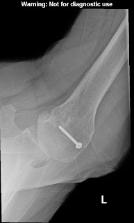


The degree of posterior subluxation can be measured as (a) the position of the center of the humeral head in relation to the plane of the scapula, (b) the position of the center of the humeral head in relation to the glenoid face or (c) the point of contact of the humeral articular surface on the glenoid articular surface. We prefer the latter because it is this point of contact that reflects the degree of centering of the net humeral joint reaction force on the glenoid. It is the malcentering of this joint reaction force that leads to posterior instability, posterior glenoid wear and to rocking horse loosening of prosthetic glenoid components. The standardized axillary view also enables the surgeon to see the shape of the glenoid surface. Three main types have been described: concentric wear (type A)


eccentric posterior wear (type B),

and dysplastic (type C)


In actual practice, there are so many intermediate types of glenoid pathoanatomy that rigorous separation into a few distinct classes is difficult. 


 An important aspect of glenoid pathology is the amount of the glenoid that is involved in the pathologic concavity, known as the ‘neoglenoid. Finally, the standardized axillary view enables the measurement of the degree of glenoid retroversion in relation to the body of the scapula. Thus, on the standardized axillary view, the surgeon can usually determine the major important characteristics of glenohumeral arthritic pathoanatomy: the amount of joint space narrowing, the degree of retroversion, the degree of posterior subluxation with the arm in a functional position, the glenoid shape, the percentage of the glenoid involved in the pathologic concavity and the angle of retroversion.


 Because of their low cost and freedom from metal artifacts, standardized axillary views provide a practical and reliable way to document the postoperative anatomy sequentially over time and to compare it to what was present before surgery.


A third view, the templating view, is obtained when humeral arthroplasty is being considered. This view is an anteroposterior (AP) view of the humerus taken with the arm in 30 degrees of external rotation relative to the x-ray beam with a magnification marker added. This view places the humeral neck in maximal profile and allows a comparison of proximal humeral anatomy with that of various humeral prostheses. In templating, it is important to recognize that the humeral canal is not cylindrical – the medial-lateral dimension is usually wider than the anteroposterior dimension so that the AP view may overestimate the size of the stem that will fit the diaphysis. This view is also useful for determining whether sufficient osteoporosis is present to merit special consideration at the time of arthroplasty 


Advanced imaging may be useful in the unusual case where the anatomy is distorted by prior injury or surgery, when there is concern about the amount of bone available for reconstruction, or when the standardized plain films cannot be obtained. In the great majority of cases, however, the extra cost and radiation of the CT scan can be avoided through the use of these standardized plain films. In that we can learn what we need to know about the status of the rotator cuff from physical examination and plain radiographs, shoulder MRIs are rarely needed unless indicated to exclude avascular necrosis or tumor. An MRI of the neck may be useful in evaluating patients suspected of having cervical radiculopathy, myelopathy, stenosis or a syrinx.

===

Consultation for those who live a distance away from Seattle.

Click here to see the new Shoulder Arthritis Book.

Click here to see the new Rotator Cuff Book

Information about shoulder exercises can be found at this link.

Use the "Search" box to the right to find other topics of interest to you.

You may be interested in some of our most visited web pages including:shoulder arthritis, total shoulder, ream and runreverse total shoulderreverse total shoulder patient information,  CTA arthroplasty, and rotator cuff surgery as well as the 'ream and run essentials'

See from which cities our patients come.