


One year ago he received a total shoulder arthroplasty using a standard (non-augmented) glenoid inserted without particular attempt to modify his glenoid version. Recently he returned to the office with excellent shoulder motion, an SST score of 8 out of 12, and these x-rays.
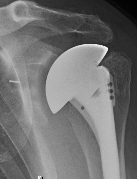
His axillary "truth" view is directly comparable to his preoperative view and shows centering of the humeral head prosthesis on a standard all-polyethylene glenoid component inserted without attempt to change glenoid version. Note the glenoid component is well supported on bone that has been minimally reamed.

Does Postoperative Glenoid Retroversion Affect the 2-Year Clinical and Radiographic Outcomes for Total Shoulder Arthroplasty?
While glenoid retroversion and posterior humeral head decentering are common preoperative features of severely arthritic glenohumeral joints, the relationship of postoperative glenoid component retroversion to the clinical results of total shoulder arthroplasty (TSA) is unclear. Studies have indicated concern for inferior outcomes when glenoid components are inserted in 15° or more retroversion.
In a population of patients undergoing TSA in whom no specific efforts were made to change the version of the glenoid, these authors asked whether at 2 years after surgery patients having glenoid components implanted in 15° or greater retroversion had (1) less improvement in the Simple Shoulder Test (SST) score and lower SST scores; (2) higher percentages of central peg lucency, higher Lazarus radiolucency grades, higher mean percentages of posterior decentering, and more frequent central peg perforation; or (3) a greater percentage having revision for glenoid component failure compared with patients with glenoid components implanted in less than 15° retroversion. They examined the records of 201 TSAs performed using a standard all-polyethylene pegged glenoid component

inserted after conservative glenoid reaming without specific attempt to modify preoperative glenoid version.

Of these, 171 (85%) patients had SST scores preoperatively and between 18 and 36 months after surgery. Ninety-three of these patients had preoperative radiographs in the database and immediate postoperative radiographs and postoperative radiographs taken in a range of 18 to 30 months after surgery. Twenty-two patients had radiographs that were inadequate for measurement at the preoperative, immediate postoperative, or latest followup time so that they could not be included. In comparison to those included in the analysis, the excluded patients did not have substantially different mean age, sex distribution, time of followup, distribution of diagnoses, American Society of Anesthesiologists class, alcohol use, smoking history, BMI, history of prior surgery or preoperative glenoid version. They analyzed the two year outcomes in the remaining 71 TSAs, comparing the 21 in the retroverted group (the glenoid component was implanted in 15° or greater retroversion (mean ± SD, 20.7° ± 5.3°)) with the 50 in the non-retroverted group ( the glenoid component was implanted in less than 15° retroversion (mean ± SD, 5.7° ± 6.9°)).
The mean (± SD) improvement in the SST (6.7 ± 3.6; from 2.6 ± 2.6 to 9.3 ± 2.9) for the retroverted group was not inferior to that for the nonretroverted group (5.8 ± 3.6; from 3.7 ± 2.5 to 9.4 ± 3.0). The percent of maximal possible improvement (%MPI) for the retroverted glenoids (70% ± 31%) was not inferior to that for the nonretroverted glenoids (67% ± 44%). The 2-year SST scores for the retroverted (9.3 ± 2.9) and the nonretroverted glenoid groups (9.4 ± 3.0) were similar (mean difference, 0.2; 95% CI, - 1.1 to 1.4; p = 0.697). No patient in either group reported symptoms of subluxation or dislocation. The radiographic results for the retroverted glenoid group were similar to those for the nonretroverted group with respect to central peg lucency (four of 21 [19%] versus six of 50 [12%]; p = 0.436; odds ratio, 1.7; 95% CI, 0.4-6.9), average Lazarus radiolucency scores (0.5 versus 0.7, Mann-Whitney U p value = 0.873; Wilcoxon rank sum test W = 512, p value = 0.836), and the mean percentage of posterior humeral head decentering (3.4% ± 5.5% versus 1.6% ± 6.0%; p = 0.223). The percentage of patients with retroverted glenoids undergoing revision (0 of 21 [0%]) was not inferior to the percentage of those with nonretroverted glenoids (three of 50; [6%]; p = 0.251).
The authors concluded that in this series of TSAs, postoperative glenoid retroversion was not associated with inferior clinical results at 2 years after surgery.
Glenoid retroversion is a relatively common finding in arthritic glenohumeral joints coming to shoulder arthroplasty. Shoulders with preoperative glenoid retroversion tend to have poorer preoperative shoulder comfort and function, posterior decentering, and glenoid biconcavity, all indicating a more severe form of the disease. There is currently great interest in methods for altering this glenoid retroversion that is commonly found in osteoarthritic glenohumeral joints. Methods used include posterior glenoid bone grafts, reaming the anterior aspect of the glenoid, and posteriorly augmented glenoid components. This study reports the two year results of a more conservative approach in which minimal glenoid bone is removed by reaming and specific attempts to alter glenoid version are not used.
Here is the two year radiographic followup on a 55 year old patient from our practice. Preoperative films show a type B2 genoid with retroversion, biconcavity and posterior humeral subluxation.


Here are the 2 year films of this shoulder after conservative shoulder arthroplasty using a standard glenoid component without attempts to modify glenoid version. The humeral head is centered in the prosthetic glenoid. At two years after surgery the patient was able to perform all 12 functions of the Simple Shoulder Test.


Note that sufficient bone stock remains to perform a revision total or a reverse total shoulder arthroplasty shoulder these procedures become necessary in the future of this young person.
Long term followup of well-characterized patients treated with the different methods for managing glenoid retroversion will be required to define the relative risks, benefits, effectiveness and durability of each of them.
=
Long term followup of well-characterized patients treated with the different methods for managing glenoid retroversion will be required to define the relative risks, benefits, effectiveness and durability of each of them.
=
We have a new set of shoulder youtubes about the shoulder, check them out at this link.
Be sure to visit "Ream and Run - the state of the art" regarding this radically conservative approach to shoulder arthritis at this link and this link
Use the "Search" box to the right to find other topics of interest to you.
You may be interested in some of our most visited web pages arthritis, total shoulder, ream and run, reverse total shoulder, CTA arthroplasty, and rotator cuff surgery as well as the 'ream and run essentials'