Surgeons and patients need to understand the anatomical characteristics of the arthritic shoulder before treatment is discussed. While there is current enthusiasm for routinely obtaining CT scans for this purpose, in the great majority of cases a pair of plain x-rays can provide all the necessary information without the time, cost and radiation exposure resulting from a CT scan.
As in all things orthopaedic, the technical detains are essential for obtaining the desired information.
The first of the pair is the anteroposterior view in the plane of the scapula. In this view, the patient's scapula is placed flat on the x-ray cassette while the x-ray beam is perpendicular to the plane of the scapula and aimed at the coracoid. The forearm of the flexed elbow is rotated 30 degrees from the x-ray beam in order to show the humeral head in profile.
The resulting view reveals such pathological features as flattening of the humeral head, humeral osteophytes, joint space narrowing, glenoid wear, and the superior/inferior relationship of the humeral head to the glenoid and the acromion.
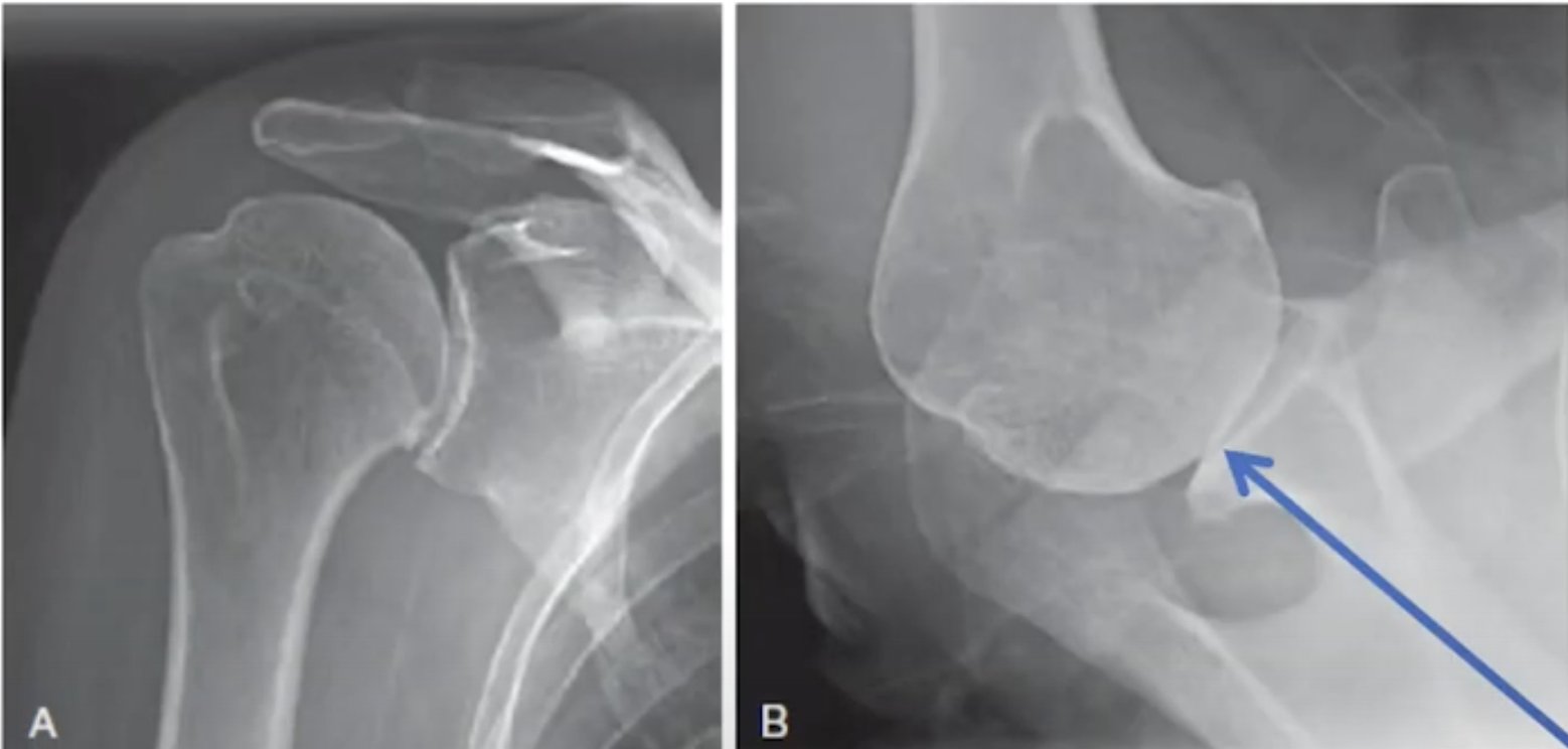
The second of the pair is the axillary "truth" view. In this view, the patient's arm is elevated in the plane of the scapula while the beam is oriented in the plane of the scapula. Proper orientation is confirmed by visualizing the "eye" of the spinoglenoid notch (blue arrow).
The resulting view reveals such pathological features as the version of the glenoid in relation to the scapular body, flattening or biconcavity of the glenoid face, medial glenoid erosion, glenoid and humeral osteophytes, and the degree of decentering of the humeral head on the face of the glenoid.
This information cannot be obtained from an improperly oriented image such as that shown below.
A properly done axillary "truth" view can be used to identify the variation in glenoid types.
Shown below are a few examples of pairs of x-rays that provided the necessary and sufficient information to plan and execute definitive surgical management of the arthritic shoulder.

One of the great values of standardized axillary "truth" views is the ability to compare preoperative and postoperative relationships, something that is not practical with CT scans. See the examples below, recognizing how comparable the views are from before and after surgery (note the "eyes" indicated by arrows).
While preoperative CT scans may be useful for characterizing complex glenohumeral arthritic anatomy, the value to the patient of routine CT scans in most cases of glenohumeral arthritis has yet to be demonstrated.
You can support cutting edge shoulder research that is leading to better care for patients with shoulder problems, click on this link.
Follow on twitter: https://twitter.com/shoulderarth
Follow on facebook: click on this link
Follow on facebook: https://www.facebook.com/frederick.matsen
Follow on LinkedIn: https://www.linkedin.com/in/rick-matsen-88b1a8133/
Here are some videos that are of shoulder interest
Shoulder arthritis - what you need to know (see this link).
How to x-ray the shoulder (see this link).
The ream and run procedure (see this link).
The total shoulder arthroplasty (see this link).
The cuff tear arthropathy arthroplasty (see this link).
The reverse total shoulder arthroplasty (see this link).
The smooth and move procedure for irreparable rotator cuff tears (see this link).
Shoulder rehabilitation exercises (see this link).