Showing posts with label X-ray. Show all posts

Thursday, July 10, 2025

Preoperative planning for reverse total shoulder using plain films.

A major component of the success of reverse total shoulder arthroplasty is the position and orientation of the base plate on the glenoid bone. While many different planning systems are available, the challenge is transferring the plan to the patient without relying on expensive and time-consuming approaches, such patient specific instrumentation or augmented reality.

Our usual target is to have the inferior edge of the baseplate at the inferior aspect of the reamed glenoid and tilted inferiorly with the central screw or peg inclined so that it is parallel to the floor of the supraspinous fossa. 

For routine cases, we plan for rTSA using plain x-rays obtained in the plane of the scapula (Grashey view). The scaled image is uploaded to the universally available PACS (Picture Archiving and Communication System). PACS tools are used to find the location of insertion point and the inclination of the drill for the central screw or post. These scaled measurements are then used to position and orient the drill on the patient's glenoid in the operating room. This generic approach does not encourage the surgeon to select implants from any particular company.

Here's an example: a 70 year old man with a massive irreparable cuff tear and pseudo paralysis. 

On the PACS screen a line segment (yellow) with a length equal to the radius of the base plate is drawn perpendicular to the floor of the supraspinatus fossa (red line) from the glenoid articular surface to the glenoid neck. The upper end of this line is the insertion point (yellow dot)


The distance of this point along the glenoid articular surface (green line) from the inferior glenoid lip is measured on the scaled PACS image. The insertion point can be found at surgery by measuring this distance using a flexible ruler.


The angle of drill insertion (dotted gold line) relative to the joint surface at the insertion point (black line) is noted on PACS and duplicated at surgery.


Favard et al have described four types of glenoid pathoanatomy in cuff tear arthropathy.  


For some cases, such as types E1 and E3, this planning approach may indicate that excessive reaming of inferior glenoid bone would be necessary to achieve the desired baseplate position and orientation. In such cases superior bone grafting or an augmented baseplate may be called for.

While more sophisticated proprietary systems can be used for more complex pathoanatomy, this generic approach can be effective for a large percentage of rTSA cases.

We use a similar plain films/PACS approach to planning a stemless anatomic arthroplasty - see this link.

Sometimes a convenient solution is staring right at us


Sooty Grouse
Mt. Rainier
July 4, 2025

You can support cutting edge shoulder research that is leading to better care for patients with shoulder problems, click on this link

Follow on twitter/X: https://x.com/RickMatsen
Follow on facebook: https://www.facebook.com/shoulder.arthritis
Follow on LinkedIn: https://www.linkedin.com/in/rick-matsen-88b1a8133/

Here are some videos that are of shoulder interest
Shoulder arthritis - what you need to know (see this link).
How to x-ray the shoulder (see this link).
The ream and run procedure (see this link).
The total shoulder arthroplasty (see this link).
The cuff tear arthropathy arthroplasty (see this link).
The reverse total shoulder arthroplasty (see this link).
The smooth and move procedure for irreparable rotator cuff tears (see this link)
Shoulder rehabilitation exercises (see this link).

Saturday, January 14, 2023

How to define the pathoanatomy of the arthritic shoulder using a pair of plain x-rays

Surgeons and patients need to understand the anatomical characteristics of the arthritic shoulder before treatment is discussed. While there is current enthusiasm for routinely obtaining CT scans for this purpose, in the great majority of cases a pair of plain x-rays can provide all the necessary information without the time, cost and radiation exposure resulting from a CT scan. 

As in all things orthopaedic, the technical detains are essential for obtaining the desired information.

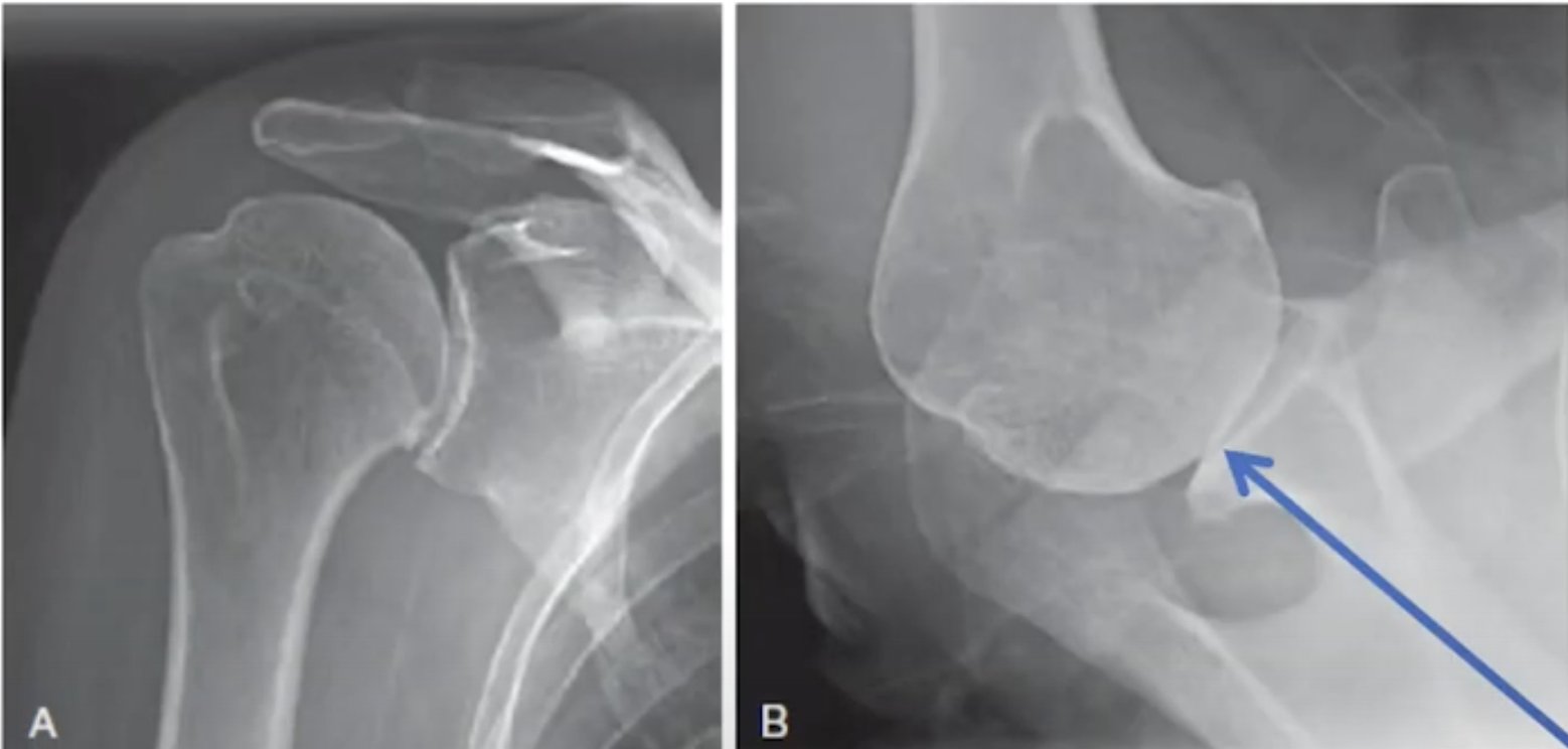
The first of the pair is the anteroposterior view in the plane of the scapula. In this view, the patient's scapula is placed flat on the x-ray cassette while the x-ray beam is perpendicular to the plane of the scapula and aimed at the coracoid.  The forearm of the flexed elbow is rotated 30 degrees from the x-ray beam in order to show the humeral head in profile.


The resulting view reveals such pathological features as flattening of the humeral head, humeral osteophytes, joint space narrowing, glenoid wear, and the superior/inferior relationship of the humeral head to the glenoid and the acromion.


The second of the pair is the axillary "truth" view. In this view, the patient's arm is elevated in the plane of the scapula while the beam is oriented in the plane of the scapula. Proper orientation is confirmed by visualizing the "eye" of the spinoglenoid notch (blue arrow). 


The resulting view reveals such pathological features as the version of the glenoid in relation to the scapular body, flattening or biconcavity of the glenoid face, medial glenoid erosion, glenoid and humeral osteophytes, and the degree of decentering of the humeral head on the face of the glenoid. 


This information cannot be obtained from an improperly oriented image such as that shown below. 


A properly done axillary "truth" view can be used to identify the variation in glenoid types.


Shown below are a few examples of pairs of x-rays that provided the necessary and sufficient information to plan and execute definitive surgical management of the arthritic shoulder.


One of the great values of standardized axillary "truth" views is the ability to compare preoperative and postoperative relationships, something that is not practical with CT scans.  See the examples below, recognizing how comparable the views are from before and after surgery (note the "eyes" indicated by arrows).


While preoperative CT scans may be useful for characterizing complex glenohumeral arthritic anatomy, the value to the patient of routine CT scans in most cases of glenohumeral arthritis has yet to be demonstrated. 

You can support cutting edge shoulder research that is leading to better care for patients with shoulder problems, click on this link.

Follow on twitter: https://twitter.com/shoulderarth
Follow on facebook: click on this link
Follow on facebook: https://www.facebook.com/frederick.matsen
Follow on LinkedIn: https://www.linkedin.com/in/rick-matsen-88b1a8133/

Here are some videos that are of shoulder interest
Shoulder arthritis - what you need to know (see this link).
How to x-ray the shoulder (see this link).
The ream and run procedure (see this link).
The total shoulder arthroplasty (see this link).
The cuff tear arthropathy arthroplasty (see this link).
The reverse total shoulder arthroplasty (see this link).
The smooth and move procedure for irreparable rotator cuff tears (see this link).
Shoulder rehabilitation exercises (see this link).

Tuesday, August 31, 2021

Is pain and function worse for shoulders with more severe arthritis?

Radiographic Severity May Not be Associated with Pain and Function in Glenohumeral Arthritis


These authors asked (1) What radiographic
findings are associated with worse pain and function in patients with glenohumeral osteoarthritis? (2) What demographic factors are associated with worse pain and function in patients with glenohumeral osteoarthritis? They conducted a retrospective study that included 3133 patients presenting for an initial office visit for primary glenohumeral osteoarthritis. 

59% (1860) had assessments of patient assessed shoulder comfort and function; 48% (893) of those had radiographs.  42% (378) of those with radiographs were excluded because of other shoulder findings, recent injection, prior surgery, or inadequate radiographs, leaving 16% (515 of 3133) who were fully analyzed in this study. 


The radiographic analysis included the joint space width, posterior humeral head subluxation, inferior humeral head osteophyte size, cystic change, and head asphericity. 


The arthritis was classified according to the Walch, Samilson-Prieto, and Kellgren-Lawrence classifications by two separate reviewers. 


Radiographic and demographic criteria as well as the presence of psychologic or mental illness were correlated with VAS Pain,, American Shoulder and Elbow Surgeons, Single Assessment Numeric Evaluation, and Simple Shoulder Test scores using univariate and multivariable regression analyses.


After accounting for age, gender, and psychologic illness in the multivariable analysis, shoulders with worse arthritis (Samilson-Prieto Grade 4 arthrosis) had lower VAS Pain scores than those with less severearthritis (Grade 0 or 1). Otherwise, the authors found no clinically important associations were found between Samilson-Prieto Grade 4, Kellgren-Lawrence Grade 3, radiographic joint space, posterior subluxation, and glenoid type; in other words, the functional levels of patients with glenohumeral arthritis did not parallel radiographic severity. 


Below is a table showing the multivariate results for the SST.


Below is a table showing the results for glenoid type.


Comment: The findings of this paper are most interesting and challenge the concept that shoulders with worse arthritis would have more pain and functional loss. This result is consistent with that reported in Prearthroplasty glenohumeral pathoanatomy and its relationship to patient's sex, age, diagnosis, and self-assessed shoulder comfort and function:Shoulders with glenoid types B1 and B2 and those with more decentering did not have worse self-assessed shoulder comfort and function than the radiographically less involved A1 types.


This leads us to some important questions:

What is the best treatment for a patient with severe pain and minimal arthritic changes on radiographs? 


Does the surgeon need to extend the workup to assure that the pain is coming from the glenohumeral joint and not from other sources? Does the patient have a low pain threshold that may portend a painful arthroplasty? What is the likely outcome for the patient having an 
arthroplasty for radiographically mild arthritis (note that in some series the postarthroplasty functional outcomes for A1 glenoids are not better than, if as good as, those for B2 glenoids - see Anatomic Total Shoulder Arthroplasty with All-Polyethylene Glenoid Component for Primary Osteoarthritis with Glenoid Deficiencies


It seems that the surgeon and patient can be most confident in the outcome when the pain and degree of functional loss match up with the degree of radiographic involvement. 


Follow on twitter: https://twitter.com/shoulderarth

Follow on facebook: https://www.facebook.com/frederick.matsen

Follow on LinkedIn: https://www.linkedin.com/in/rick-matsen-88b1a8133/


How you can support research in shoulder surgery Click on this link.


Here are some videos that are of shoulder interest
Shoulder arthritis - what you need to know (see this link)
The smooth and move for irreparable cuff tears (see this link)
The total shoulder arthroplasty (see this link).
The ream and run technique is shown in this link.
The cuff tear arthropathy arthroplasty (see this link).
The reverse total shoulder arthroplasty (see this link).
Shoulder rehabilitation exercises (see this link).

Monday, August 3, 2020

Ream and run - imaging the shoulder before and after surgery

Standardized imaging of the shoulder before and after the ream and run is important both for preoperative planning and for evaluating the arthroplasty. We use three views, as shown below: the AP in the plane of the scapula (Grashey view) on the left; the axillary "truth" view in the center, and the AP templating view on the right.


The AP templating view gives a good view of the humeral canal to anticipate any issues with placement of the stem as shown in the three examples below 


The AP in the plane of the scapula or Grashey is used for identifying medialization relative to the lateral acromial line, bone stock, glenoid inclination, foreign bodies and the size of the osteophytes.


This view is also used to evaluate proximal deformities that may require special placement of the humeral component.


And situations in which the humeral head cannot be dislocated safely so that an in situ osteotomy may be required.


The axillary "truth" view is used for evaluating the centering of the humeral head on the glenoid


Using this view, the centering can be compared before and after surgery


The axillary "truth" view is used to evaluate the glenoid bone stock and version relative to the plane of the scapula.


It can also be used to identify the glenoid type


However, we have found that the glenoid type is not an important consideration in the ream and run procedure, because in all cases the goal of reaming is to create a single glenoid concavity with maximal preservation of glenoid bone; changing glenoid version is not a priority and does not seem necessary for achieving stability or for a good functional result


Using these views we have not found that preoperative CT scans or 3D CT planning are needed.

=====
To see a YouTube video on how the ream and run is done, click on this link.

===

We have a new set of shoulder youtubes about the shoulder, check them out at this link.

Be sure to visit "Ream and Run - the state of the art" regarding this radically conservative approach to shoulder arthritis at this link and this link

Use the "Search" box to the right to find other topics of interest to you.


You may be interested in some of our most visited web pages  arthritis, total shoulder, ream and runreverse total shoulderCTA arthroplasty, and rotator cuff surgery as well as the 'ream and run essentials'


Thursday, July 20, 2017

How bad is the shoulder arthritis on x-ray?

Reliability of radiologic glenohumeral osteoarthritis classifications

These authors sought to determine the intraobserver and interobserver reliability of glenohumeral osteoarthritis classification schemes based on plain anteroposterior radiographs of 108 shoulder joints from 96 consecutive patients with glenohumeral osteoarthritis. On two occasions each of two observers graded the film into 6 classification systems. The intraobserver and interobserver reliabilities were 0.907 (observer 1), 0.965 (observer 2), and 0.851 (interobserver) for the Samilson-Prieto grading system;
0.954, 0.948, and 0.869 for the Allain modification;
0.936, 0.830, and 0.791 for the Gerbermodification;
0.887, 0.892, and 0.744 for theKellgren and Lawrence classification;
0.873, 0.964, and 0.935 for theWeinstein; and
0.854, 0.934, and 0.797 for the Guyette grading system.

Comment: Some surgeons and clinical investigators are interested using radiographic classification systems for demographic and outcomes studies.  However, radiographic evaluation is incomplete if it is based on the AP view only.

This is demonstrated by the films of a man in his mid 40s who presented to us with shoulder pain and limited motion. His anteroposterior x-ray suggested mild to moderate arthritis.

However, the 'truth' view (a standardized axillary taken with the arm in the functional position of elevation in the plane of the scapula - see this link), revealed a retroverted, biconcave glenoid with posterior humeral decentering = the bad arthritic triad (BAT - see this link), a much greater degree of pathology than what was suggested on the AP view.

Here are some other cases in which the 'truth' view demonstrated substantially greater pathoanatomy than what could be seen on the AP view


We find that the standardized AP and 'truth' axillary view taken as described in this link usually provide all the necessary imaging information to characterize the arthritic pathoanatomy and plan treatment.

===
=====
The reader may also be interested in these posts:


Information about shoulder exercises can be found at this link.

Use the "Search" box to the right to find other topics of interest to you.

You may be interested in some of our most visited web pages including:shoulder arthritis, total shoulder, ream and runreverse total shoulderCTA arthroplasty, and rotator cuff surgery as well as the 'ream and run essentials'