Showing posts with label biconcavity. Show all posts

Saturday, January 14, 2023

How to define the pathoanatomy of the arthritic shoulder using a pair of plain x-rays

Surgeons and patients need to understand the anatomical characteristics of the arthritic shoulder before treatment is discussed. While there is current enthusiasm for routinely obtaining CT scans for this purpose, in the great majority of cases a pair of plain x-rays can provide all the necessary information without the time, cost and radiation exposure resulting from a CT scan. 

As in all things orthopaedic, the technical detains are essential for obtaining the desired information.

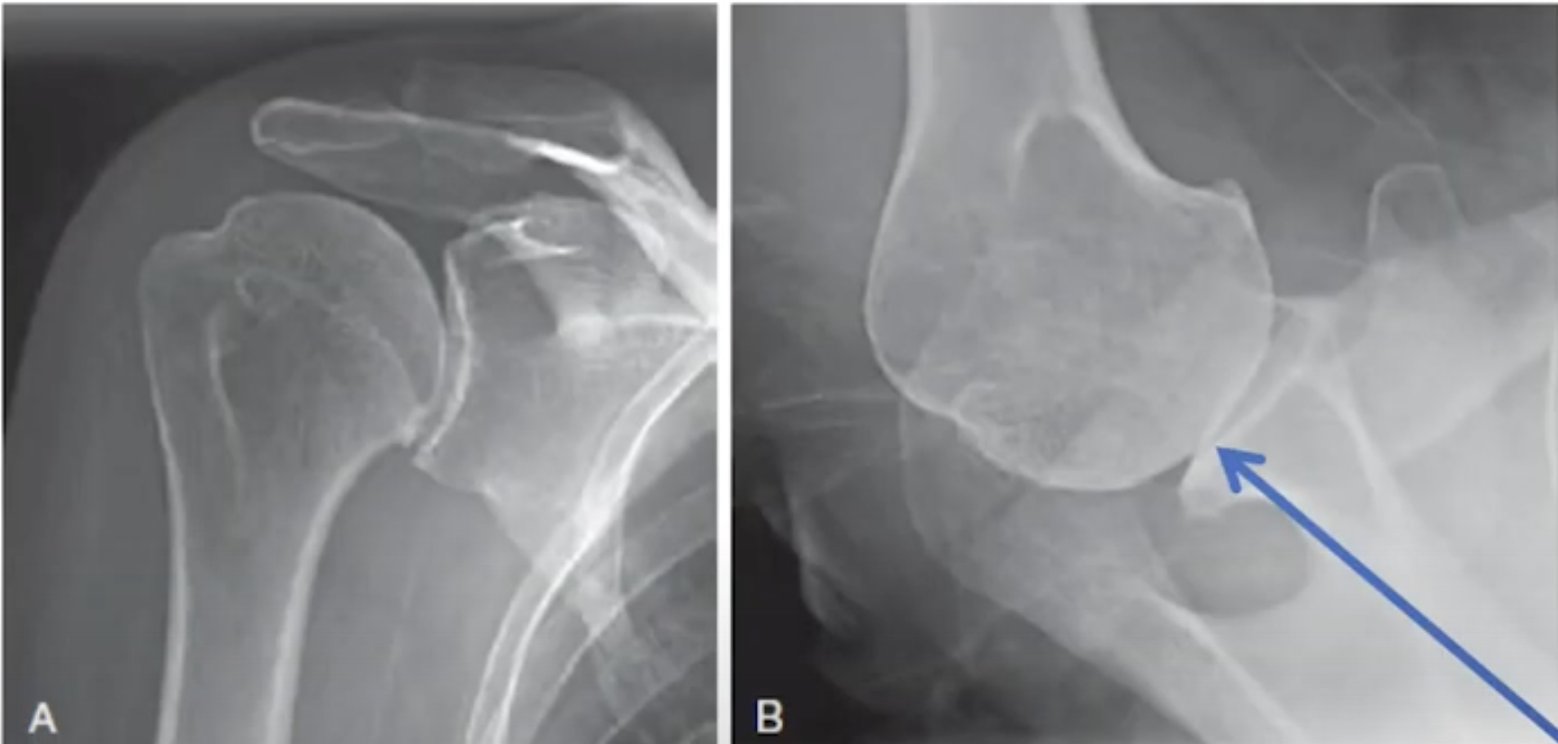
The first of the pair is the anteroposterior view in the plane of the scapula. In this view, the patient's scapula is placed flat on the x-ray cassette while the x-ray beam is perpendicular to the plane of the scapula and aimed at the coracoid.  The forearm of the flexed elbow is rotated 30 degrees from the x-ray beam in order to show the humeral head in profile.


The resulting view reveals such pathological features as flattening of the humeral head, humeral osteophytes, joint space narrowing, glenoid wear, and the superior/inferior relationship of the humeral head to the glenoid and the acromion.


The second of the pair is the axillary "truth" view. In this view, the patient's arm is elevated in the plane of the scapula while the beam is oriented in the plane of the scapula. Proper orientation is confirmed by visualizing the "eye" of the spinoglenoid notch (blue arrow). 


The resulting view reveals such pathological features as the version of the glenoid in relation to the scapular body, flattening or biconcavity of the glenoid face, medial glenoid erosion, glenoid and humeral osteophytes, and the degree of decentering of the humeral head on the face of the glenoid. 


This information cannot be obtained from an improperly oriented image such as that shown below. 


A properly done axillary "truth" view can be used to identify the variation in glenoid types.


Shown below are a few examples of pairs of x-rays that provided the necessary and sufficient information to plan and execute definitive surgical management of the arthritic shoulder.


One of the great values of standardized axillary "truth" views is the ability to compare preoperative and postoperative relationships, something that is not practical with CT scans.  See the examples below, recognizing how comparable the views are from before and after surgery (note the "eyes" indicated by arrows).


While preoperative CT scans may be useful for characterizing complex glenohumeral arthritic anatomy, the value to the patient of routine CT scans in most cases of glenohumeral arthritis has yet to be demonstrated. 

You can support cutting edge shoulder research that is leading to better care for patients with shoulder problems, click on this link.

Follow on twitter: https://twitter.com/shoulderarth
Follow on facebook: click on this link
Follow on facebook: https://www.facebook.com/frederick.matsen
Follow on LinkedIn: https://www.linkedin.com/in/rick-matsen-88b1a8133/

Here are some videos that are of shoulder interest
Shoulder arthritis - what you need to know (see this link).
How to x-ray the shoulder (see this link).
The ream and run procedure (see this link).
The total shoulder arthroplasty (see this link).
The cuff tear arthropathy arthroplasty (see this link).
The reverse total shoulder arthroplasty (see this link).
The smooth and move procedure for irreparable rotator cuff tears (see this link).
Shoulder rehabilitation exercises (see this link).

Wednesday, October 18, 2017

Managing glenoid retroversion and biconcavity simply

While it is possible to devote substantial amount of resources 
to trying to classify and quantify the great variation in glenohumeral arthritic pathoanatomy, 
to carry out extensive preoperative planning to develop patient-specific instrumentation and 
to use specialized posteriorly augmented glenoid components, 
we have found that the great majority of our patients are well served by a preoperative assessment that includes a standardized axillary view (avoiding a CT scan) and uses a standard all-polyethylene component. Here's an example of the preoperative and postoperative axillary "truth" views for a patient from yesterday's OR. On his first postoperative day he achieved 145 degrees of assisted elevation with no concern about posterior instability. 


We've included below a reprise of a prior post demonstrating that routine "correction" of glenoid version, preoperative CT scans, and patient specific instrumentation may not be necessary in total shoulder arthroplasty.

A 76 year old man presented to us with severe right shoulder pain, stiffness and the x-rays shown below. While his AP view suggested straightforward osteoarthritis

his axillary, 'truth' view showed what is known as the 'severe arthritic triad': glenoid retroversion, glenoid biconcavity, and posterior decentering of the humeral head on the glenoid. No CT needed to define the pathoanatomy!


He elected to proceed with a total shoulder using a standard glenoid component. At surgery we reamed the glenoid to a single concavity without trying to change glenoid version. We used an anteriorly eccentric humeral component and a rotator interval plication to optimize posterior stability. Immediately after surgery he was started on the standard total shoulder rehabilitation program with continuous passive motion and assisted flexion. At six weeks he started the supine press and active flexion.

At four months he has a comfortable shoulder, no problems with instability, active flexion over 120 degrees, and is continuing his rehab.

His four month films are shown below.


While this is very short term followup, it does demonstrate that immediate postoperative glenohumeral stability can be achieved with this approach. To date we've not had problems with posterior instability using a standard glenoid component inserted in retroversion.

See also:
Does Postoperative Glenoid Retroversion Affect the 2-Year Clinical and Radiographic Outcomes for Total Shoulder Arthroplasty?
That study analyzed the two year outcomes in 71 TSAs, comparing the 21 in a "retroverted" group (the glenoid component was implanted in 15° or greater retroversion (mean ± SD, 20.7° ± 5.3°)) with the 50 in the "non-retroverted group" (the glenoid component was implanted in less than 15° retroversion (mean ± SD, 5.7° ± 6.9°)). The results in the retroverted group were not inferior to those for the non-retroverted group. The mean (± SD) improvement in the SST (6.7 ± 3.6; from 2.6 ± 2.6 to 9.3 ± 2.9) for the retroverted group was not inferior to that for the nonretroverted group (5.8 ± 3.6; from 3.7 ± 2.5 to 9.4 ± 3.0). The percent of maximal possible improvement (%MPI) for the retroverted glenoids (70% ± 31%) was not inferior to that for the nonretroverted glenoids (67% ± 44%).  The 2-year SST scores for the retroverted (9.3 ± 2.9) and the nonretroverted glenoid groups (9.4 ± 3.0) were similar (mean difference, 0.2; 95% CI, - 1.1 to 1.4; p = 0.697). No patient in either group reported symptoms of subluxation or dislocation. The radiographic results for the retroverted glenoid group were similar to those for the nonretroverted group with respect to central peg lucency (four of 21 [19%] versus six of 50 [12%]; p = 0.436; odds ratio, 1.7; 95% CI, 0.4-6.9), average Lazarus radiolucency scores (0.5 versus 0.7, Mann-Whitney U p value = 0.873; Wilcoxon rank sum test W = 512, p value = 0.836), and the mean percentage of posterior humeral head decentering (3.4% ± 5.5% versus 1.6% ± 6.0%; p = 0.223). The percentage of patients with retroverted glenoids undergoing revision (0 of 21 [0%]) was not inferior to the percentage of those with nonretroverted glenoids (three of 50; [6%]; p = 0.251).

In conclusion, glenoid retroversion is a relatively common finding in arthritic glenohumeral joints coming to shoulder arthroplasty. Shoulders with preoperative glenoid retroversion tend to have poorer preoperative shoulder comfort and function, posterior decentering, and glenoid biconcavity, all indicating a more severe form of the disease. There is currently great interest in methods for managing this glenoid retroversion commonly found in osteoarthritic glenohumeral joints using posterior glenoid bone grafts, reaming the anterior aspect of the glenoid, and posteriorly augmented glenoid components. The first study reviewed above reports the result of shoulders managed by altering the glenoid version with a posterior humeral head autograft. The second study reviewed above reports the two year results of a more conservative approach in which minimal glenoid bone is removed by reaming and specific attempts to alter glenoid version are not used.

Here is the two year radiographic followup on a 55 year old patient from our practice. Preoperative films show a type B2 genoid with retroversion, biconcavity and posterior humeral subluxation.


Here are the 2 year films of this shoulder after conservative shoulder arthroplasty using a standard glenoid component without attempts to modify glenoid version. The humeral head is centered in the prosthetic glenoid. At two years after surgery the patient was able to perform all 12 functions of the Simple Shoulder Test.


Note that sufficient bone stock remains to perform a revision total or a reverse total shoulder arthroplasty shoulder these procedures become necessary in the future of this young person.

Long term followup of well-characterized patients treated with the different methods for managing glenoid retroversion will be required to define the relative risks, benefits, effectiveness and durability of each of them.


===
The reader may also be interested in these posts:


Information about shoulder exercises can be found at this link.

Use the "Search" box to the right to find other topics of interest to you.


Monday, November 16, 2015

Ream and Run for the Bad Arthritic Triad - 6 year anniversary

Many of our ream and run patients are kind enough to provide us with annual reports of their ongoing progress. Here is the six year report from a man who had a ream and run for the Bad Arthritic Triad (biconcavity, retroversion and posterior subluxation)


Post op flims at 3 months


His function several years ago is shown as the last patient in this video

Here's the report on his 6th Year Anniversary

Well after six years, I still feel my shoulder is getting stronger every year.  I feel no “wear” or joint deterioration at all in my shoulder and do not expect any.  My shoulder is only 6 years old so why should I?  
Previously on anniversaries I have tried to target a new achievement I could pursue.  This year I decided target a couple of things I had already done in the past to see if I can do better now to support my claim that a “ream and run” shoulder should continue to get stronger if the patient is persistent in staying in shape.  So today I did 30 pull-ups and recently threw a football 35 yards.  That beats my old numbers of 25 pull-ups (last year) and 30 yards (4 years ago) so hopefully that will give your other patients hope that they too should expect their new shoulders to keep going and going.


Here was his 5th Year Anniversary update:


November 16, 2015 will mark the 5th anniversary of my “ream and run” surgery on my left shoulder. As a quick review, I am left-handed and for 15 years my shoulder got worse and worse with the last 10 years being so bad I could no longer do much of anything left handed. Even combing my hair required me to hold my left elbow with my right hand in order to direct my left arm. I was resigned to just keeping my left hand in my pocket most of the time.

During the 15 year period of my shoulder getting worse and worse, I did a lot of internet research and met with a handful of doctors. I had a couple of appointments to discuss shoulder replacement surgery. Technology may have improved but I was told while my range of motion would be greatly improved with full shoulder replacement surgery, sports were not recommended as the artificial joint connected to your Glenoid joint would eventually wear out and can potentially dislocate. When I mentioned I wanted to be able to do pull-ups, both doctors I met with remarked that the pull-up exercise was the number one movement to avoid (major concern about dislocation). Therefore to commemorate my 5th anniversary of “ream and run”, I did 25 pull-ups and then did 75 push-ups.

===

Check out the new Shoulder Arthritis Book - click here.


Use the "Search" box to the right to find other topics of interest to you.

You may be interested in some of our most visited web pages including:shoulder arthritis, total shoulder, ream and runreverse total shoulderCTA arthroplasty, and rotator cuff surgery as well as the 'ream and run essentials'


Tuesday, August 4, 2015

Total shoulder arthroplasty for biconcave glenoids - choosing a glenoid component

Posterior augmented glenoid designs preserve more bone in biconcave glenoids.

These authors "virtually implanted" standard glenoid components inserted with reaming to a neutral version, stepped, and wedged components in 10 arthritic scapulae classified as Walch B2 glenoids. They calculated the volume of surgical bone removal, the maximum reaming depth, and the portion of the implant surface in contact with cancellous vs. cortical bone for each implant.
Each glenoid component was then aligned to each scapula body axis (line E-D in the diagram below).


The eroded surface made up an average of 65% ± 12% of the glenoid width. Mean surgical bone volume removed was least for the wedged (2857 ± 1618 mm(3)) compared with the stepped (4307 ± 1485 mm(3); P < .001) and standard glenoid component inserted with reaming to a neutral version (5385 ± 2348 mm(3); P < .001) designs. Maximum bone depth removed for the wedged (4.2 ± 2.0 mm) was less than for the stepped (7.6 ± 1.2 mm; P < .001) and standard glenoid component inserted with reaming to a neutral version (9.9 ± 3.2 mm; P < .001). The mean percentage of the implant's back surface supported by cancellous bone was 18.2% for the standard glenoid component inserted with reaming to a neutral version, 8.8% for the stepped (P = .02), and 4.3% for the wedged (P = .01).


Both augmented components corrected glenoid version to neutral and required less bone removal, required less reaming depth, and were supported by more cortical bone than in the standard implant. The least amount of bone removed was with the wedged design.

Comment: 
This analysis again demonstrates that the common direction of wear is not posterior, but rather posterior inferior as shown below.


From this figure, one can see that (1) this contour could be converted to a single concavity with a small amount of reaming if some retroversion was accepted, (2) substantial bone would need to be removed if the glenoid was reamed asymmetrically to 'correct' the version and  (3) prosthetic glenoids with either a wedged or stepped backside would involve more bone removal, especially if they were oriented so that the posterior augmentation was oriented in a North-South direction.

This article is yet another that is based on the precept that 'correcting' glenoid retroversion will improve the clinical results of total shoulder arthroplasty; however it is yet to be shown that techniques such as asymmetric reaming of the antero-superior high side, bone grafting or posteriorly augmented glenoid components lead to better clinical outcomes than preserving glenoid bone stock by conservative reaming to a single concavity and using anteriorly offset humeral head components and rotator interval plication to manage any tendency for posterior humeral instability. See this related post.

===

Check out the new Shoulder Arthritis Book - click here.


Use the "Search" box to the right to find other topics of interest to you.

You may be interested in some of our most visited web pages including:shoulder arthritis, total shoulder, ream and runreverse total shoulderCTA arthroplasty, and rotator cuff surgery as well as the 'ream and run essentials'


Tuesday, June 16, 2015

Fixed posterior subluxation - management with an anteriorly eccentric humeral head component

These preoperative x-rays are of the left shoulder of an active 50 year old.


While some surgeons may choose to manage this situation with a reverse total shoulder, we chose to manage this example of the BAT (bad arthritic triad) by reaming the glenoid to a single concavity and using an anteriorly eccentric humeral head component with the result shown below. At the time of surgery the shoulder was stable against posteriorly directed loads applied to the proximal humerus.


On rounds the first day post surgery she demonstrated 130 degrees of passive elevation with no sensation of posterior instability. This is in comparison to 80 degrees of elevation preoperatively.

For a related post describing our approach to the Bad Arthritic Triad see here.
===

Use the "Search" box to the right to find other topics of interest to you.


You may be interested in some of our most visited web pages including:shoulder arthritis, total shoulder, ream and run, reverse total shoulder, CTA arthroplasty, and rotator cuff surgery as well as the 'ream and run essentials'


See from which cities our patients come.


See the countries from which our readers come on this post.

Saturday, November 1, 2014

The variable biconcave glenoid, a challenge for fitting posteriorly augmented and step-cut components

Quantification of the position, orientation, and surface area of bone loss in type B2 glenoids


These authors evaluated 55 type B2 glenoids using computed tomography to create three-dimensional reconstructions.

They found the maximal erosion was usually posterior - inferior (8 o'clock in the right shoulder and 4 o'clock in the left shoulder), but there was a substantial degree of variability.  The line of erosion was curved in 35% of the cases. The pathological concavity accounted for an average of 44% ±  12% of the overall glenoid surface. The pathological concavity was flatter (radius 37mm ± 8) in comparison to the normal glenoid (radius 34mm ± 7). 

Comment: It would be of interest to know what percent of the total population of arthroplasty shoulders these 55 B2 glenoids represented.

The authors point out that the challenges in fitting the variable B2 pathology (such as that shown above) with posteriorly augmented or step-cut glenoid components.


Reaming and subsequent bone removal to accommodate posteriorly built-up glenoid components carries the risk of excess bone removal, reducing the quality and quantity of the remaining glenoid bone and leading to potentially compromised implant stability.

In our approach to prosthetic glenoid arthroplasty, we do not attempt to correct glenoid version, but rather we ream the glenoid bone as conservatively as possible to a single concavity, preserving glenoid bone stock.

As suggested in another recent related article "Glenoid component loosening in total shoulder arthroplasty may be prevented by component placement on a congruent and adequate bony surface."

===
Consultation for those who live a distance away from Seattle.

Click here to see the new Shoulder Arthritis Book

Click here to see the new Rotator Cuff Book

To see the topics covered in this Blog, click here

Use the "Search" box to the right to find other topics of interest to you.

You may be interested in some of our most visited web pages including:shoulder arthritis, total shoulder, ream and runreverse total shoulderCTA arthroplasty, and rotator cuff surgery as well as the 'ream and run essentials'


Thursday, September 25, 2014

Use of a ream and run to manage the bad arthritic triad without changing glenoid version.

Here's a brief report of a case from this week's OR. A large, strong, man in his mid sixties presented to the office requesting a ream and run for his arthritic shoulder.

His preoperative films showed synovial chondromatosis
and a retroverted, biconcave glenoid with posterior humeral subluxation on the glenoid face (= the bad arthritic triad).

 His ream and run procedure included the use of a rotator interval plication and an anteriorly eccentric humeral head.


Note that his humeral head was centered even though his glenoid version was not changed.


He was discharged on his second postop day with a comfortable 150 degrees of assisted elevation.

===
Consultation for those who live a distance away from Seattle.

Click here to see the new Shoulder Arthritis Book

Click here to see the new Rotator Cuff Book

To see the topics covered in this Blog, click here

Use the "Search" box to the right to find other topics of interest to you.

You may be interested in some of our most visited web pages including:shoulder arthritis, total shoulder, ream and runreverse total shoulderCTA arthroplasty, and rotator cuff surgery as well as the 'ream and run essentials'