Showing posts with label delayed cuff failure. Show all posts

Sunday, May 1, 2016

Rotator cuff tears after total shoulder arthroplasty in primary osteoarthritis

Rotator cuff tears after total shoulder arthroplasty in primary osteoarthritis: A systematic review

These authors conducted a systematic review of multiple databases for levels I-IV evidence clinical studies of patients with primary osteoarthritis with a minimum 2-year follow-up.

They found 15 studies with 1259 patients (1338 shoulders) were selected. 

Radiographic humeral head migration was the most commonly reported indication of rotator cuff failure. After 6.6 ± 3.1 years, 29.9 ± 20.7% of shoulders demonstrated superior humeral head migration and 17.9 ± 14.3% migrated a distance more than 25% of the head. This was associated with an 11.3 ± 7.9% incidence of postoperative superior cuff tears. 

The incidence of radiographic anterior humeral head migration was 11.9 ± 15.9%, corresponding to a 3.0 ± 13.6% rate of subscapularis tears. 

They found an overall 1.2 ± 4.5% rate of reoperation for cuff injury.


Comment:  Shoulder arthroplasty abruptly re-activates a rotator cuff that has been relatively inactive because of the reduced shoulder use imposed by the pre surgical arthritis. As a result, the cuff experiences loads to which it has not been recently exposed and becomes at risk for failure. The results of this study indicate that cuff dysfunction (as suggested by superior humeral head migration) is not uncommon six years after shoulder arthroplasty. it appears that this cuff dysfunction does not routinely lead to repeat surgery.

Being mindful of the risk of delayed cuff and subscapularis failure, we caution patients about their cuff-related activities after arthroplasty as shown in this link. It is of note that the average age of individuals having shoulder arthroplasty for arthritis is very similar to the average age of individuals with rotator cuff tears.
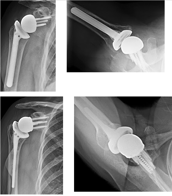
If the cuff deficiency leads to major functional loss, such as pseudo paralysis or anterosuperior escape as shown below


a reverse total shoulder can be considered (see this link). The reverse total shoulder does not depend on the rotator cuff for stability; instead its geometry enables the deltoid to function independent of the cuff.




===


Check out the new Shoulder Arthritis Book - click here.

Use the "Search" box to the right to find other topics of interest to you.

You may be interested in some of our most visited web pages including:shoulder arthritis, total shoulder, ream and runreverse total shoulderCTA arthroplasty, and rotator cuff surgery as well as the 'ream and run essentials'


Sunday, June 7, 2015

Rotator cuff repair - is the repaired tendon normal?

Delaminated rotator cuff tear: extension of delamination and cuff integrity after arthroscopic rotator cuff repair.

These authors reviewed 65 patients with delaminated rotator cuff tears. The delaminated tears were divided into full-thickness delaminated tears and partial-thickness delaminated tears. Cuff integrity was evaluated by computed tomography orthography. They found that the mean medial extension in the full-thickness and partial-thickness delaminated tears was 18.1 ± 6.0 mm and 22.7 ± 6.3 mm, respectively (P = .0084). The posterior extension into the supraspinatus and the infraspinatus was 36.9% and 32.3%, respectively, in the full-thickness delaminated tears, and it was 27.7% and 3.1%, respectively, in the partial-thickness delaminated tears (P = .0043). With regard to cuff integrity, 35 cases of anatomic healing, 10 cases of partial healing defects, and 17 cases of retear were detected. Among the patients with retear and partial healing of the defect, all the partially healed defects showed delamination. Three retear patients showed delamination.

Comment: These authors excluded traumatic tears and the average patient age was 60 years. One can surmise, therefore, that these were degenerative tears. The data shows that 35 of 65 had anatomic healing, 17 had full thickness retears and 10 had partial failure of the repair (all of which demonstrated delimitation).  The data show that cuff repair has a limited ability to restore normal rotator cuff anatomy and, presumably, strength.

---
Use the "Search" box to the right to find other topics of interest to you.

You may be interested in some of our most visited web pages including:shoulder arthritis, total shoulder, ream and runreverse total shoulderCTA arthroplasty, and rotator cuff surgery as well as the 'ream and run essentials'


Monday, May 13, 2013

Attaching the rotator cuff to bone, how does nature do it?

The attachment of a tendon to bone is called an enthesis. Here is a picture of the cuff insertion to bone in an animal a presented at the most recent Orthopaedic Research Society meeting.
We can see the zones that are so critical to managing the bending and twisting loads that are applied to the junction of flexible tendon to inflexible bone. At upper right we can see the wavy tendon fibers. At lower left we can see the solid bone. Between, stained in green, is fibrocartilage - more flexible than bone, less flexible than tendon. Nearer the bone, the fibrocartilage is calcified and nearer the tendon the fibrocartilage transitions to tendon fibers. 

This arrangement is similar to that of a modern electrical plug (see below), which has to manage the mechanical transition between the flexible wire and the rigid body of a laptop. As in the case of the normal cuff enthesis, this is accomplished by a transition zone from more flexible on the right to less flexible on the left.


When this progressive transition is lacking, the attachment is at risk for failure at the junction of the flexible to the stiff. This is, of course, where rotator cuff defects occur.


A couple of lessons may be derived from this observation:
(1) Maintaining shoulder flexibility through gentle stretching may help reduce the risk of rotator cuff failure.
(2) Surgical repairs of the rotator cuff do not, of themselves, restore this transition. 
Rather, the re-establishment of the transition zone is accomplished by progressive remodeling over time. Until the transition zone is re-established, we can suspect that the cuff repair is vulnerable to failure.
Some of the readers may remember the old-style electrical plug without the transition zone. It's easy to guess where the failure occurred.


===
Use the "Search" box to the right to find other topics of interest to you.

You may be interested in some of our most visited web pages including:shoulder arthritis, total shoulder, ream and runreverse total shoulderCTA arthroplasty, and rotator cuff surgery as well as the 'ream and run essentials'


See from which cities our patients come.


See the countries from which our readers come on this post.


Wednesday, December 26, 2012

Metal-backed glenoid component


An uncemented metal-backed glenoid component in total shoulder arthroplasty for osteoarthritis: factors affecting survival and outcome

As pointed out in a previous post and in this review article, the major issue with metal-backed glenoid components is not the fixation of the metal to bone, but rather the dissociation and wear of the polyethylene attached to the metal base plate.   Metal backed glenoid component are discussed in previous reports and here.

This article presents a minimum of 60 month followup of 33 shoulders with screw-fixed porous coated metal backed glenoid components inserted by an individual surgeon. The results were better than those reported previously for simlar components. The 10 year survivorship was 93%. Two were revised for polyethylene wear at 6 years and one for glenoid loosening at 11 years. Radiolucent zones were observed around six (19 %) glenoid components at last follow-up. These lucencies were apparent three years or more after superior subluxation was observed radiographically.

As the authors suggest, this is probably due to delayed rotator cuff failure, show in this figure from their article (radiograph B was obtained 8 years after the surgery). This phenomenon has been discussed in a previous post.


Younger patients and those with higher preoperative Constant scores experienced less improvement.

This is a relatively small series with encouraging results. It will be of great interest to follow the future results obtained with this prosthesis 
--

Use the "Search" box to the right to find other topics of interest to you.

You may be interested in some of our most visited web pages including:shoulder arthritis, total shoulder, ream and runreverse total shoulderCTA arthroplasty,  and rotator cuff surgery.


Sunday, April 1, 2012

Secondary Rotator Cuff Dysfunction Following Total Shoulder Arthroplasty for Primary Glenohumeral Osteoarthritis: Results of a Multicenter Study with More Than Five Years of Follow-up JBJS

An article, "Secondary Rotator Cuff Dysfunction Following Total Shoulder Arthroplasty for Primary Glenohumeral Osteoarthritis" was recently published in the JBJS.

The authors followed 518 shoulders having total shoulder arthroplasty. They defined 'secondary rotator cuff dysfunction' as superior subluxation of the humeral head on the glenoid revealed by anteroposterior radiographs evaluated by observers blinded to the clinical data. The incidence of this finding was 16% at 10 years after surgery and 55% at 15 years. Shoulders with this finding had worse comfort and function as well as a greater risk of glenoid component failure. Shoulders with preoperative fatty infiltration of the infraspinatus and shoulders having glenoid components inserted with a superior tilt have an increased risk of secondary cuff dysfunction. 

It is of note that the average age of the patients in this series was 68 years. This, of course, is an age in which rotator cuff failure becomes quite common, even if these defects are asymptomatic. Thus, progressive cuff failure can be expected in this patient population. In this regard it is interesting that a partial articular-sided tear of the supraspinatus was found in 48 shoulders and a complete full-thickness supraspinatus tear was found in 43 shoulders; 15% of the shoulders had cuff pathology identified at surgery. Twelve shoulders had cuff repairs at the time of arthroplasty.

It is important to keep in mind that the rotator cuff insertion to the humerus is at jeopardy when the humeral cut is made - we have seen cases in which 'secondary cuff failure' resulted from accidental cuff detachment at surgery.

Some of the cases exemplified in this study show evidence not only of superior migration and loosening, but also of glenoid component wear (thinning of the space between the humeral head and the cement at the  glenoid bone surface) and of osteolysis or even infection as shown below.


Among the lessons that can be derived from this study are
(1) inform all patients having a 'total shoulder' that the rotator cuff becomes progressively thin, weak and stiff with age, so that cuff failure is a risk - especially since the arthroplasty may enable and encourage active use of a shoulder that has been 'in irons' for quite a while before surgery
(3) avoid 'overstuffing' the shoulder with too-large components that put the cuff under increased tension
(4) assure secure and durable fixation of the glenoid component, avoiding superior tilt
(5) encourage gentle progressive rehabilitation of the shoulder after arthroplasty so that the cuff has a chance to accommodate the increased loading
(6) encourage patients to inform their surgeon if there is a loss in function of a previously functional arthroplasty
--

Use the "Search the Blog" box to the right to find other topics of interest to you.

You may be interested in some of our most visited web pages including: shoulder arthritis, total shoulder, ream and runreverse total shoulderCTA arthroplasty, and rotator cuff surgery.