
Anatomic total shoulder
B. Incomplete glenoid reaming along a normalized glenoid axis leaving the posterior aspect of the glenoid component unsupported
C. Complete glenoid reaming along a normalized glenoid axis removing sufficient anterior bone to normalize version (ATSA+HSR)
D. Conservative glenoid reaming removing a small amount of glenoid bone accepting glenoid retroversion
E. Incomplete glenoid reaming along a normalized glenoid axis supporting the posterior aspect of the glenoid component with a bone graft
F, Incomplete glenoid reaming along a normalized glenoid axis using an augmented glenoid component to accommodate the gap between the reamer and the bone
1. With glenoid reaming along a normalized glenoid axis alone

The authors of Anatomic Shoulder Arthroplasty With High Side Reaming Versus Reverse Shoulder Arthroplasty For Eccentric Glenoid Wear Patterns With An Intact Rotator Cuff: Comparing Early Versus Mid-Term Outcomes With Minimum 7 Years Of Follow Up did an interesting study comparing the two year and 7 year outcomes from anatomic arthroplasty with method "C" to a reverse total shoulder with method "G1".
After exposure of the glenoid, the anterior high side of the glenoid was preferentially reamed with the glenoid reamer to lower the high side and change glenoid version to zero degrees while minimizing the removal of the subchondral plate. In some cases up to 10 degrees of retroversion was accepted. A glenoid trial sizer was used to estimate how much of the component would be supported by bone, and what percentage was not supported posteriorly.
101 ATSA+HSR and 93 RSA had both two-year and final follow-up with a minimum of 7 years after surgery (average 8.3 years for ATSA+HSR and 7.8 years for RSA).


Comment: This study clearly points out the importance of followup longer than the traditional two years. The average time to revision for glenoid loosening and cuff failure after ATSA was over seven years.
One of the problems with high side reaming is the removal of the dense bone that has been articulating with the arthritic humeral head. Removal of this bone and "correction" of glenoid retroversion (A) leaves less bone and bone of poorer quality to support an anatomic glenoid component (B). By contrast, conservative glenoid reaming with acceptance of glenoid retroversion (C) preserves the amount of dense bone and provides bony support for a larger glenoid component (D).

As pointed out previously, a large industry has formed around the assessment of, planing for, special components to address, and surgical "correction" of glenoid retroversion to 15 degrees or less in performing an anatomic total shoulder arthroplasty.
In Do glenoid retroversion and humeral subluxation affect outcomes following total shoulder arthroplasty? the authors studied 113 patients at an average of 4 years after arthroplasty. Retroversion and humeral head subluxation before and after surgery were measured on axillary radiographs.
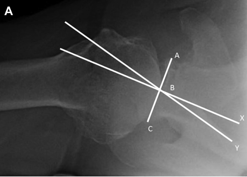

Left figure above. Assessment of preoperative glenoid retroversion. Line A-C represents the glenoid plane, which is drawn connecting the anterior (A) and posterior (C) rims of the glenoid. Line B-Y represents the scapular plane. Line B-X is the perpendicular bisector of line A-C. The retroversion of glenoid is defined as the angle between lines B-X and B-Y.
Right figure above. Assessment of preoperative subluxation. The same lines A-C and B-X are drawn. The humeral head circle is drawn with point Z at the center. Line segment D-F marks the humeral diameter, parallel to line A-C. Point E is the intersection between lines D-F and B-X. Subluxation is defined as the percentage of the humeral head posterior to line B-X, and is calculated as (E-F)/(D-F) X 100%.
At surgery, reaming of the anterior glenoid was carried out until satisfactory implant support (>80% of bony contact with the component) was achieved. There was no specific targeted amount of retroversion that was deemed acceptable.
In contrast to many other studies in which CT scans were used to measure the preoperative anatomical relationships and axillary views to make the postoperative measurements, these authors measured postoperative retroversion and subluxation in a manner identical to the preoperative measurements. As shown below.


At a mean followup of 4.2 years, the authors found no significant correlation between preoperative glenoid version or humeral head subluxation and the postoperative ASES scores.


The authors found no significant correlation between postoperative glenoid version or humeral head subluxation and the postoperative ASES scores.


There were no significant differences in preoperative or postoperative version for patients with or without glenoid lucencies.
The overall complication rate was 11.3%, including 5 periprosthetic joint infections, 3 with glenoid loosening, 2 rotator cuff failures, 2 periprosthetic fractures, 1 patient with stiffness treated with lysis of adhesions, and 1 patient with recurrent posterior instability after treatment with shoulder replacement after a locked posterior dislocation. Eleven of these patients underwent component revision. There was no observed difference between patients with or without revision surgery for either preoperative retroversion (15.2 ± 5.5 deg for failures vs. 15.3 ± 7.7 deg for non failures); or postoperative retroversion (7.1± 5.2 deg vs. 10.0 ± 6.8).
For the patients with eventual glenoid loosening, the preoperative retroversion was 15 and 17 deg in 2 of the 3 patients (third patient did not have available preoperative imaging) and the postoperative retroversion was 6, 8, and 19 deg. The preoperative subluxation was 64% and 60%, whereas postoperative subluxation was 58%, 49%, and 48%.
This study does not support the need to "correct" glenoid version to < 15 degrees in performing an anatomic total shoulder. These findings are consistent with the findings in Does Postoperative Glenoid Retroversion Affect the 2-Year Clinical and Radiographic Outcomes for Total Shoulder Arthroplasty?


The mean (± SD) improvement in the SST (6.7 ± 3.6; from 2.6 ± 2.6 to 9.3 ± 2.9) for the retroverted group was not inferior to that for the nonretroverted group (5.8 ± 3.6; from 3.7 ± 2.5 to 9.4 ± 3.0). The percent of maximal possible improvement (%MPI) for the retroverted glenoids (70% ± 31%) was not inferior to that for the nonretroverted glenoids (67% ± 44%). The 2-year SST scores for the retroverted (9.3 ± 2.9) and the nonretroverted glenoid groups (9.4 ± 3.0) were similar (mean difference, 0.2; 95% CI, - 1.1 to 1.4; p = 0.697). No patient in either group reported symptoms of subluxation or dislocation.


The authors concluded that in this series of TSAs, postoperative glenoid retroversion was not associated with inferior clinical results at 2 years after surgery.
This study evaluated the ability of shoulder arthroplasty using a standard glenoid component to improve patient self-assessed comfort and function and to correct preoperative humeral-head decentering on the face of the glenoid in patients with primary glenohumeral arthritis and type-B2 or B3 glenoids.
The authors identified 66 shoulders with type-B2 glenoids (n = 40) or type-B3 glenoids (n = 26) undergoing total shoulder arthroplasties with a non-augmented glenoid component inserted without attempting to normalize glenoid version and with clinical and radiographic follow-up that was a minimum of 2 years. The Simple Shoulder Test (SST) score (and standard deviation) improved from 3.2 ± 2.1 points preoperatively to 9.9 ± 2.4 points postoperatively (p < 0.001) at a mean time of 2.8 ± 1.2 years for type-B2 glenoids and from 3.0 ± 2.5 points preoperatively to 9.4 ± 2.1 points postoperatively (p < 0.001) at a mean time of 2.9 ± 1.5 years for type-B3 glenoids; these results were not inferior to those for shoulders with other glenoid types.

Postoperative glenoid version was not significantly different (p > 0.05) from preoperative glenoid version. The mean humeral-head decentering on the glenoid face was reduced for type-B2 glenoids from -14% ± 7% preoperatively to -1% ± 2% postoperatively (p < 0.001) and for type-B3 glenoids from -4% ± 6% preoperatively to -1% ± 3% postoperatively (p = 0.027). The rates of bone integration into the central peg for type-B2 glenoids (83%) and type-B3 glenoids (81%) were not inferior to those for other glenoid types.

The authors concluded that shoulder arthroplasty with a standard glenoid inserted without changing version can significantly improve patient comfort and function and consistently center the humeral head on the glenoid face in shoulders with type-B2 and B3 glenoids, achieving >80% osseous integration into the central peg. These clinical and radiographic outcomes for type- B2 and B3 glenoids were not inferior to those outcomes for other glenoid types
Long term followup of well-characterized patients treated with the different methods for managing glenoid retroversion will be required to define the relative risks, benefits, effectiveness and durability of each of them.

Follow on twitter: https://twitter.com/shoulderarth
Follow on facebook: click on this link
Follow on facebook: https://www.facebook.com/frederick.matsen
Follow on LinkedIn: https://www.linkedin.com/in/rick-matsen-88b1a8133/
Here are some videos that are of shoulder interest
Shoulder arthritis - what you need to know (see this link).
How to x-ray the shoulder (see this link).
The ream and run procedure (see this link).
The total shoulder arthroplasty (see this link).
The cuff tear arthropathy arthroplasty (see this link).
The reverse total shoulder arthroplasty (see this link).
The smooth and move procedure for irreparable rotator cuff tears (see this link).
Shoulder rehabilitation exercises (see this link).