Showing posts with label humeral osteotomy. Show all posts

Sunday, July 18, 2021

The importance of the humeral osteotomy in shoulder joint replacement for arthritis

A comparison of patient-specific instrumentation to navigation for conducting humeral head osteotomies during shoulder arthroplasty

These authors point out that the humeral head osteotomy during shoulder arthroplasty influences humeral component height, version and possibly neck-shaft angle. They sought to evaluate the use of patient-specific guides and surgical navigation for executing a planned humeral head osteotomy in paired 3D  polylactic models created from DICOM images of 10 shoulders (5 normal and 5 osteoarthritic).


In one of each pair a humeral osteotomy was created using a patient-specific guide; in the second a real time navigated osteotomy was performed with an optically tracked sagittal saw. 


They found no statistically significant differences between patient-specific guides and navigation for osteotomy cut height or humeral version. Navigation,  however, resulted in significantly less neck shaft angle error than the patient specific guides.


Comment: Correct positioning of the humeral component is an essential component of a successful shoulder arthroplasty. 


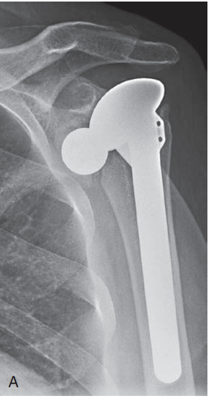
One of the most common errors is a "too-high" position, as shown below.


As shown in the examples above, too high positioning can contribute to glenoid component failure and can also result in a stiff shoulder as shown below.


as well as placing excessive tension on the rotator cuff
                                             
Too high positioning of the humeral head can result from a too high neck cut 


or from failure to properly seat the component


We like to be sure that the margin of the humeral prosthesis is just below the "berm" as shown below.


We routinely make the humeral neck cut at a 45 degree angle with the long axis of the medullary canal.

This is the case if the apparent anatomic neck is in varus
or valgus


The head cut is made in 35 degrees of retroversion using the forearm of the flexed elbow as a reference. Care is taken to avoid damage to the cuff insertion from an excessively retroverted cut.


So, as can be seen from the above, the humeral neck cut is important. It can be correctly made without special jigs, 3D planning or patient specific guides by using direct observations make at the time of surgery.


Here are some videos that are of shoulder interest
Shoulder arthritis - what you need to know (see this link).
How to x-ray the shoulder (see this link).
The total shoulder arthroplasty (see this link).
The ream and run technique is shown in this link.
The cuff tear arthropathy arthroplasty (see this link).
The reverse total shoulder arthroplasty (see this link).
Shoulder rehabilitation exercises (see this link).

Follow on twitter: Frederick Matsen (@shoulderarth)


Saturday, November 12, 2016

Humeral head osteotomy in shoulder arthroplasty - how do you slice it?

Humeral head osteotomy in shoulder arthroplasty: a comparison between anterosuperior and inferoanterior resection techniques

These authors proposed an approach to humeral osteotomy based on landmarks on the anatomic neck in an attempt to restore 3-dimensional humeral head morphology. They digitized the anatomical neck of 30 normal human cadaver shoulders.


They compared the virtual performance of two different osteotomy techniques: the traditional, following the anterosuperior anatomic neck; and a new technique, defined by the inferoanterior anatomic neck. 

Length-width difference of the anterosuperior resection area was higher than in the inferoanterior osteotomy (6 ± 2 mm vs. 3 ± 1 mm; P < .001).
Retroversion of the anterosuperior resection plane was higher than the native head (50° ± 12° vs. 37° ± 11°; P < .001), whereas retroversion after the inferoanterior osteotomy (32° ± 12°) did not differ from native (P = .057).
Inclination differed after the anterosuperior osteotomy (129° ± 5°) and the inferoanterior osteotomy (127° ± 4°) compared with the native head (134° ± 4°; P ≤ .001).

They concluded that inferoanterior referenced osteotomy generated a more circular resection area, matching the native humeral head retroversion more closely than in the anterosuperior technique.

Comment: The goal of humeral implant positioning is not to reproduce 'normal' anatomy, but rather to optimize glenohumeral stability and kinematics. This may require non-anatomic positioning of the humeral joint surface, such as in the use of anteriorly eccentric humeral heads to manage functional decentering observed at surgery.

The pathoanatomy encountered at shoulder arthroplasty varies widely, but rarely looks like the normal cadaver shown above. Here are some examples in which cases finding anteroinferior landmarks would be a challenge.


The positioning of the humeral articular surface is dictated by (a) the positioning of the prosthetic stem in the humeral canal and (b) the geometry of the humeral prosthesis placed on the prosthetic stem. In our technique, the humeral neck cut does not control the final head position.

The orthopaedic axis is defined by the largest cylindrical sizer that fits in the canal.

The cut is made at 45 degrees with this orthopaedic axis, irrespective of the proximal humeral anatomy


and in 30 degrees of retroversion.

the prosthesis is seated so that its lateral edge is just below the berm.


===


Use the "Search" box to the right to find other topics of interest to you.

You may be interested in some of our most visited web pages including:shoulder arthritis, total shoulder, ream and runreverse total shoulderCTA arthroplasty, and rotator cuff surgery as well as the 'ream and run essentials'