Showing posts with label axillary truth view. Show all posts

Saturday, November 8, 2025

Glenoid pathoanatomy: what about the B Walch types?

In his classic paper, Morphologic study of the glenoid in primary glenohumeral osteoarthritisGilles Walch identified the B1 and B2 glenoids as being common of types of arthritic glenohumeral pathoanatomy in patients presenting for anatomic shoulder arthroplasty. 

An important characteristic of these glenoid types is the posterior decentering of the humeral head on the glenoid, a critical element in the evaluation and management of glenohumeral arthritis.  Note that the degree of decentering was (and remains) defined by the relationship of the humeral head to the face of the glenoid (and not the plane of the scapula), as seen from this figure from his classic article.


The decentering of the head on the glenoid can be evaluated on the standardized axillary "truth" view, as shown in the five examples below.


By the "truth" view, we mean an axillary view obtained with the arm elevated in the plane of the scapula that shows the spinoglenoid notch or "eye" (red arrow) as shown in this Steve Lippitt illustration:


The rationale for evaluating decentering with the arm elevated to a functional position is that CTs or MRIs obtained with the arm at the side may not reveal it, as shown in the two images of the same shoulder shown below. The MRI obtained with the arm at the side does not reveal decentering, whereas dramatic posterior decentering is shown when the arm is elevated to a functional position in the axillary "truth" view.


While often considered together, the B1 and B2 are not the same. The B1 has posterior decentering of the humerus on the glenoid without biconcavity of the glenoid from bony erosion. By contrast, the B2 has posterior decentering of the humeral head on the glenoid with biconcavity of the glenoid as shown in these illustrations from the classic article by Walch.

A third B was added by the authors of A modification to the Walch classification of the glenoid in primary glenohumeral osteoarthritis using three-dimensional imaging.


As can be seen from these figures, the B3 is monoconcave (i.e. no biconcavity) with substantial retroversion and without posterior decentering of the humeral head on the glenoid, i.e. the humeral head is centered with respect to the glenoid.

This point is emphasized by the authors of Quantitative measurement of bony pathology in advanced glenohumeral osteoarthritis who use the term "humeral-glenoid alignment (HGA)" to indicate centering or decentering of the humeral head on the glenoid. HGA is measured as the position of the humeral head center relative to the perpendicular line drawn from the glenoid center point (without reference to the scapular axis). This relationship is shown in a figure from their article showing the centering of the humeral head in a B3 glenoid (i.e. the humeral head is not decentered).


How does all this relate to the practice of anatomic shoulder arthroplasty? A recent article, Why do primary anatomic total shoulder arthroplasties fail today? A systematic review and meta-analysis, is relevant. The authors reviewed a total of 44 studies involving 35,168 aTSA procedures; 2744 failures were identified. The three most prevalent types of failure were: 

(1) implant loosening (26.1%), with 21.7% of failures attributed to glenoid component loosening. 
(2) Rotator cuff insufficiency (17.3%).  
(3) Instability (10.4%) 

Another recent article compared the types of failure in the Kaiser and the Australian Orthopaedic Association databases.Early revision in anatomic total shoulder arthroplasty in osteoarthritis: a cross-registry comparison.
The most common reasons for revision in the AOA experience were instability/dislocation (31.1%), rotator cuff insufficiency (24.2%), and loosening/lysis and implant breakage glenoid insert (11.0% each). The most common reasons in Kaiser experience were rotator cuff tear (32.3%), glenoid component loosening (29.0%), and dislocation and infection (12.9% each). 

While these articles did not study the relationship of glenoid type to loosening or instability, we can venture that because B1 and B2 glenoids demonstrate preoperative posterior decentering, they would be at risk for postoperative instability. The B3, being centered preoperatively would seem less at risk for instability as long as the centering was not disrupted by the arthroplasty.

There are a number of approaches for shoulders with each of the different B types, each of which has its proponents, advantages and limitations. As emphasized in Short-term outcomes of anatomic total shoulderarthroplasty with nonaugmented glenoidcomponent for Walch B2 and B3 glenoidmorphology, all B's are not the same. In fact, each patient and their shoulder is a one of a kind combination. We like to say that each patient is an N of 1.

Here's an approach we commonly use for effectively and durably restoring stability and mobility. Note that usually we do not attempt to "correct" preoperative glenoid version (see Does postoperative glenoid component retroversion following anatomic total shoulder arthroplasty affect clinical outcomes? A systematic review and meta-analysisand judge the need for an anteriorly eccentric humeral head based on intraoperative testing at surgery with trial components in place (see Management of intraoperative posterior decentering in shoulder arthroplasty using anteriorly eccentric humeral head components).

We start with Anatomic total shoulder - preoperative planning and intraoperative decision making, recognizing the different characteristics among the 3 Bs


B1 - conservative reaming without attempting to alter version, preserving glenoid bone stock, excellent carpentry to assure perfect seating of the component, and use of an anteriorly eccentric humeral head to manage excessive posterior translation if that is evident on intraoperative examination with a concentric trial humeral head component in place.

B2 - conservative reaming - just sufficient to convert the biconcavity to a mono concavity without attempting to alter glenoid version, preserving glenoid bone stock, excellent carpentry to assure perfect seating of the component, and use of an anteriorly eccentric humeral head to manage excessive posterior translation if that is evident on intraoperative examination with a concentric trial humeral head component in place.

B3 - conservative reaming without attempt to alter glenoid version, preserving glenoid bone stock, excellent carpentry to assure perfect seating of the component. An anteriorly eccentric humeral head component is rarely necessary because of the absence of preoperative decentering.

For additional information on this approach see:


Pollination

Bumble Bee on Iris
Montlake Fill
Spring 2021

Follow on twitter/X: https://x.com/RickMatsen
Follow on facebook: https://www.facebook.com/shoulder.arthritis
Follow on LinkedIn: https://www.linkedin.com/in/rick-matsen-88b1a8133/

Here are some videos that are of shoulder interest
Shoulder arthritis - what you need to know (see this link).
How to x-ray the shoulder (see this link).
The ream and run procedure (see this link)
The total shoulder arthroplasty (see this link)
The cuff tear arthropathy arthroplasty (see this link).
The reverse total shoulder arthroplasty (see this link).
The smooth and move procedure for irreparable rotator cuff tears (see this link)
Shoulder rehabilitation exercises (see this link).

Friday, February 14, 2025

Anatomic total shoulder - preoperative planning and intraoperative decision making.

 Preoperative planning is helpful in anticipating what might be needed to reconstruct an arthritic shoulder; however the final choice of implants is determined at the time of surgery.

A 43 year old man presented with pain and stiffness of his left shoulder. He had a prior CT scan showing an arthritic humeral head centered on a somewhat retroverted glenoid.


We obtained our standard set of plain radiographs: an AP in the plane of the scapula and an axillary "truth" view taken with the arm in a functional arm position of elevation. The truth view showed posterior decentering of the humeral head that was not evident on the CT scan taken with the patient's arm at his side. No 3D CT planning was used.


At surgery, a standard glenoid component was well seated after conservative glenoid reaming. "Corrective" reaming and a posteriorly augmented glenoid component were not used.
 Trialing with an anatomic humeral head component revealed posterior instability when the arm was flexed forward. As a result, a short stemmed humeral component with an anteriorly eccentric humeral head was selected. 
Postoperatively, his shoulder is clinically and radiographically stable when the arm is elevated to a functional position (as seen on the postoperative "truth" view).


Comment: This case illustrates (1) the value of the "truth" view before and after surgery and (2) the importance of tailoring implant selection based on intraoperative testing of motion and stability. NB: when we do an arthroplasty, the shoulder we have after soft tissue releases and osteophyte resection is different from the shoulder before surgery; that's why intraoperative assessment is more important than preoperative planning.

You can support cutting edge shoulder research that is leading to better care for patients with shoulder problems, click on this link

Follow on twitter/X: https://x.com/RickMatsen
Follow on facebook: https://www.facebook.com/shoulder.arthritis
Follow on LinkedIn: https://www.linkedin.com/in/rick-matsen-88b1a8133/

Here are some videos that are of shoulder interest
Shoulder arthritis - what you need to know (see this link).
How to x-ray the shoulder (see this link).
The ream and run procedure (see this link).
The total shoulder arthroplasty (see this link).
The cuff tear arthropathy arthroplasty (see this link).
The reverse total shoulder arthroplasty (see this link).
The smooth and move procedure for irreparable rotator cuff tears (see this link)
Shoulder rehabilitation exercises (see this link).


Saturday, November 16, 2024

Can Computer Vision/AI solve a challenge in clinical shoulder arthroplasty research?

Shoulder surgeons are trying to understand the clinical importance of preoperative shoulder pathoanatomy and its modification by arthroplasty. Large-scale multicenter studies are necessary for investigating the relationship between standardized preoperative and sequential postoperative anatomical measurements and the outcome realized by the patient. 

The validity of such studies will depend on (1) standardization of the measurement methods across different centers, (2) human-observer independency avoiding the risk of inter-observer variation and observer bias, and (3) highly efficient methods that enable the evaluation of very large numbers of images. While CT scans are commonly used for characterizing preoperative shoulder anatomy, they are impractical for evaluating the postoperative anatomy and changes over time.  Standardized anteroposterior and axillary radiographs provide a practical and cost-effective approach for making preoperative and postoperative measurements using the same imaging method.

A recent study, Can computer vision / artificial intelligence locate key reference points and make clinically relevant measurements on axillary radiographs? demonstrated the potential of artificial intelligence in assessing clinically important relationships on standardized axillary x-rays. Standardized pre and post arthroplasty axillary radiographs were manually annotated by shoulder surgeons locating six reference points as shown the figure below:


The anterior and posterior edges of the glenoid face are indicated by the green dots.

The center of the glenoid face by the blue dot.

The base of the glenoid vault by the yellow dot. 

The spinoglenoid notch by black dot at tip of arrow.

The circle fitting the humerus articular surface by the blue circle.


These points were then used to measure glenoid version and humeroglenoid alignment (HGA-AP). Version was measured as the angle between the red and green lines. HGA-AP was measured as the perpendicular distance (double headed arrow) between the centre of the circle (star) and the perpendicular bisector (yellow line) of the glenoid face line (red line) divided by the diameter of the circle (dotted white line)
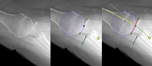


These annotated images were used to train a computer vision model that could identify these reference points and determine humeroglenoid alignment in the anterior to posterior direction and glenoid version without human guidance. 


The model's accuracy was tested on a separate test set of 52 axillary images that were not used in training the model, comparing the model's reference point locations, humero-glenoid alignment and glenoid version to the corresponding values assessed by the two surgeons. 


The model performed efficiently, allowing the rapid uploading of images and analysis of reference points, glenoid version, and humeroglenoid alignment (HGA-AP) without human participation. The model was able to produce the measurements in a matter of seconds compared to approximately two hours required for surgeon assessment of the relatively small set of 52 images.


The model was able to rapidly identify all six reference point locations to within a mean of 2 mm of the surgeon-assessed points. The mean variation in alignment and version measurements between the surgeon assessors and the model was similar to the variation between the two surgeon assessors.


The average differences between the surgeon- and the model-assessed reference points for the test set are shown below


The mean differences in glenoid version and HGA-AP between the surgeon assessors, between each surgeon assessor and the model, and between the average of the two surgeon assessors the model is shown below


The inter-observer variability between the two surgeons was similar to that between the average of the two surgeons and model 


While it will require substantial further refinement before it is ready for broad scale application, this proof-of-principle study does demonstrate the development and validation of a computer vision/artificial intelligence model that can independently identify key landmarks and determine the glenohumeral relationship and glenoid version on axillary radiographs. This observer-independent approach has the potential to enable efficient assessment of shoulder radiographs, substantially lessening the burden of manual x-ray interpretation and enabling scaling of these measurements across large numbers of patients from multiple centers so that pre- and postoperative anatomy can be correlated with patient reported clinical outcomes. 


Other studies have reported the use of artificial intelligence to classify shoulder implants 


Classifying shoulder implants in X-ray images using deep learning "In a data set containing X-ray images of shoulder implants from 4 manufacturers and 16 different models, deep learning is able to identify the correct manufacturer with an accuracy of approximately 80% in 10-fold cross validation, while other classifiers achieve an accuracy of 56% or less"


Development of a machine learning algorithm to identify total and reverse shoulder arthroplasty implants from X-ray images. "This proof of concept study demonstrates that machine learning can assist with preoperative planning and improve cost-efficiency in shoulder surgery."


A novel hybrid machine learning based system to classify shoulder implant manufacturers. "The proposed hybrid machine learning algorithms achieve the goal of low cost and high performance compared to other studies in the literature."


Deep learning to automatically classify very large sets of preoperative and postoperative shoulder arthroplasty radiographs. "We developed an efficient, accurate, and reliable AI algorithm to automatically identify key imaging features of laterality, imaging view, and implant type in shoulder radiographs. This algorithm represents the first step to automatically classify and organize shoulder radiographs on a large scale in very little time, which will profoundly enrich shoulder arthroplasty registries."


Artificial intelligence for automated identification of total shoulder arthroplasty implants. "A DL model demonstrated excellent accuracy in identifying 22 unique TSA implants from 8 manufacturers. "


EFFICACY OF ARTIFICIAL INTELLIGENCE-BASED MODELS FOR SHOULDER ARTHROPLASTY IMPLANT DETECTION AND CLASSIFICATION USING UPPER LIMB RADIOGRAPHS: A SYSTEMATIC REVIEW AND META-ANALYSIS "AI-based classification of shoulder implant types can be considered a sensitive method."


Artificial intelligence in shoulder arthroplasty


Other studies have explored the use of artificial intelligence to make measurements on x-ray images


The Development of a Yolov8-Based Model for the Measurement of Critical Shoulder Angle (CSA), Lateral Acromion Angle (LAA), and Acromion Index (AI) from Shoulder X-ray Images. "The results indicated that automatic measurement methods align with manual measurements with high accuracy and offer an effective alternative for clinical applications".


The acetabularization index: a novel measure of acromial bone loss prior to reverse shoulder arthroplasty. "AI is a reliably measurable tool on radiographs and 2D CT scans"


While others have used artificial intelligence to assess humeral fractures


Clinical validation of artificial intelligence-based preoperative virtual reduction for Neer 3- or 4-part proximal humerus fractures "The AI-based preoperative virtual reduction model showed good performance in the reduction model in proximal humerus fractures with faster working times."


Artificial intelligence versus radiologist in the accuracy of fracture detection based on computed tomography images: a multi-dimensional, multi-region analysis "The optimized AI model improves the diagnostic efficacy in detecting extremity fractures on radiographs, and the optimized AI model is significantly better than radiologists in detecting avulsion fractures, "


From the foregoing it is evident that we are on the forefront of the application of computer vision/artificial intelligence to enhance clinically important shoulder research. 


Stay tuned!


You can support cutting edge shoulder research that is leading to better care for patients with shoulder problems, click on this link

Follow on twitter/X: https://x.com/RickMatsen

Here are some videos that are of shoulder interest
Shoulder arthritis - what you need to know (see this link).
How to x-ray the shoulder (see this link).
The ream and run procedure (see this link).
The total shoulder arthroplasty (see this link).
The cuff tear arthropathy arthroplasty (see this link).
The reverse total shoulder arthroplasty (see this link).
The smooth and move procedure for irreparable rotator cuff tears (see this link).
Shoulder rehabilitation exercises (see this link). 


Thursday, January 11, 2024

Did the surgery correct the glenoid version and decentering? The value of comparable preoperative and postoperative imaging.

 A young athletic man presented with painful glenohumeral arthritis and functional posterior instability of his right shoulder as shown on the axillary "truth" view below (see Answering the critical question: "To what degree is the humeral head functionally centered on the glenoid?" in 6 easy steps.)


The surgical objective was to re-center the humeral head on the glenoid and re-establish durable functional stability of his shoulder. An axillary truth view 5 years after surgery indicated that the surgical objective was achieved. 


This type of assessment can only be made if the preoperative and postoperative imaging modalities and techniques are the same. This point is emphasized by the authors of Total shoulder arthroplasty in patients with a B2 glenoid addressed with corrective reaming: mean 8-year follow-up who used standardized axillary radiographs preoperatively and sequentially postoperatively to assess humeral head decentering relative to the glenoid vault, immediate glenoid seating, and final glenoid peg radiolucency. Interestingly, this plain radiographic study revealed that glenoid component failure was associated with poor initial glenoid component seating, with failed components having an average of 25% of the glenoid component not seated. Preoperative deformity as seen on CT scans, such as glenoid retroversion, inclination, or humeral head subluxation did not predict glenoid component failure.


This straightforward approach for determining the extent to which the procedure achieved the preoperative goal is available to all surgeons, whereas obtaining routine preoperative and sequential postoperative CT scans is impractical in most clinical settings.

Here are some other examples, demonstrating the value of comparable standardized preoperative and postoperative axillary views.


Comment: CT scans and axillary views are not only different imaging modalities, they are also obtained with the arm in different positions. Thus, it is not surprising that they yield different findings for the same shoulder, as shown below.


The point is that if surgeons wish a practical way for evaluating their success in achieving the desired postoperative glenohumeral relationships in comparison to the preoperative pathoanatomy, standardized axiallary views appear to be the best bet. Comparing preoperative CT scans to postoperative axillary views may be a bit like comparing


to


You can support cutting edge shoulder research and education that are leading to better care for patients with shoulder problems, click on this link.

Follow on twitter: https://twitter.com/RickMatsen or https://twitter.com/shoulderarth
Follow on facebook: click on this link
Follow on facebook: https://www.facebook.com/frederick.matsen
Follow on LinkedIn: https://www.linkedin.com/in/rick-matsen-88b1a8133/

Here are some videos that are of shoulder interest
Shoulder arthritis - what you need to know (see this link).
How to x-ray the shoulder (see this link).
The ream and run procedure (see this link).
The total shoulder arthroplasty (see this link).
The cuff tear arthropathy arthroplasty (see this link).
The reverse total shoulder arthroplasty (see this link).
The smooth and move procedure for irreparable rotator cuff tears (see this link).
Shoulder rehabilitation exercises (see this link).