Showing posts with label Walch classification. Show all posts

Saturday, November 8, 2025

Glenoid pathoanatomy: what about the B Walch types?

In his classic paper, Morphologic study of the glenoid in primary glenohumeral osteoarthritis, Gilles Walch identified the B1 and B2 glenoids as being common of types of arthritic glenohumeral pathoanatomy in patients presenting for anatomic shoulder arthroplasty. 

An important characteristic of these glenoid types is the posterior decentering of the humeral head on the glenoid, a critical element in the evaluation and management of glenohumeral arthritis.  Note that the degree of decentering was (and remains) defined by the relationship of the humeral head to the face of the glenoid (and not the plane of the scapula), as seen from this figure from his classic article.


The decentering of the head on the glenoid can be evaluated on the standardized axillary "truth" view, as shown in the five examples below.


By the "truth" view, we mean an axillary view obtained with the arm elevated in the plane of the scapula that shows the spinoglenoid notch or "eye" (red arrow) as shown in this Steve Lippitt illustration:


The rationale for evaluating decentering with the arm elevated to a functional position is that CTs or MRIs obtained with the arm at the side may not reveal it, as shown in the two images of the same shoulder shown below. The MRI obtained with the arm at the side does not reveal decentering, whereas dramatic posterior decentering is shown when the arm is elevated to a functional position in the axillary "truth" view.


While often considered together, the B1 and B2 are not the same. The B1 has posterior decentering of the humerus on the glenoid without biconcavity of the glenoid from bony erosion. By contrast, the B2 has posterior decentering of the humeral head on the glenoid with biconcavity of the glenoid as shown in these illustrations from the classic article by Walch.

A third B was added by the authors of A modification to the Walch classification of the glenoid in primary glenohumeral osteoarthritis using three-dimensional imaging.


As can be seen from these figures, the B3 is monoconcave (i.e. no biconcavity) with substantial retroversion and without posterior decentering of the humeral head on the glenoid, i.e. the humeral head is centered with respect to the glenoid.

This point is emphasized by the authors of Quantitative measurement of bony pathology in advanced glenohumeral osteoarthritis who use the term "humeral-glenoid alignment (HGA)" to indicate centering or decentering of the humeral head on the glenoid. HGA is measured as the position of the humeral head center relative to the perpendicular line drawn from the glenoid center point (without reference to the scapular axis). This relationship is shown in a figure from their article showing the centering of the humeral head in a B3 glenoid (i.e. the humeral head is not decentered).


How does all this relate to the practice of anatomic shoulder arthroplasty? A recent article, Why do primary anatomic total shoulder arthroplasties fail today? A systematic review and meta-analysis, is relevant. The authors reviewed a total of 44 studies involving 35,168 aTSA procedures; 2744 failures were identified. The three most prevalent types of failure were: 

(1) implant loosening (26.1%), with 21.7% of failures attributed to glenoid component loosening. 
(2) Rotator cuff insufficiency (17.3%).  
(3) Instability (10.4%) 

Another recent article compared the types of failure in the Kaiser and the Australian Orthopaedic Association databases.Early revision in anatomic total shoulder arthroplasty in osteoarthritis: a cross-registry comparison.
The most common reasons for revision in the AOA experience were instability/dislocation (31.1%), rotator cuff insufficiency (24.2%), and loosening/lysis and implant breakage glenoid insert (11.0% each). The most common reasons in Kaiser experience were rotator cuff tear (32.3%), glenoid component loosening (29.0%), and dislocation and infection (12.9% each). 

While these articles did not study the relationship of glenoid type to loosening or instability, we can venture that because B1 and B2 glenoids demonstrate preoperative posterior decentering, they would be at risk for postoperative instability. The B3, being centered preoperatively would seem less at risk for instability as long as the centering was not disrupted by the arthroplasty.

There are a number of approaches for shoulders with each of the different B types, each of which has its proponents, advantages and limitations. As emphasized in Short-term outcomes of anatomic total shoulderarthroplasty with nonaugmented glenoidcomponent for Walch B2 and B3 glenoidmorphology, all B's are not the same. In fact, each patient and their shoulder is a one of a kind combination. We like to say that each patient is an N of 1.

Here's an approach we commonly use for effectively and durably restoring stability and mobility. Note that usually we do not attempt to "correct" preoperative glenoid version (see Does postoperative glenoid component retroversion following anatomic total shoulder arthroplasty affect clinical outcomes? A systematic review and meta-analysis) and judge the need for an anteriorly eccentric humeral head based on intraoperative testing at surgery with trial components in place (see Management of intraoperative posterior decentering in shoulder arthroplasty using anteriorly eccentric humeral head components).

We start with Anatomic total shoulder - preoperative planning and intraoperative decision making, recognizing the different characteristics among the 3 Bs


B1 - conservative reaming without attempting to alter version, preserving glenoid bone stock, excellent carpentry to assure perfect seating of the component, and use of an anteriorly eccentric humeral head to manage excessive posterior translation if that is evident on intraoperative examination with a concentric trial humeral head component in place.

B2 - conservative reaming - just sufficient to convert the biconcavity to a mono concavity without attempting to alter glenoid version, preserving glenoid bone stock, excellent carpentry to assure perfect seating of the component, and use of an anteriorly eccentric humeral head to manage excessive posterior translation if that is evident on intraoperative examination with a concentric trial humeral head component in place.

B3 - conservative reaming without attempt to alter glenoid version, preserving glenoid bone stock, excellent carpentry to assure perfect seating of the component. An anteriorly eccentric humeral head component is rarely necessary because of the absence of preoperative decentering.

For additional information on this approach see:


Pollination

Bumble Bee on Iris
Montlake Fill
Spring 2021

Follow on twitter/X: https://x.com/RickMatsen
Follow on facebook: https://www.facebook.com/shoulder.arthritis
Follow on LinkedIn: https://www.linkedin.com/in/rick-matsen-88b1a8133/

Here are some videos that are of shoulder interest
Shoulder arthritis - what you need to know (see this link).
How to x-ray the shoulder (see this link).
The ream and run procedure (see this link)
The total shoulder arthroplasty (see this link)
The cuff tear arthropathy arthroplasty (see this link).
The reverse total shoulder arthroplasty (see this link).
The smooth and move procedure for irreparable rotator cuff tears (see this link)
Shoulder rehabilitation exercises (see this link).

Sunday, July 11, 2021

Progression of arthritic glenoid bone loss as revealed by the axillary "truth" view.

 Natural history of glenoid bone loss in primary glenohumeral osteoarthritis: how does bone loss progress over a decade?

These authors sought to determine how glenohumeral subluxation and glenoid bone loss changed over time in 48 shoulders that underwent arthroplasty and had been evaluated with standardized high-quality axillary radiographs 


at 1 or more time points over the 5-15 years before arthroplasty. The mean interval time between the oldest and most recent radiographs was 8.9 years (range 5-15 years). 

Below is an example of how glenoid morphology progressed over roughly an 8-year period of time from an A1 glenoid to a B3 glenoid. Note the standardization of the axillary "truth" views that enabled comparisons of the glenohumeral pathoanatomy over time. The patient was a 43-year-old male (body mass index 26.6) at initial presentation for symptomatic right shoulder osteoarthritis and went onto an anatomic total shoulder arthroplasty. From presentation to year 5, the glenoid morphology remained A1 with 3 intervening radiographs documented. At year 6, the patient was noted to have a B1 glenoid (top right), a B2 glenoid at year 7 (bottom left), and a B3 glenoid at year 8 before proceeding with surgery (bottom right).


On each axillary view, the glenoid type


and the degree of posterior humeral decentering on the face of the glenoid


were documented.


Glenoid morphology on the earliest radiograph was classified as A1 in 22, A2 in 13, B1 in 1, B2 in 9, B3 in 1, and D in 2 shoulders. 


Walch A patterns identified on early radiographs most commonly maintained an A pattern over time, but 20% developed eccentric wear with 5 of 35 becoming B type and 2 of 35 becoming a D type before arthroplasty. 


All B-type glenoids remained B type. 


Classic progression of bone loss along the same concentric or eccentric ‘‘track’’ occurred 41% of the time, with , the only B1 glenoid becoming a B2 glenoid, and 56% (5/9) of B2 glenoids becoming B3 glenoids before arthroplasty. 


Only 15% (2/13) of A2 glenoids developed eccentric wear compared with 32% (7/22) of A1 glenoids.


Comment: This study demonstrates that glenohumeral pathoanatomy can be well characterized using the axillary "truth" view without the additional expense and radiation dosage of a CT scan.


This study also demonstrates that the description of glenoid pathoanatomy cannot be constrained to discrete static "types", but rather the amount of bone loss, change in version, and humeral decentering each exist on a continuum from "none" to "a lot" with progressive transitions from one type to another.


Here are some videos that are of shoulder interest
Shoulder arthritis - what you need to know (see this link).
How to x-ray the shoulder (see this link).
The total shoulder arthroplasty (see this link).
The ream and run technique is shown in this link.
The cuff tear arthropathy arthroplasty (see this link).
The reverse total shoulder arthroplasty (see this link).
Shoulder rehabilitation exercises (see this link).

Follow on twitter: Frederick Matsen (@shoulderarth)


Saturday, June 26, 2021

The arthritic shoulder

 The Evolution of the Walch Classification for Primary Glenohumeral Arthritis

    These authors provide an interesting and comprehensive narrative of the efforts to describe glenohumeral arthritic pathoanatomy. They recognize the early work of Neer, who pointed out the widely varying patterns of glenoid erosion and the associated changes in effective version of the glenoid with respect to the body of the scapula: central erosion without change in glenoid version, anterior wear with effective glenoid anteversion, posterior wear with effective glenoid retroversion, and superior wear with effective superior inclination. 
    They acknowledge the important efforts of Walch, Friedman, Levy and others to classify the different forms of arthritic glenohumeral pathoanatomy. These efforts at classification required the postulations of "dividing lines" between the different classifications. Looking at the early Walch classification for example 
    it is necessary to decide how much medial erosion is necessary before an A1 becomes and A2, how much biconcavity is necessary before a B1 becomes a B2, how much posterior erosion is necessary before an A2 becomes a B2. And what is to be done with the in betweenners: 


    Varying methods of imaging and various applications of dividing lines have lead to inter observer variability in classification. This variability continues as an increasing number of "types" are identified: B0, B3, D, C1, C2, E1, E2, etc. As the authors point out, since modifications of the Walch classification have been introduced, the kappa scores for both interobserver and intraobserver reliabilities are not as high as initially hoped for.
    In their paragraph "What the Future Holds", the authors predict that attempts to "classify" glenohumeral pathoanatomy will give way to the characterization an arthritic glenoid using simple quantitative measurements of glenoid version, glenoid inclination, the percentage of decentering of the humeral head on the articular surface of the glenoid (as shown below in an early diagram from Walch et al).

The use of these quantitative measurements should facilitate reproducible characterization of glenohumeral pathoanatomy. 
   Critical issues remain, principally how much time and money and radiation need to be expended in characterizing the glenohumeral pathoanatomy prior to shoulder arthroplasty in the usual case. We have found that in the great majority of cases, a simple series of three plain radiographs (see this link) provides the needed information to plan anatomic and reverse total shoulder arthroplasty and to sequentially evaluate the shoulder after arthroplasty. 

 How you can support research in shoulder surgery Click on this link.

Here are some videos that are of shoulder interest
Shoulder arthritis - what you need to know (see this link).
How to x-ray the shoulder (see this link).
The total shoulder arthroplasty (see this link).
The ream and run technique is shown in this link.
The cuff tear arthropathy arthroplasty (see this link).
The reverse total shoulder arthroplasty (see this link).
The smooth and move procedure for irreparable rotator cuff tears (see this link).
Shoulder rehabilitation exercises (see this link).

Sunday, May 9, 2021

Classification of glenohumeral arthritis - how reproducible is it?

 Reliability of the modified Walch classification for advanced glenohumeral osteoarthritis using 3-dimensional computed tomography analysis: a study of the ASES B2 Glenoid Multicenter

These authors assessed the inter- and intraobserver reliability of the modified Walch classification using 3-dimensional (3D) computed tomography (CT) imaging as determined by experienced shoulder surgeons.


In Group 1 96 cases involving all modified Walch classification categories were evaluated by 12 readers.In group 2 98 cases involving posterior glenoid deformity categories [B2, B3, C1, C2] evaluated by 11 readers.


Interobserver reliability showed fair to moderate agreement for both groups (kappa of .41 to 43). Group 2 had a kappa of .37 to .38. Intraobserver reliability showed substantial agreement for group 1 (kappa of .61 to .63).For group 2, intraobserver reliability showed moderate to substantial agreement (.51 to .61).


Their  findings suggest that cases with a spectrum of posterior glenoid bone loss and/or dysplasia can be harder to distinguish by modified Walch type because of a lack of defined thresholds. 


The characteristics of the different types of glenoidhumeral arthritic pathoanatomy used in this study are shown below


A1 Centered humeral head, minor glenoid erosion.


A2 Centered humeral head, major central glenoid erosion defined by a line drawn from the anterior to posterior rims of the glenoid transecting the humeral head.


B1 Posteriorly subluxated humeral head, with no or minor posterior glenoid erosion.


B2 Posteriorly subluxated humeral head, posterior glenoid erosion with biconcavity and no dysplasia.


B3 Posteriorly worn glenoid that is monoconcave with little or no biconcavity due to posterior and central glenoid erosion, without dysplasia.


C1 Dysplastic glenoid with high degrees of retroversion due to dysplasia rather than glenoid erosion


C2 Dysplastic glenoid with acquired posterior  glenoid erosion creating glenoid biconcavity and posterior subluxation of the humeral head.


D Glenoid anteversion or anterior humeral head subluxation.


Note that the criteria above require the observer to differentiate between "minor" and "major" and "high degrees" and lesser degrees. 


The diagram below demonstrates the problem of classifying pathoanatomies that lie in between A1 and A2, between A1 and B1, between B1 and B2, and in between A2 and B2.


The fact is that arthritic glenohumeral pathoanatomy varies widely among shoulders and the attempts of a surgeon to push this variability into discrete pigeon holes is influenced by his or her own experience in looking at the images. As the authors point out "even internally consistent readers are applying criteria to select a particular modified Walch type in different ways when compared to each other".


It may be clearer to describe the pathoanatomy of each shoulder in terms of measurable characteristics: (1) glenoid retroversion in degrees 

(2) the amount of decentering of the humeral head on the glenoid as a percentage and

(3) the amount and location of glenoid bone loss.


Here are some videos that are of shoulder interest
Shoulder arthritis - what you need to know (see this link).
How to x-ray the shoulder (see this link).
The ream and run procedure (see this link).
The total shoulder arthroplasty (see this link).
The cuff tear arthropathy arthroplasty (see this link).
The reverse total shoulder arthroplasty (see this link).
The smooth and move procedure for irreparable rotator cuff tears (see this link).
Shoulder rehabilitation exercises (see this link).


Monday, December 10, 2018

Do CT scans add value to the preoperative evaluation of the arthritic shoulder?

Intraobserver and interobserver reliability of the modified Walch classification using radiographs and computed tomography 

These authors sought to evaluate the intraobserver and interobserver agreement of the modified Walch classification system using both plain radiographs and computed tomography (CT). 


Three fellowship-trained shoulder surgeons blindly and independently evaluated radiographs and CT scans of 100 consecutive shoulders (98 patients) and classified all shoulders according to the modified Walch classification in 4 separate sessions, each 4 weeks apart.

They included patients who had a diagnosis of primary osteoarthritis and who then underwent shoulder arthroplasty of some type. All patients had preoperative CT scans and axillary radiographs obtained routinely prior to surgery. There were 50 men (51%) and 48 women (49%).

The first reading by the most senior observer on the basis of CT scans was used as the gold standard (distribution: A1, 18; A2, 12; B1, 20; B2, 25; B3, 22; C, 1; and D, 2). 

The average intraobserver agreement for radiographs and CT scans was 0.73 (substantial; 0.72, 0.74, and 0.72) and 0.73 (substantial; 0.77, 0.69, and 0.72), respectively. 

The average interobserver agreement was 0.55 (moderate; 0.61, 0.51, and 0.53) for radiographs and 0.52 (moderate; 0.63, 0.50, and 0.43) for CT scans.

There was a high degree of agreement between the CT scan and the axillary views for each of the three reviewers:


This study showed that the modified classification can be applied to both CT images and axillary radiographs. It found that both axillary radiographs and CT scans can be used reliably with the modified Walch classification to deliver a reproducible assessment of glenoid morphology, as well as to broadly subcategorize the presence or absence of bone loss and eccentric wear or subluxation.This is useful for surgeons who do not, or cannot, routinely obtain CT images prior to shoulder replacement.

Comment:  This study is reassuring to those surgeons (including us) who find that an axillary view provides sufficient information to characterize the pathoanatomy and plan the surgical procedure for the great majority of patients coming to shoulder arthroplasty.

We note that CT scans expose the patient to 26 times the radiation of a standard set of plain radiographs and cost approximately $1000 more. Standardization of the axillary technique can yield highly reproducible views that can be easily analyzed for glenoid type, version, and the degree of decentering as demonstrated below


The use of standardized preoperative and postoperative axillary views provides a practical method for determining the effectiveness of surgical reconstruction.


While it can be argued that CT scans with 3D reconstructions in the plane of the scapula are more precise than an axillary view, it has not been show that patients having this more complex imaging protocol obtain the better functional outcomes necessary to justify its substantial added expense and radiation exposure.
=====

We have a new set of shoulder youtubes about the shoulder, check them out at this link.

Be sure to visit "Ream and Run - the state of the art" regarding this radically conservative approach to shoulder arthritis at this link and this link

Use the "Search" box to the right to find other topics of interest to you.


You may be interested in some of our most visited web pages arthritis, total shoulder, ream and run, reverse total shoulder, CTA arthroplasty, and rotator cuff surgery as well as the 'ream and run essentials'

Monday, January 15, 2018

Will arthritic posterior glenoid erosion get worse with time?

Progression of Glenoid Morphology in Glenohumeral Osteoarthritis

To observe how pathologic changes evolve over time, these authors identified 65 shoulders with glenohumeral osteoarthritis for which at least 2 computed tomography (CT) scans had been performed at least 24 months apart. 

The amount and location of glenoid bone loss were measured using a vault model, and rotator cuff fatty infiltration was calculated as a percentage of cross-sectional muscle area.
The initial CT scans showed 42 Walch A-type glenoids and 23 B-type glenoids. 
CT scans made at an average (± standard deviation) of 74 ± 32 months after the initial scans showed that only 8 of the 42 A1 glenoids had evidence of  pathologic progression (5 to A2 type and 3 to B type) whereas 17 of 19 B1 glenoids had progressed (15 to B2 and 2 to B3).

An example of this progression is shown below; the glenoid vault is shown in blue and the glenoid center axis is shown in white for the same glenoid at initial evaluation (A), four years later (B) and ten years later (C).


The odds of joint line medialization occurring were 8.1 times higher (95% confidence interval [CI]: 2.1 to 31.4) for B-type glenoids (red) than for A-type glenoids (blue) shown in the figure below.


The median percent fatty infiltration in the infraspinatus muscle was higher in association with B-type glenoids than in association with A-type glenoids on both the initial (14% versus 7%; p < 0.001) and the final follow-up (16% versus 10%; p = 0.003) CT scans.

Comment: More retroverted and more medially eroded glenoids are more difficult to reconstruct, this study can inform discussions regarding the timing of surgery shoulders with early stages of posterior decentering and erosion. The degeneration of the infraspinatus is of interest as well in that this muscle normally resists posterior decentering of the humeral head.

Wednesday, September 13, 2017

Shoulder arthritis - how may 'glenoid types' are there?

Quantitative Measurement of Osseous Pathology in Advanced Glenohumeral Osteoarthritis

These authors obtained preoperative 3-D CT scans on155 shoulders with primary OA with the arm at the side. The author’s glenoid vault model was used to estimate the ‘premorbid’ glenoid inclination, version, the glenoid centerpoint, and the humeroglenoid alignment. These values were compared to the pathological inclination, version, centerpoint, and humeroglenoid alignment to estimate the changes and the amount of bone loss. The presence of biconcavity was also noted.

Based on these findings they proposed 2 new glenoid subtypes: (1) a B3 glenoid with high pathologic retroversion, normal premorbid version, and acquired central and posterior bone loss that, on average, is greater than that of the B2 glenoid

 and (2) a dysplastic C2 glenoid with high pathologic retroversion, high premorbid version, and acquired posterior bone loss, giving it the appearance of a biconcave glenoid with posterior translation of the humeral head.


Comment: The value of this paper lies in its demonstrating that changes in the arthritic glenohumeral do not lend themselves into discrete classification schemes, but rather are best described by several continuous parameters: the degrees of version, the millimeters of bone loss, and the percent of decentering of the humeral head on the glenoid (which is influenced by the position of the arm when the  image is made). As suggested by the figure below, it is possible to insert an unlimited number of additional 'glenoid types' between and around the forms originally described by Gilles Walch.


In this study, glenoid version, glenoid inclination, humeral centering on the glenoid, and the degree of biconcavity were found to be continuous parameters: glenoid version varied at least from -2 to -33 degrees, joint line medialization varied at least from 0 to 8.4 mm, the humeroglenoid (centering) alignment varied at least from 0% to 26%, and biconcavity from none to a lot. This high degree of variability defies categorization, no matter how many glenoid types are described. 

Categorization depends on arbitrary lines being drawn: should the threshold for medialization be defined as 3.25 mm or 4.00 mm? should the threshold for retroversion be -15 degrees of -16.5 degrees? In actual fact, how different is a shoulder with 14 degrees of retroversion from one with 17 degrees of retroversion? Furthermore a parameter such as humeroglenoid alignment depends to a substantial degree on the position of the arm at the time of the imaging: a CT scan requires that the arm be in the rest position at the side, while the functional position of the arm is one of forward elevation – a position that may result in posterior decentering not noted on the CT scan.

The purpose of imaging of the shoulder is to help establish the diagnosis, to determine the severity of the pathoanatomy, to help in surgical planning, and to enable the surgeon to illustrate the condition of the shoulder for the patient.

Radiographs and computed tomography scans show similar observer agreement when classifying glenoid morphology in glenohumeral arthritis

 Unless a specific research protocol is in place, we resist the temptation to ‘over-image’ , i.e. obtaining scans or reconstructions that are not necessary for the care of the patient such as that shown below.


The observation that CT scans may offer a few degrees of increased precision in the measurement of glenoid version does not convince us that this precision improves the quality of the surgery or the clinical outcome. Almost always standardized plain films are sufficient to garner the needed information and, as is shown below, information can be gathered from properly taken plain films that cannot be gathered on CT scans . In that proper radiographic technique (like surgical technique) is necessary to achieve the desired outcome, we take time to assure that our x-ray technologists know what we are seeking in the images.

The first key view is the anteroposterior in the plane of the scapula taken so that the x-ray beam passes through the glenohumeral joint. This view shows the superior-inferior position of the humeral head relative to the glenoid, the presence of osteophytes on the humeral head and glenoid, joint space narrowing, the degree of medial displacement of the humerus in relation to the lateral acromial line, the quality of the humeral and glenoid bone, the presence of loose bodies, and the presence of humeral head collapse or deformity.

The second key view is the axillary view taken with the arm in the functional position of elevation in the plane of the scapula and oriented so that both the spinoglenoid notch and the scapular neck are visible. This view shows a different perspective of the humeral anatomy, the amount of glenoid bone, the shape of the glenoid, its version in relation to the plane of the scapula and the relationship of the humeral head to the glenoid fossa. We have named the axillary view taken in with the arm elevated in the plane of the scapula the ‘truth’ view. This is because it demonstrates the glenohumeral relationships in the functional position of elevation; this is in contrast to CT scans, which have the disadvantage of being taken with the arm in the adducted position


Unfortunately, many of the ‘axillary views’ sent to us on patients for consultation are taken without standardization, making it impossible to determine the important features of the glenohumeral joint as  shown below.


When taken properly, the standardized anteroposterior and axillary views indicate the thickness of the cartilage space between the humerus and the glenoid, the relative positions of the humeral head and the glenoid, the presence of osteophytes, the degree of osteopenia, and the extent of bony deformity and erosion.


Since arthritis usually involves the central aspect of the humeral head,


joint space narrowing is most evident on the truth view as opposed to images made with the arm at the side. Of even greater importance is the ability of the axillary ‘truth’ view to show posterior subluxation or ‘functional decentering’ that is not evident in images taken with the arm at the side.


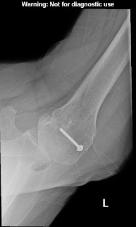


The degree of posterior subluxation can be measured as (a) the position of the center of the humeral head in relation to the plane of the scapula, (b) the position of the center of the humeral head in relation to the glenoid face or (c) the point of contact of the humeral articular surface on the glenoid articular surface. We prefer the latter because it is this point of contact that reflects the degree of centering of the net humeral joint reaction force on the glenoid. It is the malcentering of this joint reaction force that leads to posterior instability, posterior glenoid wear and to rocking horse loosening of prosthetic glenoid components. The standardized axillary view also enables the surgeon to see the shape of the glenoid surface. Three main types have been described: concentric wear (type A)


eccentric posterior wear (type B),

and dysplastic (type C)


In actual practice, there are so many intermediate types of glenoid pathoanatomy that rigorous separation into a few distinct classes is difficult. 


 An important aspect of glenoid pathology is the amount of the glenoid that is involved in the pathologic concavity, known as the ‘neoglenoid. Finally, the standardized axillary view enables the measurement of the degree of glenoid retroversion in relation to the body of the scapula. Thus, on the standardized axillary view, the surgeon can usually determine the major important characteristics of glenohumeral arthritic pathoanatomy: the amount of joint space narrowing, the degree of retroversion, the degree of posterior subluxation with the arm in a functional position, the glenoid shape, the percentage of the glenoid involved in the pathologic concavity and the angle of retroversion.


 Because of their low cost and freedom from metal artifacts, standardized axillary views provide a practical and reliable way to document the postoperative anatomy sequentially over time and to compare it to what was present before surgery.


A third view, the templating view, is obtained when humeral arthroplasty is being considered. This view is an anteroposterior (AP) view of the humerus taken with the arm in 30 degrees of external rotation relative to the x-ray beam with a magnification marker added. This view places the humeral neck in maximal profile and allows a comparison of proximal humeral anatomy with that of various humeral prostheses. In templating, it is important to recognize that the humeral canal is not cylindrical – the medial-lateral dimension is usually wider than the anteroposterior dimension so that the AP view may overestimate the size of the stem that will fit the diaphysis. This view is also useful for determining whether sufficient osteoporosis is present to merit special consideration at the time of arthroplasty 


Advanced imaging may be useful in the unusual case where the anatomy is distorted by prior injury or surgery, when there is concern about the amount of bone available for reconstruction, or when the standardized plain films cannot be obtained. In the great majority of cases, however, the extra cost and radiation of the CT scan can be avoided through the use of these standardized plain films. In that we can learn what we need to know about the status of the rotator cuff from physical examination and plain radiographs, shoulder MRIs are rarely needed unless indicated to exclude avascular necrosis or tumor. An MRI of the neck may be useful in evaluating patients suspected of having cervical radiculopathy, myelopathy, stenosis or a syrinx.

===
=====
The reader may also be interested in these posts:


Information about shoulder exercises can be found at this link.

Use the "Search" box to the right to find other topics of interest to you.

You may be interested in some of our most visited web pages including:shoulder arthritis, total shoulder, ream and run, reverse total shoulder, CTA arthroplasty, and rotator cuff surgery as well as the 'ream and run essentials'