Showing posts with label ingrowth. Show all posts

Sunday, October 22, 2017

Advantages of an all-polyethylene ingrowth glenoid component

Clinical and Radiographic Results of an All-Polyethylene Pegged Bone-Ingrowth Glenoid Component

These authors evaluated the early clinical and radiographic results in 83 shoulders having total shoulder arthroplasty with an all-polyethylene pegged glenoid component designed for hybrid (biological and cement) fixation.


At a mean followup of 46.7 months (range, 24 to 99 months), the median ASES score was 97 points (range, 43 to 100 points) and the median Oxford score was 48 points (range, 24 to 48 points). The median active forward elevation was 130 (range, 65 to 170), median external rotation was 45 (range, 5 to 80), and median internal rotation was to T11 (range, buttock to T4). Seventy-eight shoulders demonstrated a perfect Lazarus score for radiolucency (0, indicating no radiolucency). Sixty-eight shoulders demonstrated complete osseointegration, with bone ingrowth between all of the flanges seen on coronal CT.


5 demonstrated partial osseointegration; and 10 demonstrated osteolysis around the central peg. Most radiolucent lines were in the inferior pegs of the prosthesis. There were no correlations between the Yian CT scores and either the ASES or Oxford score (rho = 0.13 and 0.07, respectively). 

The glenoid inclination and version ranged from 62 to 96 degrees  and from -20 to 15 degrees, respectively. No association was found between glenoid inclination or version and the presence of radiolucent lines.

In addition to the 83 implants included in the study, 4 glenoid implants were revised, although only 1 revision was due to aseptic loosening; the others were due to rotator cuff failure, glenoid fracture, and Propionibacterium infection.

Comment: As seen in the figure below from the Australian national joint registry (see this link), all-polyethylene glenoid components have demonstrated the best survivorship. The particular all-polyethylene design discussed in this paper has the advantage of bone ingrowth without the disadvantages of a metal backing.
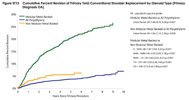


This all-poly glenoid also has the advantage of easy and bone preserving revision to a reverse, should that become necessary. In the revision. The central hole is drilled out and, if necessary, filled with bone graft followed by the usual insertion of the base plate and glenosphere.


The bone preservation with this type of conversion is in contrast to the amount of bone loss often associated with a failed metal backed component.


===

The reader may also be interested in these posts:


Information about shoulder exercises can be found at this link.

Use the "Search" box to the right to find other topics of interest to you.

You may be interested in some of our most visited web pages including:shoulder arthritis, total shoulder, ream and runreverse total shoulderCTA arthroplasty, and rotator cuff surgery as well as the 'ream and run essentials'


Monday, October 17, 2016

Failed prosthesis for fracture: the 'absent tuberosity sign'

One of the most difficult aspects of proximal humeral fracture surgery is getting the tuberosity reduced, fixed and healed. This problem is not made easier by placing a large humeral implant, which reduces the amount of bone left to reattach the displaced tuberosity.

Here's an example of this problem: a shoulder with pseudoparalysis after a post-traumatic deformity was treated with a total shoulder. Note on the AP view that the tuberosity is missing (there is no bone lateral to the arrow).

The axillary view shows the displaced, ununited greater tuberosity fragments (arrows) as well as some anterior translation of the humeral head on the glenoid.

Revision of this prosthesis to a reverse total will be complicated by (1) the bony ingrowth surface on the humeral prosthesis 


as well as (2) the bone loss that may occur when the glenoid component is removed.

Unless the tuberosity can be mobilized and fixed securely, a primary reverse total shoulder might have been a consideration at the index surgery, as shown in the x-ray below of a reverse total shoulder  (also with an 'absent tuberosity sign').

Note that, in contrast to our usual impaction grafting approach, this stem needed cement for control of prosthetic height and version.

===

Saturday, November 1, 2014

Bone ingrowth humeral components - two reasons why we avoid them. What if Propionibacterium are on them?

In some circles there is an interest in humeral components that encourage the ingrowth of bone by having a surface that is porous coated, grit blasted, or trabecular metal. We avoid such humeral components for two reasons.

First, a smooth humeral stem can be securely fixed in the humeral canal without bone ingrowth using impaction grafting as shown in this link and here and here. The advantage of impaction grafting is that a special surfaced prosthesis is not required and the need to broach the endosteum for a tight fit is eliminated.

Second, all humeral components are at potential risk for revision, whether because of malposition, infection, or the need for revision to a different prosthesis. Recently, the revision burden was reported to be 4.7% for hemiarthroplasty and 15.0% for total shoulder arthroplasty. Another recent article reported that revision cases comprised 8.8% and 8.2% of all shoulder arthroplasties in 2009 and 2011.  Yet another recent study found an 11% revision rate for total shoulders. These numbers suggest that at least 1 in 12 shoulder arthroplasties - over 6000/yr - are requiring revision!

Revision of an ingrowth humeral stem usually requires not only a split in the humeral shaft (see this link) but also circumferential osteotomization of the bone around the prosthesis with further fragmentation of the cortex and compromise of the fixation of the revision prosthesis.

Here is a recent example. After a prior surgery to remove a loose glenoid, intraoperative cultures were overwhelmingly positive for Propionibacterium in spite of the lack of serologic or intraoperative findings suggestive of infection. The ingrowth stem was not removed even though it had been placed in retroversion. The postoperative films are shown below.

However, the symptoms of infection - including pain in the arm - did not respond to a postoperative course of IV antibiotics and oral suppression. A subsequent revision was carried out to remove the humeral implant out of concern for its being a host to Propionibacterium in a biofilm. In addition to the issue of bone ingrowth into the proximal porous coated stem, close examination of the films pointed to 'spot welds' between the bone and the prosthesis (marked with lines on the x-ray below left) corresponding to the junction of the porous coated and the smooth section of the stem (below right).


 As anticipated, a humeral split and multiple circumferential cuts with a curved osteotome were required before the prosthesis could be removed with a needle nosed vice-grip and mallet.


After a thorough washout, the humerus was reconstructed with a smooth stemmed component, Vancomycin soaked allograft, and bodice reconstruction of the humeral shaft.


Motion exercises are being withheld until the humerus consolidates.
===

Consultation for those who live a distance away from Seattle.

Click here to see the new Shoulder Arthritis Book

Click here to see the new Rotator Cuff Book

To see the topics covered in this Blog, click here

Use the "Search" box to the right to find other topics of interest to you.

You may be interested in some of our most visited web pages including:shoulder arthritis, total shoulder, ream and runreverse total shoulderCTA arthroplasty, and rotator cuff surgery as well as the 'ream and run essentials'

Tuesday, September 9, 2014

Failure of a bone ingrowth total shoulder + cuff repair: revision to reverse total shoulder

A 60 year old patient had a simultaneous total shoulder and rotator cuff repair at an outside institution one year ago. A bone ingrowth prosthesis and a large humeral head were used as shown in the x-rays taken immediately after the procedure as shown below.


After surgery she presented to our service because of inability to raise her arm and pain. Her physical examination demonstrated pseudoparalysis (the inability to raise the arm away from the side in spite of a good passive range of motion). Our x-rays are shown below. The AP view shows upwards displacement of the humeral head relative to the glenoid and excessive lateralization of the tuberosity with respect to the glenoid and lateral acromion.


 The axillary view suggests loosening of the metal+polyethylene glenoid component.


At the time of our revision surgery the superior rotator cuff was deficient and the subscapularis was absent. Cultures were taken before antibiotics were administered. The loose glenoid component was removed. An attempt was made to convert to a reverse total shoulder without removing the extant bone ingrowth stem. However, the components could not be made to fit properly. 

It was, therefore, necessary to osteotomize the proximal humeral metaphysis to extract the bone ingrowth stem. With the stem removed, a reverse total shoulder could be accomplished.  Bone graft was required around the central screw to compensate for the bone lost from the prior glenoid implantation. Excellent fixation of the glenoid component was achieved.


Comment: This case illustrates a number of important considerations. First, the combination of cuff deficiency and shoulder arthritis can be difficult to manage with a cuff repair and anatomic total shoulder - the cuff repair is at risk for failure as occurred here. A large humeral head component places additional load on the repaired cuff. Second, glenoid components consisting of both metal and polyethylene may be at higher risk of failure due to the dissimilar material properties as explained here. All polyethlene glenoid components have a lower revision rate. Third, bone ingrowth components complicate revision and in our practice are not used because impaction grafting provides excellent fixation without complicating the possible need for subsequent component removal. Fourth, while it seems attractive to 'simply' convert an anatomic stem to a reverse, this is often not possible because of the need to place the stem of the reverse more distally to accommodate the position of the glenosphere.

===
Consultation for those who live a distance away from Seattle.

Click here to see the new Shoulder Arthritis Book

Click here to see the new Rotator Cuff Book

To see the topics covered in this Blog, click here

Use the "Search" box to the right to find other topics of interest to you.

You may be interested in some of our most visited web pages including:shoulder arthritis, total shoulder, ream and runreverse total shoulderCTA arthroplasty, and rotator cuff surgery as well as the 'ream and run essentials'


Sunday, August 24, 2014

Failed glenoid component, lessons and questions

Our readers are aware that the glenoid component is the 
 in total shoulder arthroplasty.

Recently we were consulted on a patient from elsewhere whose post operative x-ray taken two years ago had this appearance
Here's a closer view of his glenoid fixation showing lucency's around the pegs of the glenoid component
The dark areas between the pegs and the bone in postoperative films are filled with fluid and/or soft tissue so that the component is not well fixed in the bone. As shown in an earlier post, we use a CO2 jet to remove fluid and soft tissue from the bone/cement interface. This enables pressurized cement to completely fill the space between the pegs and the bone, optimizing component fixation. This practice has essentially eliminated the issue of postoperative periprosthetic lucent lines.

Two years later, we met this shoulder for the first time. At the time of presentation to us for consultation the patient had been experiencing increasing pain in the shoulder since his original total shoulder arthroplasty. The x-rays taken at our first visit are shown below. Note the osteolysis around the glenoid component pegs on the AP view
 and the 'watermelon seed' of cement between the glenoid component and the bone posteriorly on the axillary view. The problem with this 'watermelon seed' is shown in this prior post.
 After discussion of the alternatives and considering the possibility of Propionibacterium infection, the patient desired to have a revision with complete prosthesis removal, harvest of specimens for culture, thorough debridement, insertion of a new prosthesis with Vancomycin allograft fixation, and the red antibiotic protocol.

At surgery, the joint fluid was turbid and the glenoid component was completely loose; the 'watermelon seed' was floating free in the joint space. Removal of the trabecular metal stem was very difficult because of bone ingrowth; multiple osteotomies around the component were required. A modified bodice repair was needed to close the humeral osteotomies made for component removal. A glenoid component was not used in the reconstruction.

Note that bone ingrowth is not necessary for humeral component fixation and greatly complicates revision.

The postoperative films are shown below.


Immediate assisted range of motion exercises were started after surgery. IV antibiotics will be continued for 6 weeks and transitioned to oral antibiotics if the cultures (which are now pending) become positive. 

The patient reports that the shoulder is already more comfortable than in the prior two years. We are hopeful.

===
Consultation for those who live a distance away from Seattle.

Click here to see the new Shoulder Arthritis Book

Click here to see the new Rotator Cuff Book

To see the topics covered in this Blog, click here

Use the "Search" box to the right to find other topics of interest to you.

You may be interested in some of our most visited web pages including:shoulder arthritis, total shoulder, ream and runreverse total shoulderCTA arthroplasty, and rotator cuff surgery as well as the 'ream and run essentials'


To see other similar posts, click on the label of interest below.

Friday, November 30, 2012

Ingrowth glenoid component

Fixation and durability of a bone-ingrowth component for glenoid bone loss JSES


This article report the use of a metal backed glenoid in the management of glenohumeral arthritis in the face of central or peripheral glenoid bone deficiencies from either arthritic wear or failure of prior arthroplasty. The authors point out to the common deficiency of posterior glenoid bone in shoulder arthroplasty and the challenges associated with reconstruction in this context. While many authors are fixated on the 'need for normalizing glenoid version' there is another view as pointed out in a recent post. Recent enthusiasm for the use of step or asymmetrical glenoid components to achieve stability does not seem to be supported by the clinical results.

This article presents an attempt to achieve a robust glenoid arthroplasty using a a metal backed glenoid. By 11 years after surgery 7 of the 21 arthroplasties required revision. Among the unrevised shoulders  4 were at risk for glenoid loosening, and 1 was at risk for humeral loosening.  Common problems included accelerated polyethylene wear, metal wear, bone osteolysis, and a high frequency of clinical glenoid component loosening.

These results with a metal backed glenoid component are consistent with previous reports and this prior report by the same senior authors.


----


Use the "Search" box to the right to find other topics of interest to you.

You may be interested in some of our most visited web pages including:shoulder arthritis, total shoulder, ream and runreverse total shoulderCTA arthroplasty,  and rotator cuff surgery.