Showing posts with label glenoid version. Show all posts

Sunday, March 29, 2026

Glenoid component version in anatomic total shoulder arthroplasty- does it matter?


Almost 30 years ago Gilles Walch called our attention to arthritic glenoid retroversion. Morphologic study of the glenoid in primary glenohumeral osteoarthritis

From that point on, there has been great interest in the version of the arthritic glenoid: what is it?, how should it be measured?. 

And in the execution of an anatomic total shoulder (aTSA),  should the version of the glenoid component be "corrected" to some particular value?, if so what value?, how should this correction be achieved? and does changing the preoperative version affect the clinical outcome of aTSA?

It goes without saying that a lot of time and money can go into the evaluation and management of arthritic glenoid retroversion. Perhaps it's time to see how much glenoid component matters to the patient.

About 15 years ago, Patterns of loosening of polyethylene keeled glenoid components after shoulder arthroplasty for primary osteoarthritis: results of a multicenter study with more than five years of follow-up pointed out that posterior tilting of the glenoid component was associated with preoperative posterior decentering and with excessive reaming. The authors suggested that preserving subchondral bone may be important for long-term longevity of the glenoid component.

Glenoid component retroversion is associated with osteolysis found that osteolysis around the center peg of a glenoid component was correlated with component retroversion of ≥15°, the paper clearly stated that "the presence of osteolysis around the center peg was not correlated with a worse clinical outcome defined by shoulder scores or a reoperation due to glenoid loosening". 

Nevertheless, achieving component retroversion of <15° has become a goal for many surgeons and an opportunity for orthopaedic companies who have made substantial investments in three-dimensional planning platforms, patient specific instrumentation, navigation, augmented / virtual reality and robotic assisted glenoid preparation.

A recent paper,  Does postoperative glenoid component retroversion following anatomic total shoulder arthroplasty affect clinical outcomes? A systematic review and meta-analysis assessed the clinical importance of implanting the glenoid component in <15° of retroversion. After screening 2,457 articles, 15 studies comprising 1,190 shoulders met inclusion criteria. Patients were stratified by whether postoperative glenoid component retroversion was <15° or ≥15°The principal finding was that no clinically significant differences were observed between the two groups in patient-reported outcome scores, range of motion, or complications. 

An unexpected and important finding in this meta-analysis was that shoulders with ≥15° of postoperative retroversion were actually more likely to have no radiolucency (a Lazarus grade 0 radiographic score) than those with <15° retroversion (76.9% vs. 55.6%; P = .00021). This finding argues against the presumption that retroversion promotes loosening. 

Does Postoperative Glenoid Retroversion Affect the 2-Year Clinical and Radiographic Outcomes for Total Shoulder Arthroplasty? directly addressed the question in a case-control study. At 2-year follow-up, patients with glenoid components implanted in ≥15° retroversion had similar improvement in Simple Shoulder Test (SST) scores, similar final outcome scores, and similar radiographic findings compared to those with <15° retroversion. Notably, none of the patients with retroverted glenoid components underwent revision surgery, compared to 3 of 50 patients in the non-retroverted group.

Anatomic Total Shoulder Arthroplasty with All-Polyethylene Glenoid Component for Primary Osteoarthritis with Glenoid Deficiencies reported outcomes of aTSA with conservative, noncorrective reaming in shoulders with glenoid deficiencies, including those with significant retroversion. Mean postoperative retroversion in this series was 16°, yet mean postoperative SST was 9 out of 12, consistent with the excellent results achieved in shoulders without glenoid deformity. The revision rate was zero in 143 shoulders at mean follow-up of 34 months.

Anatomic total shoulder arthroplasty for posteriorly eccentric and concentric osteoarthritis: a comparison at a minimum 5-year follow-up compared outcomes of aTSA for posteriorly eccentric (Walch B) versus concentric osteoarthritis at minimum 5-year follow-up, finding no significant difference in ASES scores, revision rates, or radiographic loosening between groups. These durable results were achieved without attempting retroversion correction.

Does glenoid version and its correction affect outcomes in anatomic shoulder arthroplasty? A systematic review analyzed 16 studies and 1,211 shoulders finding that 8 of 11 reports found no significant association between pre- or postoperative glenoid retroversion and clinical results, including patient-reported outcomes, range of motion, and revision rates.

Total shoulder arthroplasty outcomes after noncorrective, concentric reaming of B2 glenoids reported a 95% implant survivorship at a mean of 4.9 years in a series treated with noncorrective reaming with a mean postoperative retroversion of 19°.

The Effect of Version Correction Techniques

If clinical outcomes are equivalent regardless of postoperative retroversion, the question becomes whether correction efforts add value without adding risk.  Does postoperative glenoid component retroversion following anatomic total shoulder arthroplasty affect clinical outcomes? A systematic review and meta-analysis compared three correction strategies: posteriorly augmented glenoids, eccentric (“high-side”) reaming, and conservative noncorrective reaming. Eccentric reaming was associated with a significantly higher complication rate than noncorrective reaming (9.3% vs. 3.1%; P = .043, OR 3.22) and a significantly higher revision rate (7.4% vs. 1.2%; P = .015, OR 6.18). 

Loss of the dense subchondral bone layer from corrective reaming may result in reduced glenoid component support, increased micromotion, and greater loosening risk over time. 


Implications for 3D CT Planning and Technology Transfer

CT-based three-dimensional preoperative planning, patient specific instrumentation, intraoperative navigation, augmented/virtual reality, and robotic glenoid preparation are all predicated on the same clinical logic: that achieving a glenoid component position closer to neutral version will improve patient outcomes. As reviewed above, the available evidence indicates that differences in postoperative glenoid component retroversion do not produce clinically detectable differences in pain relief, function, or implant survival at the follow-up intervals studied.

Use of Preoperative CT Scans and Patient-Specific Instrumentation May Not Improve Short-Term Adverse Events After Shoulder Arthroplasty: Results from a Large Integrated Health-Care System compared aTSA with and without preoperative CT scanning and PSI. These technologies expose patients to additional radiation from CT scanning and incur substantially greater costs of care. Use of CT scans and PSI did not reduce the rate of short-term adverse events following shoulder arthroplasty. Patients receiving PSI may be at greater risk of deep vein thrombosis or deep infection, possibly reflecting the additional operative time this technology requires. 

Three-dimensional computed tomography analysis of pathologic correction in total shoulder arthroplasty based on severity of preoperative pathology analyzed 152 shoulders with 3D CT postoperatively and found that while glenoid component shift occurred in 51% of patients, neither component shift nor central peg osteolysis was associated with worse clinical outcomes at minimum 2-year follow-up. 

What the Evidence Does Suggest About Optimizing aTSA outcomes?

The available evidence points to factors other than retroversion that drive aTSA results. 

See: Below left-poor glenoid seating with cement interposed between the glenoid component and the bone. 

Below right-posterior decentering due to poor glenoid preparation and ill-advised use of a posteriorly eccentric humeral head component.


Surgeons may wish to consider 6 aspects of aTSA that are relevant to the glenoid side of the arthroplasty.

(1) conservative reaming to retain the maximal amount of quality host bone

(2) rather than "correcting" glenoid retroversion (A, below), consider "accepting" it (B, below)


(3) component seating — good carpentry with complete backside contact of the glenoid implant against prepared bone; no cement between the component and the bone.


(4) humeral head centering on the glenoid achieved through soft tissue balancing and the possible us of an anteriorly eccentric humeral component.


(5) awareness that technologies can lead surgeons to prioritize postoperative glenoid component retroversion <15° retroversion (without acknowledging the potential risks)


(6) recognition that specialized (e.g. augmented) glenoid components may have downstream risks (see chart below from the 2025 AOANJRR)


Here is a thought provoking case in which substantial glenoid retroversion was accepted


Function at 14 years


Conclusion:
Glenoid component version may not be as critical to the outcome of aTSA as secure seating of the glenoid component on quality host bone and centering of the humeral head on the prosthetic glenoid. 

Rather than improving outcomes, there is evidence that "corrective reaming" can be associated with a significantly higher complication rate than noncorrective reaming (9.3% vs. 3.1% ) and a significantly higher revision rate (7.4% vs. 1.2%).

Seating and balance


Yellow-headed blackbird
Malheur
2024


Follow on twitter/X: https://x.com/RickMatsen

Follow on facebook: https://www.facebook.com/shoulder.arthritis

Follow on LinkedIn: https://www.linkedin.com/in/rickmatsenmd


Friday, October 11, 2024

Does postoperative glenoid version matter in anatomic total shoulder arthroplasty? The jury is in.


Much innovation, time, money, and technology are being spent on "correcting" glenoid version in anatomic total shoulder arthroplasty. Are these resources being well spent? Is "correcting" glenoid retroversion better than "accepting" it (see this link)?

The published evidence indicates that postoperative prosthetic glenoid retroversion is not associated with worse patient outcomes after anatomic total shoulder arthroplasty. 

Rather than the amount of retroversion, the quality of the seating of the glenoid component appears to be the primary driver of the clinical outcome. 

The importance of seating of the glenoid component is demonstrated in 
Edge displacement and deformation of glenoid components in response to eccentric loading. The effect of preparation of the glenoid bone

The use of augmented glenoid components may make it more difficult to achieve perfect seating (see this link and this link).

Let's look at some recent articles from the peer-reviewed literature

The authors of Anatomic Total Shoulder Arthroplasty with All-Polyethylene Glenoid Component for Primary Osteoarthritis with Glenoid Deficiencies used standard all polyethylene non-augmented glenoid components inserted without attempt to "correct" glenoid retroversion in managing glenohumeral arthritis in patients with types A1, A2, B1, B2, and B3 glenoid pathoanatomy. 
The average postoperative Simple Shoulder Test Scores tended to be higher for the glenoid components inserted in more retroversion.


Postoperative glenoid version was not significantly different from preoperative glenoid version. The mean humeral-head decentering on the glenoid face was reduced for type-B2 glenoids from -14% ± 7% preoperatively to -1% ± 2% postoperatively (p < 0.001) and for type-B3 glenoids from -4% ± 6% preoperatively to -1% ± 3% postoperatively (p = 0.027). The rates of bone integration into the central peg for type-B2 glenoids (83%) and type-B3 glenoids (81%) were not inferior to those for other glenoid types

The authors of Anatomic total shoulder arthroplasty for posteriorly eccentric and concentric osteoarthritis: a comparison at a minimum 5-year follow-up compared the clinical and radiographic outcomes of 210 TSAs using conservative glenoid reaming with no attempt at version correction for patients with and without eccentric wear patterns.There were no differences in outcome measures between patients with postoperative retroversion of more and less than 15 degrees . On multivariable analysis, glenoid component radiolucencies were most strongly associated with incomplete component seating rather than with postoperative glenoid component version.

The authors of Does Postoperative Glenoid Retroversion Affect the 2-Year Clinical and Radiographic Outcomes for Total Shoulder Arthroplasty? reported on patients undergoing anatomic TSA in whom no specific efforts were made to change the version of the glenoid. They compared the outcomes for 21 patients in which the glenoid component was implanted in 15 degrees or greater retroversion to those for the 50 in which it was implanted in less than 15 degrees retroversion. The improvement in the Simple Shoulder Test (6.7 ) for the retroverted group was not inferior to that for the nonretroverted group (5.8)). The percent of maximal possible improvement (%MPI) for the retroverted glenoids (70%) was not inferior to that for the nonretroverted glenoids (67%). No patient in either group reported symptoms of subluxation or dislocation. The radiographic results for the retroverted glenoid group were similar to those for the nonretroverted group with respect to central peg lucency, average Lazarus radiolucency scores, and the mean percentage of posterior humeral head decentering. None of the patients with retroverted glenoids underwent revision in comparison to 3 of the 50 patients with nonretroverted glenoids who required revision. They concluded that postoperative glenoid retroversion was not associated with inferior clinical results at 2 years after surgery.

The authors of Does glenoid version and its correction affect outcomes in anatomic shoulder arthroplasty? A systematic review evaluated studies on the effect of preoperative and postoperative glenoid retroversion on clinical functional and radiologic outcomes in patients who underwent anatomic TSA. They concluded that there is currently insufficient evidence that pre- or postoperative glenoid version influences postoperative outcomes independent of other morphologic factors such as joint line medialization. Given that noncorrective reaming demonstrated favorable postoperative outcomes, and postoperative glenoid version was not significantly and consistently found to impact outcomes, these authors find that there is inconclusive evidence that correcting glenoid retroversion is routinely required.

The authors of Factors associated with functional improvement after posteriorly augmented total shoulder arthroplasty pointed out that posteriorly augmented glenoid components in anatomic total shoulder arthroplasty attempt to address posterior glenoid bone loss but have inconsistent clinical results. They performed a retrospective review of 50 patients who underwent TSA with a step-type augmentation performed by a single surgeon between 2009 and 2018. Glenoid morphology included type B2 glenoids in 41 patients and type B3 glenoids in 9. Postoperative ROM and function showed no clinically important associations with postoperative glenoid retroversion. Component loosening was frequent among shoulders with 7 mm augmentation.


The authors of Do glenoid retroversion and humeral subluxation affect outcomes following total shoulder arthroplasty? investigated in 113 patients whether glenoid retroversion and humeral head subluxation were associated with inferior outcomes after TSA and whether change of retroversion influences outcomes after TSA. They found no correlation between postoperative glenoid version or humeral head subluxation and ASES scores. For patients with preoperative retroversion of >15 degrees , there was no difference in outcome scores based on postoperative retroversion. There were no differences in preoperative or postoperative version for patients with or without glenoid lucencies. They observed no significant relationship between postoperative glenoid retroversion or humeral head subluxation and clinical outcomes in patients following TSA. 

The authors of Glenoid component retroversion is associated with osteolysis found that postoperative glenoid component retroversion was correlated with osteolysis around the glenoid center peg but that the presence of osteolysis around the center peg was not correlated with a worse clinical outcome defined by shoulder scores or a reoperation due to glenoid loosening.

The authors of Total shoulder arthroplasty in patients with a B2 glenoid addressed with corrective reaming: mean 8-year follow-up reviewed 59 patients finding that glenoid component failure was associated with worse initial glenoid component seating but that there was no association between glenoid component failure and preoperative retroversion, inclination, or humeral head subluxation.

The authors of Total shoulder arthroplasty outcomes after noncorrective, concentric reaming of B2 glenoids reviewed their outcomes for 51 patients with B2 glenoids having a mean retroversion of 19.1 degrees (range 5.4 degrees -38 degrees ) who were treated with non-corrective reaming. These patients had significant improvement in clinical outcome scores, high patient satisfaction, and high survivorship ( implant survivorship rate was 95% at a mean follow-up of 4.9 years).

Glenoid retroversion does not impact clinical outcomes or implant survivorship after total shoulder arthroplasty with minimal, noncorrective reaming in 151 anatomic total shoulder arthroplasties the mean preoperative retroversion was 15.6 degrees. Total shoulder arthroplasty was performed without corrective reaming. Higher values of retroversion were not associated with early deterioration of clinical outcomes, revisions, or failures.

As we see from the above measurements of glenoid version are important to understanding and managing glenohumeral pathoanatomy. A recent article reviewed glenoid version measurements before and after shoulder arthroplasty:Inconsistencies in Measuring Glenoid Version in Shoulder Arthroplasty: A Systematic Review. They considered 61 studies encompassing 17,070 shoulder arthroplasties. Less than half (44%) of these described explicitly how glenoid version was measured. Often different methods were used to measure version before and after arthroplasty: preoperartive glenoid version was assessed using computed tomography in 75% of the cases; by contrast, over 50% of the studies that measured postoperative version used axillary radiographs. If we are to understand the preoperative to postoperative changes in glenoid version, we need for the measurements to be made using the same imaging modality.

As shown in Accuracy and reliability of postoperative radiographic measurements of glenoid anatomy and relationships in patients with total shoulder arthroplasty the positions of the humerus and scapula are quite different for axillary and CT images.


Comment: It seems that the "most important thing" in anatomic total shoulder arthroplasty is excellent seating of the glenoid component, rather than whether the glenoid component is inserted in more or less than fifteen degrees. 


You can support cutting edge shoulder research that is leading to better care for patients with shoulder problems, click on this link

Follow on twitter/X: https://x.com/RickMatsen
Follow on facebook: https://www.facebook.com/shoulder.arthritis
Follow on LinkedIn: https://www.linkedin.com/in/rick-matsen-88b1a8133/

Here are some videos that are of shoulder interest
Shoulder arthritis - what you need to know (see this link).
How to x-ray the shoulder (see this link).
The ream and run procedure (see this link).
The total shoulder arthroplasty (see this link).
The cuff tear arthropathy arthroplasty (see this link).
The reverse total shoulder arthroplasty (see this link).
The smooth and move procedure for irreparable rotator cuff tears (see this link).
Shoulder rehabilitation exercises (see this link). 


Saturday, January 6, 2024

"Posterior humeral subluxation" is a result of glenoid retroversion; is it a clinical problem?

A recent article, Total Shoulder Arthroplasty for Primary Glenohumeral Osteoarthritis: does the posterior humeral subluxation persist after correction of the glenoid version at 5 years minimum?, used the term "posterior humeral subluxation" to refer to the percentage (a/D (%)) of the humeral head posterior to the scapular axis. 


  1. The authors observed that "posterior humeral subluxation" was highly correlated (p<0.0001) with glenoid version both preoperatively 

    and postoperatively


    This result was noted in a prior post Arthritic subluxation of the shoulder - what does it mean?.


    In this diagram from that post, the percentage of posterior displacement of the humeral head relative to the scapular axis (blue symbols) was strongly correlated with the glenoid retroversion, irrespective of Walch glenoid type. By contrast, the percentage of posterior displacement (decentering) of the humeral head relative to the glenoid face (red symbols) was not strongly related to glenoid retroversion, but was strongly related to Walch glenoid type (note differences between type A1 and A2 glenoids in comparison to types B1, B2, B3 and C).

  2. The reason for the strong relationship between glenoid retroversion and the posterior displacement of the humeral head relative to the scapular axis can be seen by comparing the two diagrams below; in each the humeral head is centered in the glenoid fossa. 
    The first shows that 5 degrees of retroversion results in 6% posterior displacement of the humeral head in relation to the scapular axis (equivalent to 56% "posterior humeral subluxation").

  1. The second shows that 25 degrees of retroversion results in 38% posterior displacement of the humeral head in relation to the scapular axis (equivalent to 88% "posterior humeral subluxation").
    Note that the values obtained from these simple diagrams (green dots) are highly consistent with actual clinical values reported above.


  1. Other studies have also pointed out that posterior displacement of the humeral head in relation to the plane of the scapula is determined primarily by the degree of glenoid retroversion.  Importance of a three-dimensional measure of humeral head subluxation in osteoarthritic shoulders found that the correlation between retroversion and posterior displacement of the humeral head relative to the body of the scapula had an R2  of 0.8634. A similar relationship can be seen in the data for 707 shoulders from Identification of threshold pathoanatomic metrics in primary glenohumeral osteoarthritis. Qualitative and quantitative analysis of glenoid bone stock and glenoid version: inter-reader analysis and correlation with rotator cuff tendinopathy and atrophy in patients with shoulder osteoarthritis found that the correlation between retroversion and posterior displacement of the humeral head relative to the plane of the scapula had an R2of 0.8161. CT assessment of the relationship between humeral head alignment and glenoid retroversion in glenohumeral osteoarthritis found “a nearly perfect linear relationship between glenoid retroversion and humeral-scapular alignment, with a Pearson correlation coefficient of 0.90 (R2 = 0.81, p < 0.001).” 

  2. From all these studies we can conclude that >80% of the variance in the alignment of the humeral head to the plane of the scapula is determined by glenoid retroversion.

    Two prior posts discuss whether or not it is important to "correct" glenoid retroversion when the humeral head is centered in the glenoid socket. "Subluxation of the shoulder", why does it matter? and Does glenoid version need to be corrected in anatomic shoulder arthroplasty? Interestingly, Static posterior humeral head subluxation and total shoulder arthroplasty found that re-centering of the humeral head on the glenoid was not correlated with glenoid version or its correction.

    You can support cutting edge shoulder research and education that are leading to better care for patients with shoulder problems, click on this link.

Follow on twitter: https://twitter.com/RickMatsen or https://twitter.com/shoulderarth
Follow on facebook: click on this link
Follow on facebook: https://www.facebook.com/frederick.matsen
Follow on LinkedIn: https://www.linkedin.com/in/rick-matsen-88b1a8133/

Here are some videos that are of shoulder interest
Shoulder arthritis - what you need to know (see this link).
How to x-ray the shoulder (see this link).
The ream and run procedure (see this link).
The total shoulder arthroplasty (see this link).
The cuff tear arthropathy arthroplasty (see this link).
The reverse total shoulder arthroplasty (see this link).
The smooth and move procedure for irreparable rotator cuff tears (see this link).
Shoulder rehabilitation exercises (see this link).

Saturday, January 14, 2023

How to define the pathoanatomy of the arthritic shoulder using a pair of plain x-rays

Surgeons and patients need to understand the anatomical characteristics of the arthritic shoulder before treatment is discussed. While there is current enthusiasm for routinely obtaining CT scans for this purpose, in the great majority of cases a pair of plain x-rays can provide all the necessary information without the time, cost and radiation exposure resulting from a CT scan. 

As in all things orthopaedic, the technical detains are essential for obtaining the desired information.

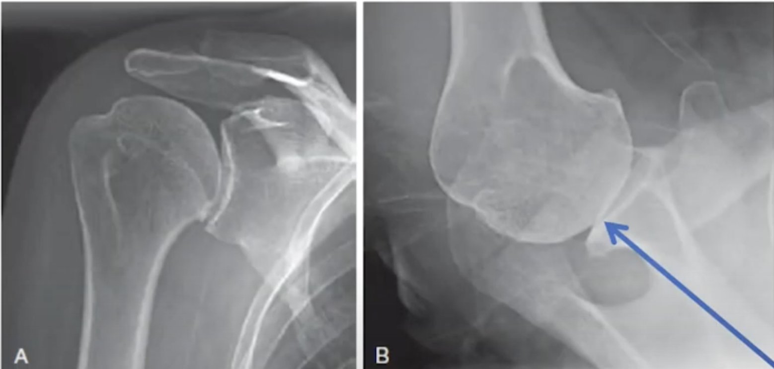
The first of the pair is the anteroposterior view in the plane of the scapula. In this view, the patient's scapula is placed flat on the x-ray cassette while the x-ray beam is perpendicular to the plane of the scapula and aimed at the coracoid.  The forearm of the flexed elbow is rotated 30 degrees from the x-ray beam in order to show the humeral head in profile.


The resulting view reveals such pathological features as flattening of the humeral head, humeral osteophytes, joint space narrowing, glenoid wear, and the superior/inferior relationship of the humeral head to the glenoid and the acromion.


The second of the pair is the axillary "truth" view. In this view, the patient's arm is elevated in the plane of the scapula while the beam is oriented in the plane of the scapula. Proper orientation is confirmed by visualizing the "eye" of the spinoglenoid notch (blue arrow). 


The resulting view reveals such pathological features as the version of the glenoid in relation to the scapular body, flattening or biconcavity of the glenoid face, medial glenoid erosion, glenoid and humeral osteophytes, and the degree of decentering of the humeral head on the face of the glenoid. 


This information cannot be obtained from an improperly oriented image such as that shown below. 


A properly done axillary "truth" view can be used to identify the variation in glenoid types.


Shown below are a few examples of pairs of x-rays that provided the necessary and sufficient information to plan and execute definitive surgical management of the arthritic shoulder.


One of the great values of standardized axillary "truth" views is the ability to compare preoperative and postoperative relationships, something that is not practical with CT scans.  See the examples below, recognizing how comparable the views are from before and after surgery (note the "eyes" indicated by arrows).


While preoperative CT scans may be useful for characterizing complex glenohumeral arthritic anatomy, the value to the patient of routine CT scans in most cases of glenohumeral arthritis has yet to be demonstrated. 

You can support cutting edge shoulder research that is leading to better care for patients with shoulder problems, click on this link.

Follow on twitter: https://twitter.com/shoulderarth
Follow on facebook: click on this link
Follow on facebook: https://www.facebook.com/frederick.matsen
Follow on LinkedIn: https://www.linkedin.com/in/rick-matsen-88b1a8133/

Here are some videos that are of shoulder interest
Shoulder arthritis - what you need to know (see this link).
How to x-ray the shoulder (see this link).
The ream and run procedure (see this link).
The total shoulder arthroplasty (see this link).
The cuff tear arthropathy arthroplasty (see this link).
The reverse total shoulder arthroplasty (see this link).
The smooth and move procedure for irreparable rotator cuff tears (see this link).
Shoulder rehabilitation exercises (see this link).

Sunday, November 22, 2020

Total shoulder arthroplasty for type B2 and B3 glenoid using standard glenoid components without version correction

Anatomic Total Shoulder Arthroplasty with All-Polyethylene Glenoid Component for Primary Osteoarthritis with Glenoid Deficiencies

These authors evaluated the ability of shoulder arthroplasty using a standard all-polyethylene glenoid component 


to improve patient self-assessed comfort and function and to correct preoperative humeral-head decentering on the face of the glenoid in patients with primary glenohumeral arthritis and type-B2 or B3 glenoids.


They identified 66 shoulders with type-B2 glenoids (n = 40) or type-B3 glenoids (n = 26) undergoing total shoulder arthroplasties with a non-augmented glenoid component inserted without attempting to normalize glenoid version and with clinical and radiographic follow-up that was a minimum of 2 years. 


The Simple Shoulder Test (SST) score improved from 3.2 ± 2.1 points preoperatively to 9.9 ± 2.4 points

postoperatively at a mean time of 2.8 ± 1.2 years for type-B2 glenoids and from 3.0 ± 2.5 points preoperatively to 9.4 ± 2.1 points postoperatively at a mean time of 2.9 ± 1.5 years for type-B3 glenoids; these results were not inferior to those for shoulders with other glenoid types. 


Postoperative glenoid version was not significantly different from preoperative glenoid version. 


The mean humeral-head decentering on the glenoid face was reduced for type-B2 glenoids from -14%  preoperatively to -1% postoperatively and for type-B3 glenoids from -4% preoperatively to -1% postoperatively.


The rates of bone integration into the central peg for type-B2 glenoids (83%) and type-B3 glenoids (81%) were not inferior to those for other glenoid types (A1 - 67%, A2 - 85%, B1 - 74%, C - 75%).


The authors concluded that shoulder arthroplasty with a standard glenoid inserted without changing version can significantly improve patient comfort and function and consistently center the humeral head on the glenoid face in shoulders with type-B2 and B3 glenoids, achieving >80% osseous integration into the central peg. These clinical and radiographic outcomes for type- B2 and B3 glenoids were not inferior to those outcomes for other glenoid types.


Another interesting aspect was the comparison of data from this study obtained with axillary x-rays to published data for CT scans for the typical glenoid version and decentering seen with different glenoid types.


Our approach to total shoulder arthroplasty can be viewed by clicking here.
To support our research to improve outcomes for patients with shoulder problems, click here.
To subscribe to this blog, enter your email in the box to your right that looks like the below


===
How you can support research in shoulder surgery Click on this link.

We have a new set of shoulder youtubes about the shoulder, check them out at this link.

Be sure to visit "Ream and Run - the state of the art" regarding this radically conservative approach to shoulder arthritis at this link and this link

Use the "Search" box to the right to find other topics of interest to you.

You may be interested in some of our most visited web pages  arthritis, total shoulder, ream and runreverse total shoulderCTA arthroplasty, and rotator cuff surgery as well as the 'ream and run essentials'