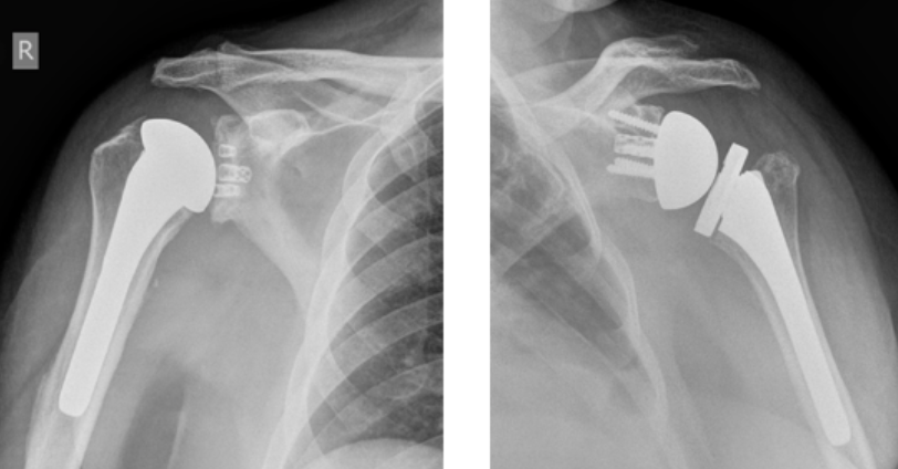
Showing posts with label reverse total shoulder complications. Show all posts

Saturday, December 13, 2025

Preventing Acromial/Scapular Spine Fractures in Reverse Shoulder Arthroplasty: Defining what the surgeon can control.


I'll start out with a few conclusions:

(1) Acromial and scapular spine stress fractures are clinically important complications of reverse total shoulder arthroplasty (RSA), occuring in 3-11% of cases and often resulting in poor functional outcomes such as persistent pain and limited active motion. 

(2) The principal risk factors - female sex, advanced age, poor bone quality, inflammatory arthropathy, cuff deficiency, corticosteroid use, thinned acromion from prior surgery or erosion, proximal humeral migration - are not under the control of the surgeon, except as they affect the decision to proceed with RSA surgery. 

(3) Surgeons do control humeral and glenosphere component selection and positioning. However the evidence guiding practice is not robust due to the lack of standardized nomenclature and what parameters should be measured in future clinical research. 

Here are a set of four easy to make measurements the importance of which is supported by the review below. These address the problem of uncertainty and inconsistency found in published studies regarding RSA geometry. Such measurements will be important in answering the key questions surgeons have: what component positions provide the best function and which minimize the risk of complications such as scapular stress fractures for my patients?

(1) Acromio-humeral distance measured from the acromion to the greater tuberosity along a line parallel to the bony glenoid face (both post-op and pre-op to post-op change).


(2) The perpedicular distance between the glenosphere center of rotation (COR yellow dot) and the glenoid bony face (yellow line)

(3) The perpendicular distance between the lateral extent of the glenosphere and the glenoid bony face (blue line)

(4) The perpendicular distance between the tuberosity and the glenoid bony face (long black line).


Consistent use of these measurements would address much of the current ambiguity in the literature, as illustrated by the following review.

Glenosphere lateralization 

One of the issues in reviewing the literature on "glenosphere lateralization" is a failure of many articles to define the term. Are the authors talking about lateralization of the center of rotation in relation to the glenoid bone (yellow line) or lateralization of the lateral aspect of the glenosphere in relation to the glenoid bone (blue line)? The former affects the deltoid moment arm and the range of impingement-free range of motion, while the latter contributes to the global lateralization of the humeral tuberosity (black line) which affects the soft tissue tension that is important for stabilizing the articulation through concavity compression. As seen in Know Your Glenospheres these two dimensions can be varied independently by changing the diameter of curvature of the glenosphere. The effect of the humerus on the global lateralization is the difference between the black and blue lines. 


Biomechanical studies

Implant positioning in reverse shoulder arthroplasty has an impact on acromial stresses and The effect of load and plane of elevation on acromial stress after reverse shoulder arthroplasty found that glenosphere lateralization, but not humeral lateralization, increased acromial stress.

Factors Influencing Acromial and Scapular Spine Strain after Reverse Total Shoulder Arthroplasty: A Systematic Review of Biomechanical Studies found glenoid lateralization was consistently associated with increased acromial and scapular spine strain. 

In addition, transection of the coracoacromial ligament resulted in significantly increased strains. Although preserving the integrity of the CAL is not an implant-related factor, it is a surgeon-controlled variable. Its importance is demonstrated in two basic science papers: Scapular Ring Preservation: Coracoacromial Ligament Transection Increases Scapular Spine Strains Following Reverse Total Shoulder Arthroplasty and Coracoacromial ligament integrity influences scapular spine strain after reverse shoulder arthroplasty and finally the clinical study Does Preservation of Coracoacromial Ligament Reduce the Acromial Stress Pathology Following Reverse Total Shoulder Arthroplasty? Transection of the coracoacromial ligament consistently increased scapular spine strain in biomechanical studies and was associated with higher clinical fracture rates in the 265-patient study (29.4% vs 13.2% with CAL section vs. preservation).

Clinical evidence

There is a lack of clinical studies that have actually measured the radiographic glenosphere COR lateralization and correlated it with acromial fracture risk.  

Up to 8 mm of glenoid-sided lateralization does not increase the risk of acromial or scapular spine stress fracture following reverse shoulder arthroplasty with a 135° inlay humeral component examined RSA patients categorized based on implant specifications (metallic offset from baseplate and glenosphere selection). The amount of glenoid-sided lateralization varied from 0 to 8 mm in 2-mm increments. The actual glenosphere COR lateralization with respect to the glenoid bone was not measured radiographically. 

Does isolated glenosphere lateralization affect outcomes in reverse shoulder arthroplasty? compared shoulders with the COR 2 mm lateral to the glenoid bone to those with the COR 6 mm lateral to the glenoid bone. Acromion and spine fractures were found it 3% of the 2 mm group and in 1% in the 6 mm group.

Lateralized versus nonlateralized glenospheres in reverse shoulder arthroplasty: a systematic review with meta-analysis found no difference in acromion/ spine fracture rates between RSAs catagorized as "lateralized" and "nonlateralized". Data on the difference in COR to glenoid bone distance for the two groups are not presented.

The risk of postoperative scapular spine fracture following reverse shoulder arthroplasty is increased with an onlay humeral stem did not find a fracture rate difference between lateralized and non-lateralized glenospheres. Data on the difference in COR to glenoid bone distance for the two groups are not presented

Implant-Positioning and Patient Factors Associated with Acromial and Scapular Spine Fractures After Reverse Shoulder Arthroplasty found that "excessive" glenoid-sided and global lateralization were associated with higher fracture rates; "excessive" is not defined. "Total glenoid lateral offset" was defined as the sum of lateralization contributed by the glenosphere, baseplate, and bone graft if present. Data on the relation of the glenosphere COR to the humeral bone are not presented.

Humeral position

The humerus can be moved distally by the glenosphere (inferior positioning on the glenoid bone, inferior tilt, inferior offset) and by the humerus (using an onlay component, high positioning of an inlay component). Humerus distalization can be documented in terms of postoperative position or as the change in preoperative to postoperative position. 

Different methods have been used to characterize humeral distalization making it difficult to compare studies. It seems most intuitive to directly measure acromiohumeral distance: the distance from the lateral acromion to the lateral prominence of the tuberosity along a line parallel to the glenoid face. This approach can be used both before and after RSA.


Up to 8 mm of glenoid-sided lateralization does not increase the risk of acromial or scapular spine stress fracture following reverse shoulder arthroplasty with a 135° inlay humeral component found that the change in acromiohumeral distance (delta AHD) was significantly higher in the stress fracture group. For every 1cm increase in delta AHD, there was a 121% increased risk for fracture. For every 1mm increase in inferior glenosphere overhang, there was a 19% increase in fracture risk.

The risk of postoperative scapular spine fracture following reverse shoulder arthroplasty is increased with an onlay humeral stem found "Increased postoperative distalization is associated with an increased risk of SSF after RSA." While the authors also concluded that  "An onlay stem resulted in a 10 mm increase in distalization compared with an inlay stem, and a 2.5 times increased risk of SSF. " it is apparent that what's important is not only the component design (inlay vs onlay) but also on the amount of distalization, which is influenced by both design and implant position. An onlay component can be inset in the humerus while an inlay component can be placed high with respect to the tuberosity.

Acromial Fractures in Reverse Shoulder Arthroplasty: A Clinical and Radiographic Analysis  greater arm lengthening was more common in the fracture group 

Predictive factors of acromial fractures following reverse total shoulder arthroplasty: a subgroup analysis of 860 shoulders  showed that a significant association of higher postoperative lateralization (by lateralization shoulder angle), lower distalization (by distalization shoulder angle), a lower acromiohumeral distance, and higher age were predictive only for Levy type III fractures. These result are  contradictory to other data, possibly because of small numbers (only 16 Levy III fractures) and the confounder of older age. Notably among the 860 shoulders the fracture types most clearly related to deltoid tension (Levy I and II) showed no association with any measured parameter.

The data on the effect of humeral component lateralization on acromial/spine fractures is inconclusive.

Summary: The current data are incomplete and, in many cases, inconclusive. Humeral distalization beyond 20-25mm appears to increase fracture risk (biomechanical threshold ~25mm; clinical data showing 121% increased risk per 10mm increase in delta acromiohumeral distance). Transection of the coracoacromial ligament consistently increased scapular spine strain in biomechanical studies and was associated with higher clinical fracture rates in the 265-patient study (29.4% vs 13.2% acromial pathology with CAL section vs. preservation).

Basic science data suggest that glenosphere COR lateralization may increase fracture risk, but clinical studies using categorical classifications ('lateralized' vs 'non-lateralized') without actual measurements are inconclusive. 

The effect of humeral lateralization remains unclear. 

Thus for high-risk patients (elderly women, inflammatory arthropathy, prior acromioplasty, thin acromion), limiting the change in acromiohumeral distance (delta AHD) to <20mm,  preserving the CAL, and avoiding excessive glenoid lateralization may be prudent pending better evidence—which will require standardized measurements such as those proposed above.

See also Preventing Scapular Spine Fractures: The Superior Baseplate Screw Question

Fractures


Mt. Rainier National Park

July 2024

Follow on twitter/X: https://x.com/RickMatsen
Follow on facebook: https://www.facebook.com/shoulder.arthritis
Follow on LinkedIn: https://www.linkedin.com/in/rick-matsen-88b1a8133/

Here are some videos that are of shoulder interest
Shoulder arthritis - what you need to know (see this link).
How to x-ray the shoulder (see this link).
The ream and run procedure (see this link)
The total shoulder arthroplasty (see this link)
The cuff tear arthropathy arthroplasty (see this link).
The reverse total shoulder arthroplasty (see this link).
The smooth and move procedure for irreparable rotator cuff tears (see this link)
Shoulder rehabilitation exercises (see this link).


Friday, May 30, 2025

How can we prevent acromial and spine fractures after reverse total shoulder?

As emphasized in a recent post, acromial and scapular spine fractures continue to be a major cause of poor results after reverse total shoulder arthroplasty.

A couple of examples to kick things off.

Case 1: A 78 year old man had recognized risk factors for these fractures: osteoporosis and the diagnosis of cuff tear arthropathy. He has been treated for his osteoporosis with Alendronate.

His preoperative and post reverse total shoulder radiographs are shown below.


.


A month after surgery, while his arm was still in a sling immobilizer, he developed pain in his lateral shoulder. Examination revealed a localized spot of exquisite tenderness on the lateral acromion. On an axillary view a non-displaced crack is seen in his acromion at the site of his tenderness.


Case 2: An 82 year old woman had symptomatic cuff tear arthropathy and this AP radiograph


A reverse total shoulder was performed as shown below


Two months after an uneventful recovery, she developed pain on use of the arm and point tenderness over the acromion posteriorly. While plain x-rays were unremarkable, a CT scan documented her stress fracture at the point of her tenderness


While these patients' age, sex, diagnoses of cuff tear arthropathy and osteoporosis were not modifiable, the question is whether there are modifiable risk factors, such as the geometry of the prosthetic RSA reconstruction.

A review of much of the current literature on this topic can be found in this post.

A recent publication, Shoulder Geometry After Reverse Total Shoulder Arthroplasty with a Medialized Glenoid and a Lateralized Humerus Predicts Subacromial Notching and Acromial or Scapular Spine Fractures, attempted to assess (1) whether the difference between the acromion to glenosphere center of rotation distance (DA) and the greater tuberosity to glenosphere center distance (DGT) influences the incidence of subacromial notching (SaN) in shoulders following reverse total shoulder arthroplasty (rTSA) and (2) whether this relationship is associated with the incidence of acromion or scapular spine fractures.


They  conducted a retrospective cohort study of 526 patients who underwent RSA with a medialized glenoid and a lateralized humerus.

After propensity score matching, 360 shoulders were analyzed (240 in the DA ≥ DGT group and 120 in the DA < DGT group). Both groups showed similar improvements in clinical outcomes postoperatively.

The DA ≥ DGT group exhibited a significantly lower incidence of SaN (0%) compared to the DA < DGT group (10.8%, P < 0.001). Additionally, the DA ≥ DGT group had a lower rate of acromion or scapular spine fractures (0.4%) compared to the DA < DGT group (5.0%, P = 0.006) [although a larger sample size will be necessary to achieve statistical power]. 

If we go back to Case 1, the 78 year old man with the acromial fracture, his distance to acromion (green arrow) was ≥ distance to greater tuberosity (yellow arrow).


  1. If we revisit Case 2, the 82 year old lady, her distance to acromion (green arrow) was ≥ the distance to greater tuberosity (yellow arrow).


These cases remind us that age, diagnosis of cuff tear arthropathy and osteoporosis are more strongly associated with the occurrence of acromion/spine fractures than component design or position. That said, patient demographics (female sex, age, rheumatoid arthritis) and shoulder diagnosis (cuff tear arthropathy, massive irreparable cuff tears with pseudoparalysis) are not modifiable, so we need to continue to research modifiable factors that may reduce the rate of these factors especially in high risk patients. 

Possible candidates to be studied are (1) assuring that osteoporosis is under optimal management, (2) minimizing global lateralization of the humerus in RSA, (3) defining the optimal degree of glenosphere tilt and inferior placement, (4) burring down the lateral aspect of the greater tuberosity to make sure that there is no tuberosity/acromial contact when the arm is abducted and rotated, (5) slowing the return to activity after surgery, (6) prophylactic calcitonin, (7) considering a cuff tear arthropathy prosthesis rather than a RSA in high risk patients.

Jon Levy kindly responded to this post stating that his big three for minimizing the acromial / spine fracture risk are 

(1) optimizing glenoid component fixation

(2) avoiding early arc abduction impingement (he currently uses 70 degrees as his goal post).

(3) avoiding lateralizing the final humerus position more than the preop position.

We need to continue explore better methods for preventing these fractures: they are disabling for the patients that sustain them; they are too common. 


Common Yellowthroat
Montlake Fill, April 2020

You can support cutting edge shoulder research that is leading to better care for patients with shoulder problems, click on this link

Follow on twitter/X: https://x.com/RickMatsen
Follow on facebook: https://www.facebook.com/shoulder.arthritis
Follow on LinkedIn: https://www.linkedin.com/in/rick-matsen-88b1a8133/

Here are some videos that are of shoulder interest
Shoulder arthritis - what you need to know (see this link).
How to x-ray the shoulder (see this link).
The ream and run procedure (see this link).
The total shoulder arthroplasty (see this link).
The cuff tear arthropathy arthroplasty (see this link).
The reverse total shoulder arthroplasty (see this link).
The smooth and move procedure for irreparable rotator cuff tears (see this link)
Shoulder rehabilitation exercises (see this link).


Sunday, November 12, 2023

The reverse total shoulder - some news

Here are some recent topics and articles of interest regarding the reverse total shoulder.

*The fixation of single piece (non-modular, monobloc) reverse total shoulder humeral components is more stable than that of modular components when the proximal humeral bone is deficient.

The authors of Torsional stability of modular and non-modular reverse shoulder humeral components in a proximal humeral bone loss model tested the torsional stability of three reverse humeral stem designs (two modular and one monobloc) in Sawbones humeri prepared to simulate intact and proximal humeral bone loss. All fixation failures, whether in intact or bone loss humeri, were in modular implants. In the bone loss model, all of the modular humeral components failed at the connection between the humeral socket and the humeral stem. None of the single piece (non-modular, monobloc) (shown below) humeral components failed in either the intact or bone loss humeri.


In cases of proximal bone deficiency, the addition of allograft may add to stability; however, as shown in the case below, the humeral socket-stem junction of a modular humeral component is still at risk for failure.


*Who gets acromial/spine fractures and in what part of the scapula do they occur?


From Predictive factors of acromial fractures following reverse total shoulder arthroplasty: a subgroup analysis of 860 shoulders, from Acromion Fractures after Reverse Shoulder Arthroplasty Occur in Predictable Clusters and from Predictors of acromial and scapular stress fracture after reverse shoulder arthroplasty: a study by the ASES Complications of RSA Multicenter Research Group we see that the great majority (80%) of the patients with  acromial fractures complicating reverse total shoulder had their RSA performed for rotator cuff deficiency. This observation suggests that the deltoid origin on the acromion/scapular spine is at increased risk of fatigue fracture following RSA if the stabilizing and supporting function of the rotator cuff is absent.


CT analysis showed the fracture locations were evenly distributed among four locations on the scapula (Acromion Fractures after Reverse Shoulder Arthroplasty Occur in Predictable Clusters)


Outcomes of conservative treatment of acromial and scapular spine stress fracture post reverse shoulder arthroplasty – a systematic review with meta-analysis noted that non-operative treatment of Type 3 fractures tended to have worse clinical outcomes than non-operative treatment for the other types of fractures.


*Does constraint offer stability?

Since instability is an important risk after reverse total shoulder arthroplasty, one might think that a deeper humeral socket (i.e. a "constrained" liner) would improve the stability of the articulation. 


However, the range of glenohumeral motion after a reverse total shoulder relies on freedom from unwanted contact (impingement) between the humeral and scapular elements. In contrast to the hip, which is stabilized by a deep socket, the reverse glenohumeral joint has a shallow socket and is stabilized by concavity compression; see Understanding the dislocating reverse total shoulder: concavity compression. Impingement can not only restrict the range of motion, but can also cause instability as the articular surfaces are levered apart when the humeral cup contacts the scapula.


The authors of Impact of constrained humeral liner on impingement-free range of motion and impingement type in reverse shoulder arthroplasty using a computer simulation aimed to determine the influence of humeral liner constraint (depth) on impingement-free ROM utilizing a computer simulation model. They found that impingement-free ROM was reduced during abduction, external rotation, and internal rotation with the combination of a standard glenosphere and constrained humeral liner. Abduction was limited by contact between the constrained liner and the superior glenoid neck (see figure below). This effect was less with a lateralized glenosphere.


Retentive (constrained) liners can also risk unwanted liner-glenoid contact inferiorly, posteriorly and anteriorly.


*Are we asking the right question?


The authors of Reverse shoulder arthroplasty for primary glenohumeral osteoarthritis: significantly different characteristics and outcomes in shoulders with intact versus torn rotator cuff found that at 2 years following reverse total shoulder arthroplasty, Constant scores were significantly better for primary osteoarthritis (OA) with intact rotator cuff, compared to either primary OA with rotator cuff tears or cuff tear arthropathy (OA secondary to cuff tears). 


Notably, one in ten patients having OA with intact rotator cuff experienced a complication (intraoperative humeral fracture, intraoperative glenoid fracture, glenoid loosening, perioperative fracture, neurologic injury). 


The point is that we can't change a patient's diagnosis, so this study is unlikely to change treatment. The question that needs to be answered is, "in matched patients with primary osteoarthritis and an intact rotator cuff, how do the results compare between anatomic and reverse total shoulders?" How likely is it that an anatomic total shoulder is complicated by humeral fracture, glenoid fracture, neurologic injury, acromial fracture, or dislocation?


You can support cutting edge shoulder research and education that are leading to better care for patients with shoulder problems, click on this link.

Follow on twitter: https://twitter.com/RickMatsen or https://twitter.com/shoulderarth
Follow on facebook: click on this link
Follow on facebook: https://www.facebook.com/frederick.matsen
Follow on LinkedIn: https://www.linkedin.com/in/rick-matsen-88b1a8133/

Here are some videos that are of shoulder interest
Shoulder arthritis - what you need to know (see this link).
How to x-ray the shoulder (see this link).
The ream and run procedure (see this link).
The total shoulder arthroplasty (see this link).
The cuff tear arthropathy arthroplasty (see this link).
The reverse total shoulder arthroplasty (see this link).
The smooth and move procedure for irreparable rotator cuff tears (see this link).
Shoulder rehabilitation exercises (see this link).


Monday, June 19, 2023

Reverse total shoulder outcomes in female patients, are they worse than for male patients? Fractures in 26%

A recent post, What is the outcome of shoulder joint replacement arthroplasty? - need to consider all three dimensions  emphasized the importance of understanding the three different dimensions of measuring the outcome of shoulder arthroplasty: the final scores for comfort and function, the improvement in the comfort and function scores, and the patient satisfaction. In that post, it was pointed out that these three dimensions often yielded different impressions of the surgical outcome.

This point is demonstrated by a recent series of 693 patients receiving an Anatomical Shoulder Inverse/Reverse prosthesis (Zimmer-Biomet).


The authors of Why is female gender associated with poorer clinical outcome after reverse total shoulder arthroplasty? reported the absolute Constant scores (aCS), relative Constant scores (rCS) and Subjective Shoulder Values (SSV) for 422 female patients and 271 male patients. The results are shown below:

final score for comfort and function scores (better in males)
females: aCS  63.5, rCS 77.7, SSV 78.4%
males:    aCS  67.6, rCS 81.4, SSV 79.1%

the improvement in the comfort and function scores (better in females)
females: ΔaCS 31.9, ΔrCS 38.3, ΔSSV 47.8%
males:    ΔaCS 29.6, ΔrCS 33.8, ΔSSV 46.8% 

patient satisfaction (no difference between males and females)
"the subjective satisfaction after RTSA is on average the same in men and women. The two groups also do not differ significantly in terms of postoperative pain and limitations in terms of activities of daily living"

Yet another dimension was revealed by the observation that one out of four (111 out of 422) female patients sustained an intraoperative or postoperative fracture, where as the fracture rate was half that for males.


Comment: The results of this study exemplify the three dimensions of outcome assessment. Women were equally satisfied after RTSA, they improved by a greater amount than males, but had lower final scores. 

The high fracture rate in this study is a concern. The report does not clarify if the fracture rate is related to implant design, implant size, surgical technique or to patient characteristics, such as bone mineral density.

You can support cutting edge shoulder research that is leading to better care for patients with shoulder problems, click on this link.

Follow on twitter: https://twitter.com/shoulderarth
Follow on facebook: click on this link
Follow on facebook: https://www.facebook.com/frederick.matsen
Follow on LinkedIn: https://www.linkedin.com/in/rick-matsen-88b1a8133/

Here are some videos that are of shoulder interest
Shoulder arthritis - what you need to know (see this link).
How to x-ray the shoulder (see this link).
The ream and run procedure (see this link).
The total shoulder arthroplasty (see this link).
The cuff tear arthropathy arthroplasty (see this link).
The reverse total shoulder arthroplasty (see this link).
The smooth and move procedure for irreparable rotator cuff tears (see this link).
Shoulder rehabilitation exercises (see this link).


Thursday, April 20, 2023

Reverse total shoulder or anatomic total shoulder for osteoarthritis?


A recent article from the Journal of Shoulder and Elbow Surgery, Total shoulder replacement stems in osteoarthritis—short, long, or reverse? An analysis of the impact of crosslinked polyethylene, presented data from the Australian Orthopaedic Association National Joint Replacement Registry on the long-term survivorship for patients with osteoarthritis having reverse total shoulders (RTSA) performed for osteoarthritis in comparison to those having anatomic total shoulders (TSA) with modern polyethylene for the same diagnosis.

In this population-based registry, RTSA had significantly higher short and long term revision rates than TSA with cross linked polyethylene.


The incidences over time for the different indications for revision of RTSA are shown below


The incidences over time for the different indications for revisions of TSA with cross linked polyethylene are shown below


A recent short term (one-year) followup study in the JAAOS compared the demographics, surgical complications, and revision procedures between RTSA and TSA: Surgical Complications After Reverse Total Shoulder Arthroplasty and Total Shoulder Arthroplasty in the United States.


The one year revision rates were higher for RTSA in both the AOA/JSES and the JAAOS studies.


In the JAAOS studhy, patients younger than 50 years had higher one year rates of surgical complications. Male patients had higher RTSA complication rates, whereas female patients had higher TSA complication rates. History of tobacco use, depression, and obesity were risk factors for higher complication rates.


As shown below, the one year rates of infection/drainage, dislocation and fracture were higher for RTSA while the rate of rotator cuff tear was higher for TSA.


From these data it can be seen that the rate of rotator cuff tears after TSA was higher than the rate of revision for TSA, suggesting that patients with cuff tears after TSA may have had sufficiently preserved function and tolerated the cuff defect so they did not want to have a revision to a RSA.


Comment: In spite of the widespread use of RTSA for patients with osteoarthritis and an intact cuff, these two studies do not provide data showing that RTSA has better outcomes than TSA. Further well-controlled population-based studies that include data on patient comfort and function will be necessary to illuminate the value of the two procedures.

You can support cutting edge shoulder research that is leading to better care for patients with shoulder problems, click on this link.

Follow on twitter: https://twitter.com/shoulderarth
Follow on facebook: click on this link
Follow on facebook: https://www.facebook.com/frederick.matsen
Follow on LinkedIn: https://www.linkedin.com/in/rick-matsen-88b1a8133/

Here are some videos that are of shoulder interest
Shoulder arthritis - what you need to know (see this link).
How to x-ray the shoulder (see this link).
The ream and run procedure (see this link).
The total shoulder arthroplasty (see this link).
The cuff tear arthropathy arthroplasty (see this link).
The reverse total shoulder arthroplasty (see this link).
The smooth and move procedure for irreparable rotator cuff tears (see this link).
Shoulder rehabilitation exercises (see this link).