Showing posts with label seating. Show all posts

Thursday, May 29, 2025

Learning from surgical failure

 On 22 October 1895, there was a particularly spectacular railway accident at the Gare de l'Ouest in Paris. The No.56 train arriving from Grandville hurtled into the station at a speed of 40 to 60 kilometres an hour, and, unable to stop, plowed through the buffers at the end of the platform. Its engine crashed through the façade of the station building, and fell down on to the Place de Rennes.

Hopefully the responsible railway system, Chemins de fer de l'Ouest, learned more from studying this failure than from studying all their on time, safe arrivals.

The May 26, 2025 post, "Do higher case volumes make us safer and more effective surgeons?", pointed out that it is not the number of cases we do that make us better, but rather the study of our failures that provide a path to improvement. This post also pointed out that the surgeon performing the procedure is, in many instances, the most important determinant of the outcome: the surgeon is the method. 

I found the recent article: Anatomic Total Shoulder Arthroplasty using a Short Humeral Stem and a Non-Augmented Minimally Cemented All-Polyethylene Glenoid: Minimum 2-Years Outcome and Predictors of Clinical Failure of interest in this regard because - in contrast to most other clinical reports -  it presents the experience of an individual senior shoulder surgeon. In the words of Kahneman, this filters out the pattern noise when multiple surgeons are included in the report (see the striking chart in the May 26 post).

The authors reported excellent average outcomes for 128 consecutive anatomic total shoulder arthroplasty (aTSA) using non-augmented glenoid implants and short humeral stems for glenohumeral arthritis with an intact rotator cuff. In spite of the average success, they sought to identify factors associated with adverse outcomes.

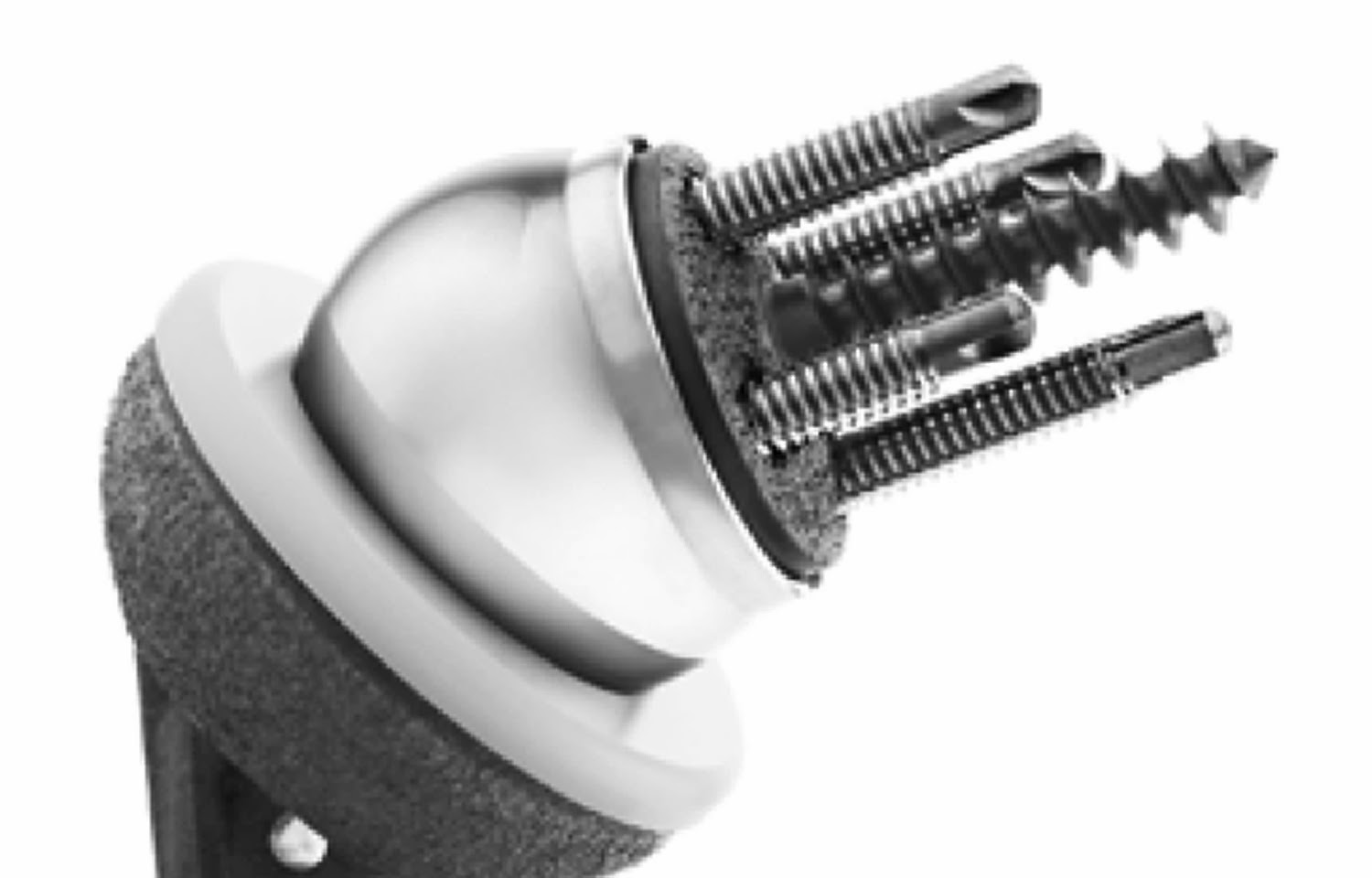
The surgeon's technique included minimal glenoid reaming to achieve >90% backside contact of a pegged, all polyethylene standard glenoid implant, inserted with minimal cement. The surgeon used a subscapularis peel which was repaired after insertion of the components using 6 to10 high-strength #2 braided sutures passed through transosseous drill holes. After the implants were placed, the subscapularis tendon was closed with these sutures and with closure of the distal rotator cuff interval with additional braided sutures to reinforce the repair.

104 of the 128 aTSAs were available for evaluation at minimum 2 years follow-up (range 2-5.6 years) (see the challenge of longer term followup). In comparison to many published reports, this represents a high percent followup. The authors included information on the remaining 24 - a few had passed on or became invalid, a few were revised and only a dozen or so could not be tracked. In clinical studies the denominator counts big and the reasons for loss to followup are important.

As stated in A Study in Hospital Efficiency (Codman, E.A. 1917). "All patients should be followed long enough to determine whether or not the treatment has been successful, and to ask 'if not, why not?'".

Overall, mean ASES scores improved from 41 to 89, SST from 4.5 to 10, and VAS-pain from 5.5 to 0.7. [Note that these average postoperative scores are within the minimal clinically important differences of perfect scores.  Thus it would be statistically impossible for any change in implant or technique (for example, routine use of preoperative 3D CT planning) to make a clinically significant improvement in the average outcomes.]

Fifty-nine patients had preoperative posterior glenoid wear patterns (Walch B2 or B3) and 47 had concentric or minimal glenoid wear (Walch A1, A2, or B1). The presence of posterior glenoid wear was not found to influence any outcome score

There were three modes of failure identified by the authors
(1) Subscapularis failure: 5 patients underwent revision to reverse shoulder arthroplasty for subscapularis insufficiency and one underwent open subscapularis repair with graft augmentation following traumatic injury. Three of the subscapularis tears arose because of trauma and/or patient reported noncompliance with postoperative immobilization or restrictions. All but one of the subscapularis failures reported here occurred within the first 12 months postoperatively. None of the patients with subscapularis failure requiring surgery had undergone prior open capsulorrhaphy or subscapularis repair

(2) ASES score <70Ten patients had a final ASES score < 70, which was associated with a history of previous surgery (overall, twenty-four shoulders had undergone previous surgery). 

(3) Glenoid osteolysis10 patients (13%) had radiographs  demonstrating glenoid osteolysis (defined as "radiolucency extending at least two mm from the center of peripheral pegs"). Glenoid osteolysis was not found to be associated with preoperative posterior glenoid erosion. The presence of glenoid osteolysis was not found to have an adverse effect on clinical outcome with the period of followup in this study. 

Comment: These authors report overall excellent results with their surgical technique. They have also defined three adverse outcomes that merit further study so that they and we can learn to avoid them.

Subscapularis failure: It would be of interest to know the rehabilitation program used by the surgeon. I am using a "slow roll" approach to range of motion exercises during the first six weeks and spend time cautioning the patient about falls and about avoiding external rotation stretching and active internal rotation during this period. Perhaps this surgeon does the same.

Prior surgery: It's a fact that young patients with arthritis commonly have had prior surgery. While the patient's history is not modifiable, we should try to learn if these patients have particular issues that could be addressed at surgery, such as (a) stiffness that might drive consideration of more aggressive soft tissue releases or smaller components or (b) difficulty in centering the humeral head on the glenoid that may drive use of eccentric humeral head components.

Glenoid osteolysis: While glenoid osteolysis was not noted to have an adverse effect on clinical outcomes in this minimum two year followup study, longer term followup is needed, although such studies are challenging.  These authors took care to optimize glenoid bone preparation. It would be of interest to know how well the components were seated, in that glenoid seating may be a major factor in achieving durable fixation.

This is a fine example of an individual surgeon study that has identified three adverse outcomes from anatomic arthroplasty. While it is unlikely that modifications in the surgical technique will lead to clinically significant improvement in the average outcome scores (because the average scores reported are so good), it is possible that additional study of patients with these failure modes may show us how to lower their frequency. 

The first step is to define the problems.


Fish with a problem
Montake Fill, Seattle June 2019

You can support cutting edge shoulder research that is leading to better care for patients with shoulder problems, click on this link

Follow on twitter/X: https://x.com/RickMatsen
Follow on facebook: https://www.facebook.com/shoulder.arthritis
Follow on LinkedIn: https://www.linkedin.com/in/rick-matsen-88b1a8133/

Here are some videos that are of shoulder interest
Shoulder arthritis - what you need to know (see this link).
How to x-ray the shoulder (see this link).
The ream and run procedure (see this link).
The total shoulder arthroplasty (see this link).
The cuff tear arthropathy arthroplasty (see this link).
The reverse total shoulder arthroplasty (see this link).
The smooth and move procedure for irreparable rotator cuff tears (see this link)
Shoulder rehabilitation exercises (see this link).


Friday, February 14, 2025

Anatomic total shoulder - preoperative planning and intraoperative decision making.

 Preoperative planning is helpful in anticipating what might be needed to reconstruct an arthritic shoulder; however the final choice of implants is determined at the time of surgery.

A 43 year old man presented with pain and stiffness of his left shoulder. He had a prior CT scan showing an arthritic humeral head centered on a somewhat retroverted glenoid.


We obtained our standard set of plain radiographs: an AP in the plane of the scapula and an axillary "truth" view taken with the arm in a functional arm position of elevation. The truth view showed posterior decentering of the humeral head that was not evident on the CT scan taken with the patient's arm at his side. No 3D CT planning was used.


At surgery, a standard glenoid component was well seated after conservative glenoid reaming. "Corrective" reaming and a posteriorly augmented glenoid component were not used.
 Trialing with an anatomic humeral head component revealed posterior instability when the arm was flexed forward. As a result, a short stemmed humeral component with an anteriorly eccentric humeral head was selected. 
Postoperatively, his shoulder is clinically and radiographically stable when the arm is elevated to a functional position (as seen on the postoperative "truth" view).


Comment: This case illustrates (1) the value of the "truth" view before and after surgery and (2) the importance of tailoring implant selection based on intraoperative testing of motion and stability. NB: when we do an arthroplasty, the shoulder we have after soft tissue releases and osteophyte resection is different from the shoulder before surgery; that's why intraoperative assessment is more important than preoperative planning.

You can support cutting edge shoulder research that is leading to better care for patients with shoulder problems, click on this link

Follow on twitter/X: https://x.com/RickMatsen
Follow on facebook: https://www.facebook.com/shoulder.arthritis
Follow on LinkedIn: https://www.linkedin.com/in/rick-matsen-88b1a8133/

Here are some videos that are of shoulder interest
Shoulder arthritis - what you need to know (see this link).
How to x-ray the shoulder (see this link).
The ream and run procedure (see this link).
The total shoulder arthroplasty (see this link).
The cuff tear arthropathy arthroplasty (see this link).
The reverse total shoulder arthroplasty (see this link).
The smooth and move procedure for irreparable rotator cuff tears (see this link)
Shoulder rehabilitation exercises (see this link).


Monday, September 9, 2024

What's important in anatomic total shoulder arthroplasty?

Anatomic total shoulder arthroplasty reliably provides long term comfort and function for patients with osteoarthritis and an intact rotator cuff, even in patients with deficiencies in glenoid bone (see Anatomic Total Shoulder Arthroplasty with All-Polyethylene Glenoid Component for Primary Osteoarthritis with Glenoid Deficiencies). While some surgeons attempt to "correct" the patient's glenoid version, others "accept" it (see Glenoid Version, Acceptors and Correctors). It either case, one of the most important technical goals of anatomic total shoulder arthroplasty is excellent seating of the glenoid component (see Edge displacement and deformation of glenoid components in response to eccentric loading. The effect of preparation of the glenoid bone).

Good seating is achieved by reaming the glenoid bone to a single concavity


that matches the backside of the glenoid component
so that no cement is placed between the bone and the backside of the prosthesis to fill in the gaps,


backside cement indicates poor seating and risks cement failure


below is an example of a well-seated glenoid component. The only cement visible is in the peg holes.


To further investigate the technical factors associated with anatomic total shoulder (aTSA) outcomes, the authors of Anatomic total shoulder arthroplasty for posteriorly eccentric and concentric osteoarthritis: a comparison at a minimum 5-year follow-up evaluated 210 patients at a minimum of five years after aTSAs performed with conservative glenoid reaming with no attempt at version correction. 

Preoperatively, 98 (47%) had posteriorly decentered humeral heads and 108 (51%) had centered humeral heads. There were 77 shoulders with Walch type A glenoids and 122 with Walch type B glenoids. 

At a mean 8-year follow-up, the final SST score, change in SST score, and percentage of maximal improvement were not correlated with preoperative or postoperative humeral head centering, Walch classification, or glenoid version. 

Two patients (1%) underwent open reoperations during the study period. 

In patients with Walch B1 and B2 glenoids (n = 110), there were no differences in outcome measures between patients that had postoperative retroversion of more or less than 15°. 

Although 15 of 51 patients (29%) with minimum 5-year radiographs had glenoid radioluciences, these radiographic findings were not associated with inferior clinical outcomes. 

On multivariable analysis, glenoid component radiolucencies were most strongly associated with incomplete component seating.


Careful preparation of the glenoid bone

From the authors of Edge displacement and deformation of glenoid components in response to eccentric loading. The effect of preparation of the glenoid bone we learn that the wobble and warp of the polyethylene component is minimized by spherically reaming the bone to precisely match the back of the component.


Glenoid bone stock is preserved by reaming only enough to create a single concavity, rather than trying to "correct" glenoid version.


The adequacy of glenoid reaming can be evaluated by using a pegless trial with the same backside curvature as the actual component and assuring that there is no rocking with eccentric loading. The goal is complete congruency.


Assuring optimal seating and cementing of the component. 

From the authors of The radiographic evaluation of keeled and pegged glenoid component insertion we learn that poor seating and poor cement technique contribute to poor fixation as evidenced by radiolucent lines on the immediate postoperative x-rays. 

Poor seating is evidenced by the presence of cement between the glenoid bone and the backside of the component. 


Good seating is indicated by the absence of cement between the component and bone (below top), rather than using cement as putty in an attempt to compensate for inadequate reaming.


A thin layer of cement between the bone and component is brittle and subject to cracking, displacement and loss of support for the glenoid implant.

Optimal cementing is reflected by the absence of radiolucent lines on postoperative radiographs.


We have learned that this can be achieved by drying each fixation hole with a CO2 spray


Immediately before pressurizing the cement into the hole


Comment: The survivorship of cemented, pegged, all-polyethylene glenoid components has yet to be surpassed by other types of glenoid implants (see Total shoulder replacement stems in osteoarthritis-short, long, or reverse? An analysis of the impact of crosslinked polyethylene). Attention to the details of bone-preserving bone preparation, complete seating of the component and modern cement technique may further improve the clinical outcomes of anatomic total shoulder arthroplasty. Our technique for this procedure is shown in this link.


Tuesday, October 11, 2022

The glenoid component in total shoulder arthroplasty: getting it done right.

Glenoid component loosening has been and remains an important cause of failure of anatomic total shoulder arthroplasty (data below from AOANJRR)


However, the survivorship of a pegged, cemented, cross linked, all-polyethylene glenoid component is excellent, with a 13 year cumulative percent revision rate of less than 5%. No metal backed or hybrid glenoid component has matched this outcome.


This type of glenoid arthroplasty serves shoulders across the range of glenoid types, with the lowest revision rates being for the B1 and B2 glenoids.


So, how might surgeons make the good results with the cemented, pegged all-polyethylene component even better? Here are some important concepts that can be put into action.

Careful preparation of the glenoid bone

From the authors of Edge displacement and deformation of glenoid components in response to eccentric loading. The effect of preparation of the glenoid bone we learn that the wobble and warp of the polyethylene component is minimized by spherically reaming the bone to precisely match the back of the component.


Glenoid bone stock is preserved by reaming only enough to create a single concavity, rather than trying to "correct" glenoid version.


The adequacy of glenoid reaming can be evaluated by using a pegless trial with the same backside curvature as the actual component and assuring that there is no rocking with eccentric loading. The goal is complete congruency.


Assuring optimal seating and cementing of the component. 

From the authors of The radiographic evaluation of keeled and pegged glenoid component insertion we learn that poor seating and poor cement technique contribute to poor fixation as evidenced by radiolucent lines on the immediate postoperative x-rays. 

Poor seating is evidenced by the presence of cement between the glenoid bone and the backside of the component. 


Good seating is indicated by the absence of cement between the component and bone (below top), rather than using cement as putty in an attempt to compensate for inadequate reaming.


A thin layer of cement between the bone and component is brittle and subject to cracking, displacement and loss of support for the glenoid implant.

Optimal cementing is reflected by the absence of radiolucent lines on postoperative radiographs.


We have learned that this can be achieved by drying each fixation hole with a CO2 spray


Immediately before pressurizing the cement into the hole


Comment: The survivorship of cemented, pegged, all-polyethylene glenoid components has yet to be surpassed by other types of glenoid implants (see Total shoulder replacement stems in osteoarthritis-short, long, or reverse? An analysis of the impact of crosslinked polyethylene). Attention to the details of bone-preserving bone preparation, complete seating of the component and modern cement technique may further improve the clinical outcomes of anatomic total shoulder arthroplasty. Our technique for this procedure is shown in this link.


To add this blog to your reading list in Google Chrome, click on the reading list icon


You can support cutting edge shoulder research that is leading to better care for patients with shoulder problems, click on this link.


Follow on twitter: https://twitter.com/shoulderarth

Follow on facebook: click on this link

Follow on facebook: https://www.facebook.com/frederick.matsen

Follow on LinkedIn: https://www.linkedin.com/in/rick-matsen-88b1a8133/

Here are some videos that are of shoulder interest
Shoulder arthritis - what you need to know (see this link).
How to x-ray the shoulder (see this link).
The ream and run procedure (see this link).
The total shoulder arthroplasty (see this link).
The cuff tear arthropathy arthroplasty (see this link).
The reverse total shoulder arthroplasty (see this link).
The smooth and move procedure for irreparable rotator cuff tears (see this link).
Shoulder rehabilitation exercises (see this link).

Monday, August 1, 2016

Reverse total shoulder - is the glenosphere completely seated?

Since we posted on this article in April, we have checked the number of turns of the set screw in the fully seated glenosphere of the DJO reverse shoulder prosthesis. In each case 4.5 turns were necessary to fully tighten this screw. Should it not be possible to turn the screw 4.5 times, we would be concerned about incomplete seating of the glenosphere. This check is now a routine part of our procedure.


The April post is reproduced below


These authors point to the uncommon but important risk of glenophere dissociation reverse shoulder arthroplasty.  A mechanically compromised Morse taper is thought to be the main cause of this complication, with bony abutment and soft tissue interposition being cited as the most important problems along with cantilevered engagement of the glenosphere due to impaction under a slight angle and incomplete engagement due to proud or cross-threaded baseplate screws.

They suggest that current methods for assessing the security of the Morse taper assembly require applying considerable torque to the glenosphere which may damage the quality of the taper. They proposed measuring the implant-specific angular rotation–torque curve while engaging the Morse taper by tightening the central locking screw. 

As can be seen from their graph below, in comparison to the desired (baseline) seating, the torque increase with screw tightening occurs with fewer rotations of the screw driver when there is interposition or abutment.


This is shown diagrammatically below; note that the insertion of the screw was blocked with fewer turns of the screw in cases B and C when the glenosphere is incompletely seated.


Although small interpositioning and impingement defects are difficult to detect without using an instrumented tool, such as the one presented in this study, large defects could probably be detected without an instrumented tool. The 1000-μ m and 2000-μ m defects locked the screw for both tested implants more than 1 full turn before their normal angular rotation end point. Thus the authors propose the following:  By applying moderate pressure on the glenosphere central screw while screwing in a counterclockwise direction before the start of engagement, a “click” can be sensed that indicates the starting point. From that point on, the number of turns can be counted before a considerable amount of torque needs to be applied to the screw to further tighten it. If the screw begins to tighten 1 or more full turns less than the number of turns known to completely seat the screw (approximately 6.4 full turns for the Delta CTA or Delta Xtend implant), it is time to check for peripheral bony abutment or soft tissue interposition. Note that the number of full rotations before reaching the angular rotation end point is prosthesis specific.

Comment: By virtue of its constrained kinematics, the reverse total shoulder transmits loads directly from the humeral component to the glenoid component, without the suppleness of a normal or an anatomic arthroplasty. Thus, the fixation and integrity of reverse total shoulder components may be challenged by impacts that would be unlikely to affect a conventional total shoulder. Because the reverse total shoulder components are often modular and held together by Morse tapers and because they can be loaded in directions that can challenge the Morse taper, there is a risk of dissociation with impact loading.


Glenoid component dissociation has been reported with various designs of reverse total shoulders (Sirveaux 2004)(Ekelund 2011) (Zumstein 2011) (Middernacht 2008)(Farshad 2010)(Kempton 2011)(Clark 2012).

A recent article on Glenosphere dissociation after reverse shoulder arthroplasty is of interest. These authors reviewed their reverse total shoulder arthroplasty database and identified 13 patients with glenosphere dissociation between 1999 and 2013; dissociation occurred 0.5 months to 7 years postoperatively.Incidence of dissociation was correlated to glenosphere size (p < .001). Dissociated glenosphere size distribution was as follows: 32 mm (n = 1), 36 mm (n = 4), 40 mm (n = 6), and 44 mm (n = 2).The authors noted that improper taper engagement reduced the torsional capacity of the glenosphere-baseplate interface.


The risk of dissociation can be reduced by considering the geometry of the specific implant and the instruments, by specific surgical steps, by vigorous intraoperative testing and by cautioning the patient to avoid impact loading after surgery. 

As is the case with any Morse taper, incomplete seating - even by a fraction of a millimeter - can reduce the security of the cold weld between the two parts assembled by the taper. Complete seating can be prevented by fluid in the well of the female aspect of the assembly, by tissue or bone that block complete seating, or by insufficient force applied to impact the two components together.

In the design shown below, the glenoid head (glenosphere) fits over the baseplate but does not completely cover it. thus it may be difficult to see whether or not the glenoid head is completely seated. 


Instruments, such as the rim reamer shown below help remove potentially interfering bone that may prevent the glenoid head from being completely seated.

This works well for the small glenoid head, because the outside diameter of the rim reamer is greater than that of the collar of the small glenoid head.


The rim reamer may be less effective when the collar of the glenoid head has an outside diameter greater than that of the rim reamer.


The principal method by which the seating can be verified is to pull vigorously on the glenoid head after it has been impacted into position, attempting to dissociate it from the baseplate. With some designs, vigorous traction can be applied using a t-handled instrument. An even better test can be performed by attempting to twist the glenosphere using the t-handle: if it twists on the base plate, it is not securely seated. 

After the glenosphere has been securely impacted into position, the retaining screw is inserted. The retaining screw should be tightened to the maximum number of turns that the Torque Driver allows. The Torque Driver will limit the torque to 22.5 in-lbs +/- 2.5 in-lbs. It will typically be about four full turns for all glenospheres.
After surgery, patients need to be reminded that impact loading is to be avoided.

Our current reverse total shoulder technique is shown in this link.


====