Showing posts with label x-rays. Show all posts

Sunday, September 22, 2024

Can Artificial Intelligence solve one of the big problems in shoulder arthroplasty research?.

We now recognize that most of our questions about shoulder arthroplasty cannot be answered using a few hundred shoulders. We also know that plain anteroposterior and axillary radiographs (not CT scans) are the most practical images for evaluating the geometry of shoulder arthroplasty.

 If we want to find out what geometric parameters determine the clinical result of anatomic or reverse total shoulder arthroplasty, we need BIG data, i.e. an assessment of tens of thousands of preoperative and postoperative plain radiographs to identify the characteristics that affect the patient's outcome. 


If performed manually by a human,  this labor would be prohibitive in time, cost and effort, even for Hercules. However, the task 
is well suited to artificial intelligence by which algorithms can be trained to rapidly categorize and - eventually - to analyze very large numbers of anteroposterior and axillary plain x-rays before and after shoulder arthroplasty so that the geometry of the reconstruction can be correlated with the clinical outcome.

As a step in this direction, the authors of Deep learning to automatically classify very large sets of preoperative and postoperative shoulder arthroplasty radiographs developed an artificial intelligence (AI) algorithm to classify preoperative and postoperative radiographs from shoulder arthroplasty patients according to laterality, radiographic projection, and implant type.

They used 2303 plain x-rays from 1724 shoulder arthroplasty patients. Two observers manually labeled all radiographs according to (1) laterality (left or right), (2) projection (anteroposterior, axillary, or lateral), and (3) whether the radiograph was a preoperative radiograph or showed an anatomic total shoulder arthroplasty or a reverse shoulder arthroplasty. The 18 classes of x-rays are shown below.


The labeled plain x-rays were randomly split into developmental and testing sets. 

A deep-learning algorithm was trained on the developmental set to classify the 3 characteristics described above. 


The trained algorithm was then evaluated on the testing set.

The trained algorithm perfectly classified laterality (right vs left).

The algorithm achieved accuracy scores of 99.2%, 100%, and 100% in identifying anteroposterior, axillary, and Y views

The algorithm achieved accuracy scores of 100%, 95.2%, and 100% in identifying preoperative radiographs, anatomic total shoulder arthroplasty radiographs, and reverse shoulder arthroplasty radiographs 

It took the algorithm 20.3 seconds to analyze 502 images.

The model focused on certain relevant pixel regions. For example, for the classification of prosthesis type, the model selected the pixels around the edge of the implant as shown by the green dots shown below.


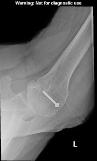
From the examples provided, it appears that the imaging protocols for the different x-ray views were well standardized; i.e. all of the axillary views show the same angle of the beam and position of the arm. It would be of interest to know whether there needs to be a step that excludes non-standard projections such as that shown below.

Comment: For any of our readers it would be straightforward and quick to classify one shoulder radiograph regarding laterality, view, preoperative vs. postoperative, and anatomic vs. reverse arthroplasty. However, classifying and recording this information for thousands of radiographs would become tedious and time-consuming. The ability to complete these tasks automatically, quickly and precisely is an important step in the use of artificial intelligence to classify shoulder x-rays. 

You can support cutting edge shoulder research that is leading to better care for patients with shoulder problems, click on this link

Follow on twitter/X: https://x.com/RickMatsen
Follow on facebook: https://www.facebook.com/shoulder.arthritis
Follow on LinkedIn: https://www.linkedin.com/in/rick-matsen-88b1a8133/


Here are some videos that are of shoulder interest
Shoulder arthritis - what you need to know (see this link).
How to x-ray the shoulder (see this link).
The ream and run procedure (see this link).
The total shoulder arthroplasty (see this link).
The cuff tear arthropathy arthroplasty (see this link).
The reverse total shoulder arthroplasty (see this link).
The smooth and move procedure for irreparable rotator cuff tears (see this link).
Shoulder rehabilitation exercises (see this link). 


Monday, February 14, 2022

Evaluating the glenohumeral joint with standardized plain x-rays

In the normal shoulder, the ball of the humeral head and the glenoid socket of the scapula are covered with smooth articular cartilage. This cartilage gives the joint a coefficient of friction less than that of an ice skate on ice. It is this cartilage that is destroyed in shoulder arthritis.

Cartilage cannot be seen on X-rays, but the thickness of cartilage can be detected by observing the separation of the bones, the 'radiographic joint space'.


Let's take a look at how the shoulder should be x-rayed to diagnose shoulder arthritis and to plan a joint replacement. 


In the shoulder, two X-ray views are essential for determining the 'joint space', the AP (anteroposterior) and the axillary views.
An AP view of a normal shoulder looks like this:
and an axillary view of a normal shoulder looks like this:
In each view one can see the separation of the ball from the socket due to the presence of cartilage in this normal right shoulder.
These views must be taken carefully to make sure that poor technique does not hide the true condition of the joint.

More about x-rays in arthritis can be found at this link.

Follow on twitter: https://twitter.com/shoulderarth

Follow on facebook: click on this link

Follow on facebook: https://www.facebook.com/frederick.matsen

Follow on LinkedIn: https://www.linkedin.com/in/rick-matsen-88b1a8133/


Here are some videos that are of shoulder interest
Shoulder arthritis - what you need to know (see this link).
How to x-ray the shoulder (see this link).
The ream and run procedure (see this link).
The total shoulder arthroplasty (see this link).
The cuff tear arthropathy arthroplasty (see this link).
The reverse total shoulder arthroplasty (see this link).
The smooth and move procedure for irreparable rotator cuff tears (see this link).
Shoulder rehabilitation exercises (see this link).

Wednesday, June 30, 2021

The power of the "truth" view in the detection of early shoulder arthritis

 A young man presented with pain and stiffness after prior procedures on his clavicle, biceps, and subscapularis.

His examination confirmed mild-moderate stiffness of his glenohumeral joint.

His Grashey view was not remarkable


However, his axillary "truth" view revealed decentering of the humeral head on the glenoid and moderate glenohumeral osteoarthritis.

Our thoughts on how to get the most information out of three simple plain films is shown in this link.


How you can support research in shoulder surgery Click on this link.

Here are some videos that are of shoulder interest
Shoulder arthritis - what you need to know (see this link).
The total shoulder arthroplasty (see this link).
The ream and run technique is shown in this link.
The cuff tear arthropathy arthroplasty (see this link).
The reverse total shoulder arthroplasty (see this link).
The smooth and move procedure for irreparable rotator cuff tears (see this link).
Shoulder rehabilitation exercises (see this link).


Tuesday, March 16, 2021

Reverse total shoulder - why take x-rays in the recovery room?

 Clinical Utility of Immediate Postoperative Radiographs following Uncomplicated Primary Reverse Shoulder Arthroplasty

These authors reviewed the the recovery room (PACU) x-rays and other 90 postoperative films of 157 patients having uncomplicated primary reverse total shoulders. 

Immediate postoperative radiographs were read by the radiologist as normal/unremarkable for 100% of patients.  


13 patients had abnormal findings determined by the surgeon during the initial 3 month postoperative period.  

Ten out of 13 patients had radiographic findings at the first postoperative clinic visit that differed from the initial radiologist interpretation documented on the PACU radiograph.

The most common complication was instability and dislocation, which occurred in 9 patients. Additional complications included baseplate failure,  glenosphere dissociation, and humeral stem subsidence. Twelve of the 13 patients underwent revision surgery.


The authors concluded, "Based on our findings we see utility for an intra-operative or immediate postoperative radiograph following uncomplicated, primary reverse shoulder arthroplasty, especially when done as a salvage procedure for indications such as proximal humerus non-union. However, there is low utility for obtaining an immediate postoperative radiograph in the PACU when indications include osteoarthritis or rotator cuff tear arthropathy."


Comment: In contrast to these authors, we believe that it is important to obtain good quality postoperative x-rays in each case of reverse total shoulder arthroplasty, regardless of indication. The first reason is that these films provide the surgical team with immediate feedback on whether or not they realized their preoperative plan with respect to component positioning, screw placement, baseplate seating, humeral component positioning and humeral component fixation. 


Second, a "normal/unremarkable" reading by a radiologist may overlook abnormal findings of importance to the shoulder surgeon, such as the incomplete glenosphere seating shown below. Such a finding, even in a routine case may prompt a return to the OR from the PACU.


A third reason for taking a PACU x-ray is shown by the table from the article:


Case 5 (RSA for cuff tear arthropathy) was found to have baseplate failure 2 weeks after the procedure
Case 8 (RSA for massive cuff tear) was found to have a glenosphere dissociation a 3 weeks after the procedure.
Case 12 (RSA for massive cuff tear) was found to have a dislocation at two weeks after surgery
Case 13 (RSA for avascular necrosis) was found to have a greater tuberosity at 6 weeks after surgery.

In each of these cases of early failure it seems likely that there was a technical problem at surgery and that a careful examination of good quality PACU films might have identified the issue in a more timely manner.

Subscribe to this blog by entering your email in the "subscribe" box to the right.

Here are some videos that are of shoulder interest
Shoulder arthritis - what you need to know (see this link).
How to x-ray the shoulder (see this link).
The ream and run procedure (see this link).
The total shoulder arthroplasty (see this link).
The cuff tear arthropathy arthroplasty (see this link).
The reverse total shoulder arthroplasty (see this link).
The smooth and move procedure for irreparable rotator cuff tears (see this link).
Shoulder rehabilitation exercises (see this link).

Wednesday, February 1, 2017

Total shoulder arthroplasty - costs of imaging

Utility of postoperative radiography in routine primary total shoulder arthroplasty

These authors sought to identify the medical relevance and cost-benefit of routine radiographs after primary anatomic total shoulder arthroplasty (TSA). They reviewed 160 consecutive patients undergoing anatomic TSA by an individual fellowship-trained surgeon who ordered radiographs obtained at 2 weeks, 6 weeks, 4 months, and 1 year postoperatively.

The authors reviewed the clinic notes to determine if a change in postoperative care occurred as a result of radiographic findings. Cost data and amount billed were obtained.

 Patients underwent radiography at 1.8 ± 0.2 weeks, 6.5 ± 1.2 weeks, 14.9 ± 2.9 weeks, and 46.8 ± 19.5 weeks postoperatively. Findings on the radiologist’s reading were normal/unremarkable for 100.0%, 96.8%, 95.9%, and 95.2%, respectively, at each visit. 

Review of the radiographs yielded no change in management based on these parameters. The amount billed for radiographs was $284,281 ($1776.76 per patient).

Comment: This study is a model for considering the value of tests that are ordered.  The key question is the one posed: how often does the test obtained change the management of the patient?

This is indeed the question we asked in this post:
Preoperative imaging of the glenoid - what information is needed? In that post we raise similar questions regarding the utility of routine preoperative CT scans.

Saturday, February 6, 2016

Preoperative CT scan measurements - are they worth the trouble?

The effect of sagittal rotation of the glenoid on axial glenoid width and glenoid version in computed tomography scan imaging.

These authors point out that computed tomography (CT) scans of the shoulder are often not well aligned to the axis of the scapula and glenoid and that these malalignments can affect axial measurements of anterior-posterior (AP) glenoid width and glenoid version.

They studied 30 CT scans of the shoulder that were reformatted using OsiriX software multiplanar reconstruction. The uncorrected (UNCORR) and corrected (CORR) CT scans were compared for measurements of both (1) axial AP glenoid width and (2) glenoid version at 5 standardized axial cuts.

They found the mean difference in glenoid version was 2.6% (2° ± 0.1°; P = .0222) and the mean difference in AP glenoid width was 5.2% (1.2 ± 0.42 mm; P = .0026) in comparing the CORR and UNCORR scans. The mean angle of correction required to align the sagittal plane was 20.1° of rotation (range, 9°-39°; standard error of mean, 1.2°).

They concluded that uncorrected CT scans of the glenohumeral joint do not correct for the sagittal rotation of the glenoid, and this affects the characteristics of the axial images. Failure to align the sagittal image to the 12-o'clock to 6-o'clock axis results in measurement error in both glenoid version and AP glenoid width. Use of UNCORR CT images may have notable implications for decision-making and surgical treatment.

Comment: While there is no question that measurements of pre and postoperative glenoid orientation are of interest, we find that it is uncommon to need CT scans for 'decision-making and surgical treatment.'

The cost of using corrected scans to change the measurement of version by two degrees or the width by 1.2 mm is not provided.

We find that the necessary and sufficient information for the planning and conduct of shoulder arthroplasty can be gained from the economical standardized AP view in the plane of the scapula


and the standardized axillary view taken with the arm elevated 90 degrees in the plane of the scapula (position of function).

===

Check out the new Shoulder Arthritis Book - click here.


You may be interested in some of our most visited web pages including:shoulder arthritis, total shoulder, ream and runreverse total shoulderCTA arthroplasty, and rotator cuff surgery as well as the 'ream and run essentials'


Wednesday, June 3, 2015

Shoulder joint replacement arthroplasty - x-ray evaluation

The purpose of imaging of the shoulder is to help establish the diagnosis, to determine the severity of the pathoanatomy, to help in surgical planning, and to enable the surgeon to illustrate the condition of the shoulder for the patient. Unless a specific research protocol is in place, we resist the temptation to ‘over-image’ , i.e. obtaining scans or reconstructions that are not necessary for the care of the patient such as that shown below.


The observation that CT scans may offer a few degrees of increased precision in the measurement of glenoid version does not convince us that this precision improves the quality of the surgery or the clinical outcome. Almost always standardized plain films are sufficient to garner the needed information and, as is shown below, information can be gathered from properly taken plain films that cannot be gathered on CT scans . In that proper radiographic technique (like surgical technique) is necessary to achieve the desired outcome, we take time to assure that our x-ray technologists know what we are seeking in the images.

The first key view is the anteroposterior in the plane of the scapula taken so that the x-ray beam passes through the glenohumeral joint. This view shows the superior-inferior position of the humeral head relative to the glenoid, the presence of osteophytes on the humeral head and glenoid, joint space narrowing, the degree of medial displacement of the humerus in relation to the lateral acromial line, the quality of the humeral and glenoid bone, the presence of loose bodies, and the presence of humeral head collapse or deformity.

The second key view is the axillary view taken with the arm in the functional position of elevation in the plane of the scapula and oriented so that both the spinoglenoid notch and the scapular neck are visible. This view shows a different perspective of the humeral anatomy, the amount of glenoid bone, the shape of the glenoid, its version in relation to the plane of the scapula and the relationship of the humeral head to the glenoid fossa. We have named the axillary view taken in with the arm elevated in the plane of the scapula the ‘truth’ view. This is because it demonstrates the glenohumeral relationships in the functional position of elevation; this is in contrast to CT scans, which have the disadvantage of being taken with the arm in the adducted position


Unfortunately, many of the ‘axillary views’ sent to us on patients for consultation are taken without standardization, making it impossible to determine the important features of the glenohumeral joint as  shown below.


When taken properly, the standardized anteroposterior and axillary views indicate the thickness of the cartilage space between the humerus and the glenoid, the relative positions of the humeral head and the glenoid, the presence of osteophytes, the degree of osteopenia, and the extent of bony deformity and erosion.


Since arthritis usually involves the central aspect of the humeral head,


joint space narrowing is most evident on the truth view as opposed to images made with the arm at the side. Of even greater importance is the ability of the axillary ‘truth’ view to show posterior subluxation or ‘functional decentering’ that is not evident in images taken with the arm at the side.




The degree of posterior subluxation can be measured as (a) the position of the center of the humeral head in relation to the plane of the scapula, (b) the position of the center of the humeral head in relation to the glenoid face or (c) the point of contact of the humeral articular surface on the glenoid articular surface. We prefer the latter because it is this point of contact that reflects the degree of centering of the net humeral joint reaction force on the glenoid. It is the malcentering of this joint reaction force that leads to posterior instability, posterior glenoid wear and to rocking horse loosening of prosthetic glenoid components. The standardized axillary view also enables the surgeon to see the shape of the glenoid surface. Three main types have been described: concentric wear (type A)


eccentric posterior wear (type B),

and dysplastic (type C)


In actual practice, there are so many intermediate types of glenoid pathoanatomy that rigorous separation into a few distinct classes is difficult. 


 An important aspect of glenoid pathology is the amount of the glenoid that is involved in the pathologic concavity, known as the ‘neoglenoid. Finally, the standardized axillary view enables the measurement of the degree of glenoid retroversion in relation to the body of the scapula. Thus, on the standardized axillary view, the surgeon can usually determine the major important characteristics of glenohumeral arthritic pathoanatomy: the amount of joint space narrowing, the degree of retroversion, the degree of posterior subluxation with the arm in a functional position, the glenoid shape, the percentage of the glenoid involved in the pathologic concavity and the angle of retroversion.


 Because of their low cost and freedom from metal artifacts, standardized axillary views provide a practical and reliable way to document the postoperative anatomy sequentially over time and to compare it to what was present before surgery.


A third view, the templating view, is obtained when humeral arthroplasty is being considered. This view is an anteroposterior (AP) view of the humerus taken with the arm in 30 degrees of external rotation relative to the x-ray beam with a magnification marker added. This view places the humeral neck in maximal profile and allows a comparison of proximal humeral anatomy with that of various humeral prostheses. In templating, it is important to recognize that the humeral canal is not cylindrical – the medial-lateral dimension is usually wider than the anteroposterior dimension so that the AP view may overestimate the size of the stem that will fit the diaphysis. This view is also useful for determining whether sufficient osteoporosis is present to merit special consideration at the time of arthroplasty 


Advanced imaging may be useful in the unusual case where the anatomy is distorted by prior injury or surgery, when there is concern about the amount of bone available for reconstruction, or when the standardized plain films cannot be obtained. In the great majority of cases, however, the extra cost and radiation of the CT scan can be avoided through the use of these standardized plain films. In that we can learn what we need to know about the status of the rotator cuff from physical examination and plain radiographs, shoulder MRIs are rarely needed unless indicated to exclude avascular necrosis or tumor. An MRI of the neck may be useful in evaluating patients suspected of having cervical radiculopathy, myelopathy, stenosis or a syrinx.

Be sure to click on this link to the Shoulder Arthritis Book.

---
Use the "Search" box to the right to find other topics of interest to you.

You may be interested in some of our most visited web pages including:shoulder arthritis, total shoulder, ream and runreverse total shoulderCTA arthroplasty, and rotator cuff surgery as well as the 'ream and run essentials'

See from which cities our patients come.

See the countries from which our readers come on this post.