Showing posts with label repair. Show all posts

Wednesday, January 25, 2017

The subscapularis - taking it down and putting it back

Management of the subscapularis tendon during total shoulder arthroplasty

These authors reviewed some of the issues surrounding subscapularis tendon management at shoulder arthoplasty.  Options include tendon tenotomy, peel, lesser tuberosity osteotomy, and even subscapularis sparing techniques. Inadequate healing of the subscapularis tendon can lead to postoperative pain, weakness, and instability.

Comment: We avoid lesser tuberosity osteotomy because it compromises the fixation of the humeral component in the metaphysis and obligates sacrifice of the long head tendon of the biceps.

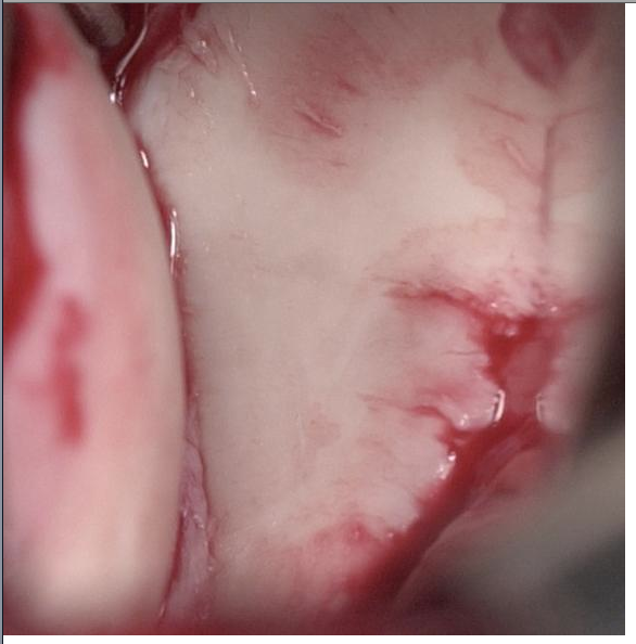
Our surgical approach involves a careful peel of the subscapularis tendon from the lesser tuberosity with attention to preserving the integrity of the biceps tendon and a 360 degree release of the capsule from the glenoid to resolve limitation of external rotation. By retaining the capsule on the deep surface of the tendon, the strength of the repair is enhanced.


At the conclusion of the case, drill holes are placed through good bone at the margin of the neck cut and six sutures of #2 non-absorbable suture are passed through these holes.

 These sutures are then passed through the tendon edge and tied securely.

A principal cause of post operative subscapularis failure is the overzealous and premature stretching of external rotation or premature initiation of internal rotation strengthening as explained in this post:
Rehabilitation after shoulder arthroplasty - cautions!

Our approach is to limit external rotation stretching to zero degrees (the hand shake position) and avoid internal rotation strengthening exercises for at least 3 months after surgery. We also caution patients about the risk of events that may suddenly externally rotate the shoulder such as a fall or a sudden pull on the arm from a leashed dog.


Other elated posts are listed below:

Subscapularis failure after arthroplasty - evaluation and management

The biomechanics of subscapularis repair - all sutures are not equal!

Subscapularis in shoulder arthroplasty


Shoulder joint replacement arthroplasty - spare the subscapularis, spoil the arthroplasty?

How well does the subscapularis work after total shoulder arthroplasty? ?Hazards of inter scalene block?

Failure of lesser tuberosity osteotomy in total shoulder joint replacement - a cautionary tale


===


To see the topics covered in this Blog, click here

Use the "Search" box to the right to find other topics of interest to you.

You may be interested in some of our most visited web pages including:shoulder arthritis, total shoulder, ream and runreverse total shoulderCTA arthroplasty, and rotator cuff surgery as well as the 'ream and run essentials'

Tuesday, June 18, 2013

Bankart surgical repair for dislocating shoulders

While many surgeons prefer arthroscopic repair for shoulders with recurrent dislocations, increasingly patients are coming to our office requesting open Bankart repair because of the lower redislocation rate.
See also this post.

Here is some information regarding our approach to recurrent traumatic anterior shoulder (glenohumeral) instability and its management by anatomic open surgical repair without suture anchors.


Starting with the basics, the shoulder is normally stabilized by concavity compression - the rotator cuff pressing the humeral head into the glenoid concavity.
This labrum provides a 'suction cup' effect, adding to stability of the joint as shown in this video.

With a traumatic dislocation, the labrum is torn from the glenoid socket.

 In patients who have experienced a traumatic anterior dislocation and who have recurrent dislocations of their shoulder, it is important to assess the integrity of the bony glenoid socket. This can be accomplished with plain x-rays (no CT scan is needed). The necessary views include an AP in the plain of the scapula and an axillary view.
We also obtain an apical oblique which enables us to see the anteroinferior glenoid rim as well as the posterior-lateral aspect of the humeral head (where a Hill Sachs lesion occurs).

 The apical oblique view below shows the Hill-Sachs defect flattening the upper left aspect of the humeral head and a moderate sized bony glenoid defect of the anterior-inferior glenoid (bottom right).
In the absence of a significant bony glenoid defect, stability can be restored by placing sutures through the lip of the glenoid and using these sutures to repair the soft tissues torn away from the lip.


If however, there is a substantial bony glenoid lip defect

 It may be necessary to add back bone using a bone graft secured with screws.


Fortunately, the great majority of individuals do not require a bony procedure. Our technique for the anatomic Bankart is shown here. First the skin incision, concealed in the normal skin crease.
 Here is a diagram of the repair of the capsule and labrum as well as that of the subscapularis
Here is a close up view of the repaired capsule and labrum, restoring the "O" ring and its contribution to stability.

===
To see the topics covered in this Blog, click here

Use the "Search" box to the right to find other topics of interest to you.

You may be interested in some of our most visited web pages including:shoulder arthritis, total shoulder, ream and runreverse total shoulderCTA arthroplasty, and rotator cuff surgery as well as the 'ream and run essentials'


See from which cities our patients come.


See the countries from which our readers come on this post.


Thursday, August 16, 2012

Dallas Braden needs another shoulder surgery CBS Sports

CBS Sports reports that Dallas Braden needs another shoulder surgery. The A's pitcher who threw a perfect game in May of 2010 had a 'repair of a torn capsule in his left shoulder' one year later. Now, a little over a year after surgery, it appears that the shoulder has not responded as hoped for.

SFGate provides a bit of additional opinion from one of their staff writers:

"It’s bad news, unfortunately, for Dallas Braden, who had hoped to make a few starts in September for the A’s but whose left shoulder has not responded as hoped after surgery in May 2011 to repair the capsule. He will have more surgery; Melvin termed it “exploratory.”

As I have written before, shoulder-capsule repair is not a procedure that has been perfected, unlike elbow ligament replacement. It generally takes quite a lot of time to come back from (see: Johan Santana) and there is no guarantee of a full return. Here’s the main issue: Surgeons must basically guess on how tight to make the shoulder. There is no way to calibrate it. So some shoulders are too tight, some aren’t tight enough. The hope, of course, with any capsule repair is to get the tightness on the money the first time around. I don’t know if that’s what’s going on with Braden, but I have been told by medical experts that generally speaking, the tightness issue is the big question mark when it comes to shoulder capsules. "

Shoulder stability is very complex as explained in the text and videos explained here by my late partner Doug Harryman (while the video is a bit dated, the information is as solid now as it was when he produced it). This is especially the case for throwers who use their arm at the extremes of the range of motion.

Attempts to repair an unstable shoulder in a high performing athlete can be challenging and good followup is necessary to determine if the procedure has been successful, as pointed out in a previous post.


--

Use the "Search the Blog" box to the right to find other topics of interest to you.

You may be interested in some of our most visited web pages including:shoulder arthritis, total shoulder, ream and runreverse total shoulderCTA arthroplasty,  and rotator cuff surgery.


Saturday, October 29, 2011

Rotator cuff papers at the 2011 closed meeting of the ASES - do cuff repairs work?

Two papers caught my attention:

MacDonald et al conducted a randomized control trial comparing single row with double row fixation in arthroscopic rotator cuff repair. The outcome was evaluated in terms of clinical scoring as well as healing assessment by MRI or ultrasound. While both groups were improved from their pre-surgical status, there was no significant difference in the outcome scores. Interestingly, the rotator cuff imaging showed that between 23 and 35% of the repairs were not intact at 2 years after surgery.

McCarron et al did a most interesting study where they used the 'suture bridge' technique for cuff repair, a method that has been reported to be associated with a high rate of failure. The authors placed metal markers (1.6 mm tantalum beads) placed 2 centimeters medial to the lateral edge of the tendon. CT scans were obtained on the day of surgery and at four intervals up to 1 year after surgery as were ultrasound examinations of the cuff. They found that the tendon edge pulled away from the repair site by an average of 1.6 centimeters over the first 26 weeks after repair, even though ultrasound showed recurrent defects in only 2 of the 13 shoulders. Clinical outcome scores were improved for these shoulders in spite of the failure of the repair to remain fast to the bone.

These two studies again bring into question the degree to which arthroscopic rotator cuff repair can anatomically restore the attachment of the rotator cuff tendon to bone. As originally pointed out by my late partner, Doug Harryman, clinical improvement commonly occurs after rotator cuff surgery even if the repair of tendon to bone is not successful. This is why we consider a 'smooth and move' procedure when conditions are not optimal for cuff healing, thereby avoiding the need to protect a repair after surgery.


--


Use the "Search the Blog" box to the right to find other topics of interest to you.

You may be interested in some of our most visited web pages including: shoulder arthritis, total shoulder, ream and runreverse total shoulderCTA arthroplasty, and rotator cuff surgery.


Saturday, August 27, 2011

Rotator Cuff 4 - Mechanisms of Tear, Factors Affecting Repair, "Impingement"

Initiation of Cuff Failure


Cuff fiber failure commonly results from the sudden application of eccentric loads, for example when the muscle attempts to resist a downward force on the arm
while the cuff seems to be better able to tolerate concentric loads, for example in a controlled lift away from the side. 


An anatomic factor predisposing to deep surface failure of the cuff insertion is internal abutment, where the corner of the glenoid contacts the deep aspect of the cuff at its tuberosity insertion 
This is most likely to be a problem for throwers who have stretched out their anterior capsule, allowing increased external rotation.


It is apparent that the most common form of cuff fiber failure, that which occurs on the articular surface of the cuff tendon, cannot be attributed to scuffing of the bursal surface by the acromion: so-called subacromial ‘impingement.’ In fact, the cuff insertion is well under the acromion at relatively small angles of elevation where it is protected from contact with the coracoacromial arch. Current evidence indicates that the most rotator cuff tears arise from tension overload and age-related attrition, rather than 'impingement'.

Readers might be interested in a recent review of the literature regarding the diagnosis of "impingement syndrome".


Factors Compromising Tendon Healing


Deep surface rotator cuff fiber failure exposes the defect to joint fluid. This joint fluid prevents the formation of a fibrin clot and, thus, healing is contravened.

Furthermore, tension at the edge of the cuff tear compromises the circulation to the margin of the tendon

For these reasons, left to their own devices, cuff defects tend to progress rather than healing. An optimal cuff repair surgery will bring healthy tendon into contact with vascularized bone and exclude joint fluid from the repair site. Subsequent posts will review the principles of surgical repair in some detail.


Factors Affecting Reparability

In considering the potential for surgically restoring a durable tendon insertion to bone, the surgeon needs to consider the quality of the tissue to be used in the repair. The ability of the cuff tendon tissue to withstand tensile loads is compromised by age, disuse, steroid injections, smoking, and poor general health. These predisposing factors can dispose the cuff tendons to fail with minimal force – essentially an atraumatic fiber failure. Cuff fibers that fail atraumatically may be so constitutionally weak that they cannot hold up even if repaired back to the bone. Thus, in chronic atraumatic cuff tears there is reason to consider a non-operative approach to improving shoulder function by rehabilitating the muscle–tendon units that remain intact.


Acute, traumatic cuff detachments that result from major force application are likely to be repairable

If acute traumatic cuff tears are not repaired promptly, the muscle may undergo intramuscular contracture, atrophy, and fatty degeneration and the tendon may become progressively reabsorbed. These degenerative changes compromise the opportunity for surgical repair. Thus, as with any other tendon avulsion from bone, time is of importance in the repair of acute tears of the rotator cuff. 


Loss of the rotator cuff subjects the superior glenoid to increased loads that can contribute to its erosion 


Progressive upward displacement of the humeral head produces secondary changes in the coracoacromial arch 

Once the humeral head has ascended so that its equator is above the residual cuff, contraction of the cuff muscles lock the humeral head in the superiorly displaced position

Chronic upwards displacement of the humeral head from cuff deficiency and superior glenoid erosion can result in cuff tear arthropathy,

--


Use the "Search the Blog" box to the right to find other topics of interest to you.

You may be interested in some of our most visited web pages including: shoulder arthritis, total shoulder, ream and runreverse total shoulderCTA arthroplasty, and rotator cuff surgery.