Showing posts with label glenoid bone loss. Show all posts

Friday, October 13, 2023

Is it necessary to use an augmented component to address the B2 glenoid?

As the figures at the top of this post indicate, the B2 glenoid can managed with a ream and run procedure.

As the figures below demonstrate, the B2 glenoid can also be managed with a standard all polyethylene glenoid and anatomic humeral head.

The preoperative axillary view shows the humeral head to be posteriorly decentered on the retroverted glenoid.


The two highly comparable axillary "truth" views enable verification of the postoperative centering of the humeral head on the prosthetic glenoid.


Comment: While preoperative 3D CT based planning software may suggest the need for augmented glenoid components to "correct" joint geometry, most arthritic shoulders with B2 glenoid pathoanatomy can be durably managed with anatomic arthroplasty using standard components, accepting the glenoid retroversion (see this related post on correctors and acceptors).

You can support cutting edge shoulder education that is leading to better care for patients with shoulder problems, click on this link.

Follow on twitter: https://twitter.com/RickMatsen or https://twitter.com/shoulderarth
Follow on facebook: click on this link
Follow on facebook: https://www.facebook.com/frederick.matsen
Follow on LinkedIn: https://www.linkedin.com/in/rick-matsen-88b1a8133/

Here are some videos that are of shoulder interest
Shoulder arthritis - what you need to know (see this link).
How to x-ray the shoulder (see this link).
The ream and run procedure (see this link).
The total shoulder arthroplasty (see this link).
The cuff tear arthropathy arthroplasty (see this link).
The reverse total shoulder arthroplasty (see this link).
The smooth and move procedure for irreparable rotator cuff tears (see this link).
Shoulder rehabilitation exercises (see this link).


Thursday, January 14, 2021

Progression of arthritic glenoid bone loss as shown by the axillary "truth" view.

Natural history of glenoid bone loss in primary glenohumeral osteoarthritis: how does bone loss progress over a decade?

These authors sought to determine how glenohumeral subluxation and glenoid bone loss changed over time in 48 shoulders that underwent arthroplasty and had been evaluated with standardized high-quality axillary radiographs 


at 1 or more time points over the 5-15 years before arthroplasty. The mean interval time between the oldest and most recent radiographs was 8.9 years (range 5-15 years). 

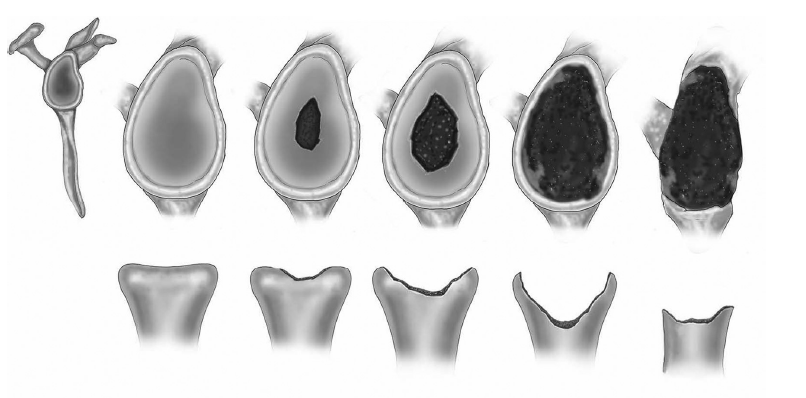
Below is an example of how glenoid morphology progressed over roughly an 8-year period of time from an A1 glenoid to a B3 glenoid. Note the standardization of the axillary "truth" views that enabled comparisons of the glenohumeral pathoanatomy over time. The patient was a 43-year-old male (body mass index 26.6) at initial presentation for symptomatic right shoulder osteoarthritis and went onto an anatomic total shoulder arthroplasty. From presentation to year 5, the glenoid morphology remained A1 with 3 intervening radiographs documented. At year 6, the patient was noted to have a B1 glenoid (top right), a B2 glenoid at year 7 (bottom left), and a B3 glenoid at year 8 before proceeding with surgery (bottom right).


On each axillary view, the glenoid type


and the degree of posterior humeral decentering on the face of the glenoid


were documented.


Glenoid morphology on the earliest radiograph was classified as A1 in 22, A2 in 13, B1 in 1, B2 in 9, B3 in 1, and D in 2 shoulders. 


Walch A patterns identified on early radiographs most commonly maintained an A pattern over time, but 20% developed eccentric wear with 5 of 35 becoming B type and 2 of 35 becoming a D type before arthroplasty. 


All B-type glenoids remained B type. 


Classic progression of bone loss along the same concentric or eccentric ‘‘track’’ occurred 41% of the time, with , the only B1 glenoid becoming a B2 glenoid, and 56% (5/9) of B2 glenoids becoming B3 glenoids before arthroplasty. 


Only 15% (2/13) of A2 glenoids developed eccentric wear compared with 32% (7/22) of A1 glenoids.


Comment: This study demonstrates that glenohumeral pathoanatomy can be well characterized using the axillary "truth" view without the additional expense and radiation dosage of a CT scan.


This study also demonstrates that the description of glenoid pathoanatomy cannot be constrained to discrete static types, but rather the amount of bone loss, change in version, and humeral decentering each exist on a continuum from "none" to "a lot" with progressive transitions from one type to another.


Finally, in considering the case example provided, it seems that a standard approach to anatomic arthroplasty would have served the patient in each of the 4 different stages of his disease.


===

Our approach to total shoulder arthroplasty can be viewed by clicking here.


To support our research to improve outcomes for patients with shoulder problems, click here.
To subscribe to this blog, enter your email in the box to your right that looks like the below


===
How you can support research in shoulder surgery Click on this link.

We have a new set of shoulder youtubes about the shoulder, check them out at this link.

Be sure to visit "Ream and Run - the state of the art" regarding this radically conservative approach to shoulder arthritis at this link and this link

Use the "Search" box to the right to find other topics of interest to you.

You may be interested in some of our most visited web pages  arthritis, total shoulder, ream and runreverse total shoulderCTA arthroplasty, and rotator cuff surgery as well as the 'ream and run essentials'


Saturday, December 5, 2020

Reverse total shoulder arthroplasty when the glenoid bone is deficient.

Is bone grafting always necessary in revision reverse total shoulder arthroplasty with uncontained glenoid bone defects?

Patients with an uncontained glenoid bone defect are being treated with a reverse total shoulder arthroplasty (RTSA). Currently there is a tendency towards a reconstruction using bone grafts. These authors investigated whether central peg positioning in the spine pillar (CPPSP) is a feasible alternative to the use of bone grafts.


They reviewed 60 revisions to a RTSA with uncontained glenoid bone defects ; bone grafts were used in 29 cases and the CPPSP technique in 31 cases. For the CPPSP technique, the K-wire was used to find the longest bony pillar of the spine (the direction was always downward tilted and in anteversion to the native glenoid). Once the pillar was identified, the proximal cortex was perforated with this K-wire. Next, reaming of the glenoid surface was performed until a minimum of 50% of contact area with the baseplate could be reached.


In the CPPSP group the Constant score changed from 42 to 69 points. In the bone graft group it changed from 47 to 60 points. This difference in increase in Constant score was significant, due to a significant difference in strength, in favor of the CPPSP group. 


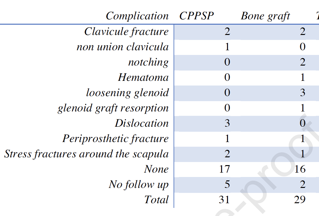
The overall complication rate was 37,7% (20/53) with a reoperation rate of 18,9% (10/53).


Dislocations occurred only in the CPPSP group (n=3) and loosening of the glenoid occurred only in the bone graft group (n=3).


Comment: Two critical elements in the stability of the glenosphere are (1) bony support of the baseplate and (2) secure screw fixation into native bone.


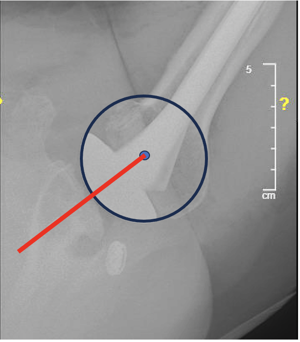
The use of the alternative center line is discussed this link


To see our technique for reverse total shoulder, click on this link.

To support our research to improve outcomes for patients with shoulder problems, click here.

To subscribe to this blog, enter your email in the box to your right

=====
How you can support research in shoulder surgery Click on this link.

To see our new series of youtube videos on important shoulder surgeries and how they are done, click here.

Be sure to visit "Ream and Run - the state of the art"  regarding this radically conservative approach to shoulder arthritis at this link and this link. Also see the essentials of the ream and run.

Use the "Search" box to the right to find other topics of interest to you.

You may be interested in some of our most visited web pages   arthritis, total shoulder, ream and runreverse total shoulderCTA arthroplasty, and rotator cuff surgery 


Friday, March 27, 2020

Does a preoperative CT scan improve patient outcomes?

Comparing the Use of Axillary Radiographs and Axial Computed Tomography Scans to Predict Concentric Glenoid Wear

These authors sought to compare the use of axillary radiographs and midglenoid axial CT scans to identify glenoid wear.

Five independent examiners with differing levels of experience characterized the glenoid morphology as either concentric or eccentric on preoperative axillary radiographs and mid-glenoid axial CT scans for 330 patients who underwent anatomic total shoulder arthroplasty.


Intraobserver consistency averaged 75% for radiographs and 73% for CT scans. There was significant interobserver consistency, as higher levels of training corresponded with greater consistency between imaging analyses (p < 0.001).


Comment: Especially in these times with huge strains on the nation's medical budget, we need to be thoughtful about how our health care dollars are spent.

As the authors state, "Although CT scans are associated with greater financial cost and exposure to radiation than radiographs, the literature has yet to describe the additional clinical value and/or potential cost-value benefit as a result of improved outcomes provided by the use of CT scans in patients undergoing total shoulder arthroplasty, even when integrated with virtual planning software and generation of patient specific instrumentation."

We agree that CT scans without or with 3D reconstructions can provide additional detail regarding glenohumeral pathoanatomy in comparison to plain radiographs, it remains to be seen whether this increment in information leads to significantly better clinical outcomes for the patient. Any benefit would need to be balanced against the increased costs and the 200 to 1,000 fold increase in radiation exposure of the CT scans (2.06 mSV)75 (10.83 mSV)76 in comparison to plain radiographs (0.01 mSV). In addition to the cost, physicians are increasingly concerned about the relationship between radiation dosage and the risk of the patient developing cancer.

Further research is needed to show that preoperative CT scans contribute to improved outcomes for the patient.

Meanwhile, standardized preoperative views can provide the information needed to evaluate and manage the arthritic shoulder (see article below).

Prearthroplasty glenohumeral pathoanatomy and its relationship to patient’s sex, age, diagnosis, and self-assessed shoulder comfort and function

These authors examined 544 patients within 6 weeks before shoulder joint replacement arthroplasty with the goals of characterizing the radiographic characteristics of the arthritic joint and the relationship of these pathologic changes to the patients' age, sex and diagnosis. They also studied the inter-relationships among glenoid type, glenoid version, and amount of decentering of the humeral head on the glenoid; as well as the relationships of the pathoanatomy to the patient’s self-assessed shoulder comfort and function.

Examples of the different types of glenoid pathoanatomy are shown below.


They found that male patients had a higher frequency of type B2 glenoids and a lower frequency of A2 glenoids.


The arthritic shoulders of men were more retroverted and had greater amounts of posterior decentering.


Patients with types A1 and C glenoids were younger than those with other glenoid types. 

Shoulders with osteoarthritis were more likely to be type B2 and to be retroverted. 

Types B2 and C had the greatest degree of retroversion, whereas types B1 and B2 had the greatest amounts of posterior decentering. 


Shoulders with glenoid types B1 and B2 and those with more decentering did not have worse preoperative self-assessed shoulder comfort and function.


==
To see a YouTube of our technique for total shoulder arthroplasty, click on this link.

===

How you can support research in shoulder surgery Click on this link.

To see our new series of youtube videos on important shoulder surgeries and how they are done, click here.

Use the "Search" box to the right to find other topics of interest to you.

You may be interested in some of our most visited web pages arthritis, total shoulder, ream and run, reverse total shoulder, CTA arthroplasty, and rotator cuff surgery as well as the 'ream and run essentials'

Wednesday, January 17, 2018

Reverse total shoulder in the face of glenoid defects

Management of glenoid bone defects with reverse shoulder arthroplasty—surgical technique and clinical outcomes

Between 2001 and 2010, these authors found that 94 of 1074 reverse total shoulders had significant glenoid bone loss. Of these patients, 17% had a centric defect 

 and 83% had an eccentric glenoid defect.


Composite glenoid grafts were required in 12 patients, 9 of whom required a glenoid baseplate with a long central peg.


92.5% (87/94) of the patients could be managed with a single-stage procedure.  If the long central peg did not purchase a minimum of 50% of its length in the native scapula, a single-stage reconstruction was aborted in favor of a 2-stage procedure: the glenoid defect was grafted with a composite graft and fixated using cortical screws as required to attain graft stability. In cases of centric defects, a modular humeral component was used and a hemiarthroplasty performed. In cases in which eccentric defects were reconstructed, a resection arthroplasty was performed. In the authors’ experience, a hemiarthroplasty for an eccentric bone defect led to poor graft incorporation and graft nonunion or resorption.

Comment: The key in such cases is that secure baseplate fixation needs to be achieved before the construct is loaded by completing the reverse total shoulder with a glenosphere and a humeral component.

====
The reader may also be interested in these posts:


Consultation for those who live a distance away from Seattle.

Click here to see the new Shoulder Arthritis Book.

Click here to see the new Rotator Cuff Book

Information about shoulder exercises can be found at this link.

Use the "Search" box to the right to find other topics of interest to you.

You may be interested in some of our most visited web pages including:shoulder arthritis, total shoulder, ream and runreverse total shoulderCTA arthroplasty, and rotator cuff surgery as well as the 'ream and run essentials'

See from which cities our patients come.

See the countries from which our readers come on this post.

Monday, June 5, 2017

Reverse total shoulder - glenoid bone grafting

Midterm outcomes of bone grafting in glenoid defects treated with reverse shoulder arthroplasty

These authors reviewed 20 patients with glenoid bone loss who underwent primary or revision surgery using a glenoid bone allo or autograft.

The majority of grafts incorporated.  There was a 20% postoperative complication rate: 1 case of aseptic glenoid component loosening, 1 surgical wound hematoma, 1 acromial fracture, and a symptomatic grade 3 scapular notching.

Comment : Several key factors deserve consideration before proceeding with a bone graft reconstruction. (1) Is the patient a suitable candidate for a major and potentially complex surgery? (2) Is a bone graft necessary or can the glenosphere be stabilized in the residual glenoid bone (see technique described later in this post)? (3) Can the very strong construct shown in the figure above be securely fixed to the often weak bone of the residual scapula? (4) How worried to we need to be about graft resorption? The answers to these questions are individualized for each patient and each surgeon. To put this small series in perspective, we've reposted some previous posts on the subject below.

As an aside, it is of interest that the case shown in this article demonstrates a revision of a failed metal back glenoid and a platform humeral component. The former resulted in severe glenoid bone loss and the 'promise' of easy conversion using a platform prosthesis was not realized as shown by the postoperative film below.


Here is some previously posted content on this topic:

Glenoid bone grafting in primary reverse total shoulder arthroplasty

These authors sought to determine results, complications, and rates of failure of glenoid bone grafting in 41 shoulders having primary reverse shoulder arthroplasty.

These surgeons used bone grafting to achieve between 30% and 50% contact  between the implant and bone. In specific instances of superior bone loss, graft was used to promote inferior tilt of the implant. In the setting of posterior or anterior defects, graft was used to  restore glenoid version. Cancellous graft was used in the setting of  lesser defects. In larger glenoid deficiencies, the use of structural grafts was considered.

Thirty-four received corticocancellous grafts and 7 structural grafts. At a minimum follow-up of 2 years, they found that preoperative severe glenoid erosion and increasing body mass index were significantly associated with worse American Shoulder and Elbow Surgeons scores.

Two sets of x-rays were presented.


On radiographic evaluation, 7 patients had grade 1 or grade 2 glenoid lucency. Glenoid bone graft incorporation was observed in 31 patients. Twelve patients suffered from grade 1 or grade 2 scapular notching. All of the patients with structural grafts showed graft incorporation and no signs of glenoid lucency. None of the patients needed revision surgery. 

Comment: As the authors point out, major glenoid erosion is an indication of severe disease. Patients with severe disease are more likely to have poor outcomes.

The decision to graft and what type of graft to use depends on a number of factors, including the type of baseplate fixation of the particular implant, the experience of the surgeon, the pathoanatomy, and the quality of the residual bone.

In our practice, we use a small drill inserted into the central glenoid vault as a 'dip stick' to determine the adequacy of the extant bone. If a bone depth of approximately 3 cm is present, we conclude that there is sufficient bone to stabilize the implant and ream around a tap placed along the path of this drill.

The use of bone grafting in reverse total shoulder is also discussed in this link.

Our standard reverse technique is shown in this link.

A bit more about bone grafting is detailed in the articles and discussion below

Management of glenoid bone loss in primary and revision reverse total shoulder arthroplasty

These authors reviewed the literature on the management of glenoid defects in reverse shoulder arthroplasties and presented their approach.


They present a classification system:

 For contained defects, they have used impaction allografting beneath the base plate


For eccentric defects, they have used structural grafts


or allograft composites.


If there is concern about the stability of the construction, the authors consider a two stage procedure in which the  glenoid augmentation is performed without implanting the base plate combined with a humeral hemiarthroplasty. The reverse arthroplasty is done as a secondary procedure after confirming graft incorporation with CT scans.

The system used by the authors includes a central bone ongrowth peg. For cases of glenoid deficiency they use an extended length peg.


Comment: This is a useful review of the challenges posed by defects in glenoid bone when performing a reverse total shoulder.

It can be compared to anther recent publication:


These authors point out that large glenoid defects pose difficulties in shoulder arthroplasty. They reviewed 44 patients (20 men and 24 women; mean age, 69 years) having a bulk structural graft behind the baseplate of a reverse total shoulder to manage structure defects.  The grafts consisted of a humeral head autograft in 29, iliac crest autograft in 1, or femoral head allograft in 14.

These cases used a prosthesis with a bone ingrowth stem on the baseplate


inserted over the bone graft as shown below.


Postoperative scores for the bone graft cohort were significantly lower than those in a cohort without grafting. Complete or partial incorporation was shown radiographically in 81% of grafts. 

Six baseplates were considered loose. The major (clinically significant) complication rate was 13.6%. There were 2 graft failures that caused clinical loosening of the baseplate and required revision. Two infections occurred (1 autograft and 1 allograft). One was revised in a 2-stage procedure, and 1 elected to retain the antibiotic spacer. One patient with clinical humeral loosening required revision (autograft). One postoperative dislocation (autograft) occurred. The patient was treated with a closed reduction in the operating room and immobilized for 6 weeks. No further dislocations occurred. Other minor (not clinically significant) complications included 6 patients with scapular notching of grades 1 or 2, and 4 patients with radiolucent lines of 2 mm around the humeral stem but without evidence of gross loosening. This resulted in a total complication rate of 36%.


Comment: There is no question that bone deficiency complicates shoulder arthroplasty. The question is when to use a graft and when to work with the bone stock available. That is a decision that can be made only by the operating surgeon based on the glenoid, the prosthesis used and the surgical technique.

We prefer to avoid structural grafts whenever possible because of uncertainty regarding quality, healing, and resistance to resorption. As pointed out in this paper, bone graft failures can cause clinical loosening of the baseplate.

Our preferred method for reverse total shoulder arthroplasty uses a central screw on the baseplate that provides immediate purchase in glenoid cortical bone without having to depend on bone ingrowth into a central peg and that provides lateral offset of the glenosphere to make up for bone deficiency.


In cases of glenoid bone deficiency, we use a small drill as a 'dip stick' to probe the glenoid to find the appropriate area that offers the best bone stock.  The depth of solid bone is then verified with the tap: 30 mm is ideal.


The baseplate is then screwed into position, assuring that it locks securely in place when tightened.

Here is a recent case of failed shoulder hemiarthroplaty with severe glenoid deficiency.


Secure baseplate and glenosphere fixation was achieved by the method described.

This method may be considered instead of bone grafting when there is sufficient remaining bone of quality.

===


Use the "Search" box to the right to find other topics of interest to you.