Showing posts with label glenoid component fixation. Show all posts

Wednesday, May 1, 2024

Of mousetraps and anatomic glenoid components.


Ralph Waldo Emerson is credited with the saying, "build a better mousetrap and the world will beat a path to your door". 


A similar sentiment may drive the marketing of many different types of glenoid components in anatomic shoulder arthroplasty. Almost all have a polyethylene surface for articulation with the humeral component. The difference lies in the mechanism for fixation to the bone of the glenoid. Below are a few of the many.


One innovation uses porous metal into which bone can grow for fixation. The question is "does this innovation improve outcomes for patients?"  A recently study, A multicenter, randomized controlled trial comparing a second-generation uncemented trabecular metal-backed vs. cemented polyethylene glenoid component in total shoulder arthroplasty: 5-year results compared cemented all polyethylene glenoid components to uncemented trabecular metal glenoid components.


This study found no statistical or clinically relevant advantage of the trabecular metal-backed cementless component over the all polyethylene cemented component. No glenoid implant failures were reported, and complication rates were similar between groups. Metal debris was observed in 11 (23.9%) of the patients receiving trabecular metal components (see circle on x-ray below).


In addition, on the x-ray above it is not clear how much of the original polyethylene remains between the metal backing and the humeral component (red arrows).

The problem of accelerated polyethylene wear with metal backed components was pointed out in Metal-backed glenoid implant with polyethylene insert is not a viable long-term therapeutic option, the authors of which concluded "uncemented MB [metal backed] glenoid resurfacing is not a viable long-term therapeutic option because of accelerated PE [polyethylene]wear leading to early revision surgery". See also

The authors of Comparable low revision rates of stemmed and stemless total anatomic shoulder arthroplasties after exclusion of metal backed glenoid components: a collaboration between the Australian and Danish national shoulder arthroplasty registries found that "The adjusted hazard ratio for revision of total shoulder arthroplasties with metal backed glenoid components compared to all-polyethylene glenoid components was 2.54 (95% CI 1.70-3.79, p < 0.001) in the Australian Orthopaedic Association National Joint Replacement Registry (AOANJRR) and 4.1 (95% CI 1.92-8.58, p<0.001) in the Danish Shoulder Arthroplasty Registry (DSR). The authors concluded "We advocate that metal-backed glenoid components should be used with caution and not on a routine basis."

The Australian Orthopaedic Association National Joint Replacement Registry further studied cumulative percent revision rates by glenoid type including modular metal backed (orange), non-modular metal backed (red), cemented all poly (green), and all poly with a modified central peg (blue). 


An example of an all poly glenoid component with a central peg modified to allow bone ingrowth is shown below.

It is apparent that long term, population-based studies are the key to tracking the outcome of different glenoid component designs. In the meanwhile, we should be prepared for the continued marketing of new glenoid prostheses, such as the 'inset' design shown below.


You can support cutting edge shoulder research that is leading to better care for patients with shoulder problems, click on this link.

Follow on twitter/X: https://x.com/RickMatsen
Follow on facebook: https://www.facebook.com/shoulder.arthritis
Follow on LinkedIn: https://www.linkedin.com/in/rick-matsen-88b1a8133/

Here are some videos that are of shoulder interest
Shoulder arthritis - what you need to know (see this link).
How to x-ray the shoulder (see this link).
The ream and run procedure (see this link).
The total shoulder arthroplasty (see this link).
The cuff tear arthropathy arthroplasty (see this link).
The reverse total shoulder arthroplasty (see this link).
The smooth and move procedure for irreparable rotator cuff tears (see this link).
Shoulder rehabilitation exercises (see this link).

Friday, January 26, 2024

Anatomic total shoulder - is a guidewire helpful for reaming?

As pointed out in Edge displacement and deformation of glenoid components in response to eccentric loading. The effect of preparation of the glenoid bone and The radiographic evaluation of keeled and pegged glenoid component insertion, a key to anatomic glenoid component durability is the reaming of the glenoid bone surface so that its concavity exactly fits and completely supports the convex back of the glenoid component as it is exposed to eccentric loading.


Here's an example of poor glenoid preparation.


that can result when a guide wire fixes the orientation of a cannulated reamer in an attempt to modify glenoid version (see How much does "corrective glenoid reaming" actually change version and is the version change of benefit ?)

Poor glenoid preparation cannot be remedied by placing cement between the bone and component - this cement will fracture, displace and leave the component unsupported, often in the critical posterior aspect of the joint.


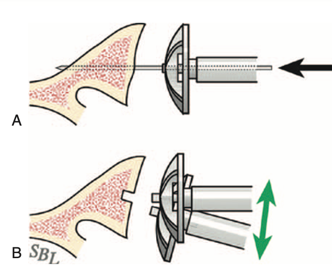
In contrast to the guide wire and cannulated reamer ("A" below), the wireless approach uses a non-cannulated nubbed reamer ("B" below) that can be angulated so that the bony glenoid face is completely reamed to a single concavity with minimal bone removal. 


While reaming, the surgeon can adjust the angle of the reamer so that blades contact and ream the entire glenoid bone surface (green arrows below).


The adequacy of the reaming can be verified by using a round backed trial to see if it tips when loaded (upper figure) or if it is stable on the reamed glenoid surface (bottom figure).


Wireless reaming can be used with any implant system offering a nubbed reamer that can be inserted into a shallow hole in the center of the glenoid.


As described above, the surgeon can adjust the angle of the reamer so that blades contact and prepare the entire glenoid bone surface (green arrows below).


And again, the completeness and adequacy of the reaming can be verified by using a round backed trial to see if it tips when loaded or if it is stable on the reamed glenoid surface.


Below are the preoperative and postoperative axillary truth views from a patient who had wireless reaming for B2 glenohumeral pathology.


The wireless preparation of the glenoid does not require a preoperative CT scan.

It also reduces the risk of over reaming with loss of the glenoid bone stock needed to support the glenoid component as shown in the upper two figures below. (see Version Correction via Eccentric Reaming Compromises Remaining Bone Quality in B2 Glenoids: A Computational Study)


Thanks as always to Steve Lippitt for his wonderful art - often used, seldom acknowledged.

You can support cutting edge shoulder research and education that are leading to better care for patients with shoulder problems, click on this link.

Follow on twitter: https://twitter.com/RickMatsen or https://twitter.com/shoulderarth
Follow on facebook: click on this link
Follow on facebook: https://www.facebook.com/frederick.matsen
Follow on LinkedIn: https://www.linkedin.com/in/rick-matsen-88b1a8133/

Here are some videos that are of shoulder interest
Shoulder arthritis - what you need to know (see this link).
How to x-ray the shoulder (see this link).
The ream and run procedure (see this link).
The total shoulder arthroplasty (see this link).
The cuff tear arthropathy arthroplasty (see this link).
The reverse total shoulder arthroplasty (see this link).
The smooth and move procedure for irreparable rotator cuff tears (see this link).
Shoulder rehabilitation exercises (see this link).

Tuesday, October 11, 2022

The glenoid component in total shoulder arthroplasty: getting it done right.

Glenoid component loosening has been and remains an important cause of failure of anatomic total shoulder arthroplasty (data below from AOANJRR)


However, the survivorship of a pegged, cemented, cross linked, all-polyethylene glenoid component is excellent, with a 13 year cumulative percent revision rate of less than 5%. No metal backed or hybrid glenoid component has matched this outcome.


This type of glenoid arthroplasty serves shoulders across the range of glenoid types, with the lowest revision rates being for the B1 and B2 glenoids.


So, how might surgeons make the good results with the cemented, pegged all-polyethylene component even better? Here are some important concepts that can be put into action.

Careful preparation of the glenoid bone. 

From the authors of Edge displacement and deformation of glenoid components in response to eccentric loading. The effect of preparation of the glenoid bone we learn that the wobble and warp of the polyethylene component is minimized by spherically reaming the bone to precisely match the back of the component.


Glenoid bone stock is preserved by reaming only enough to create a single concavity, rather than trying to "correct" glenoid version.


The adequacy of glenoid reaming can be evaluated by using a pegless trial with the same backside curvature as the actual component and assuring that there is no rocking with eccentric loading. The goal is complete congruency.


Assuring optimal seating and cementing of the component. 

From the authors of The radiographic evaluation of keeled and pegged glenoid component insertion we learn that poor seating and poor cement technique contribute to poor fixation as evidenced by radiolucent lines on the immediate postoperative x-rays. 

Poor seating is evidenced by the presence of cement between the glenoid bone and the backside of the component. 


Good seating is indicated by the absence of cement between the component and bone (below top), rather than using cement as putty in an attempt to compensate for inadequate reaming.


A thin layer of cement between the bone and component is brittle and subject to cracking, displacement and loss of support for the glenoid implant.

Optimal cementing is reflected by the absence of radiolucent lines on postoperative radiographs.


We have learned that this can be achieved by drying each fixation hole with a CO2 spray


Immediately before pressurizing the cement into the hole


Comment: The survivorship of cemented, pegged, all-polyethylene glenoid components has yet to be surpassed by other types of glenoid implants (see Total shoulder replacement stems in osteoarthritis-short, long, or reverse? An analysis of the impact of crosslinked polyethylene). Attention to the details of bone-preserving bone preparation, complete seating of the component and modern cement technique may further improve the clinical outcomes of anatomic total shoulder arthroplasty. Our technique for this procedure is shown in this link.


To add this blog to your reading list in Google Chrome, click on the reading list icon


You can support cutting edge shoulder research that is leading to better care for patients with shoulder problems, click on this link.


Follow on twitter: https://twitter.com/shoulderarth

Follow on facebook: click on this link

Follow on facebook: https://www.facebook.com/frederick.matsen

Follow on LinkedIn: https://www.linkedin.com/in/rick-matsen-88b1a8133/

Here are some videos that are of shoulder interest
Shoulder arthritis - what you need to know (see this link).
How to x-ray the shoulder (see this link).
The ream and run procedure (see this link).
The total shoulder arthroplasty (see this link).
The cuff tear arthropathy arthroplasty (see this link).
The reverse total shoulder arthroplasty (see this link).
The smooth and move procedure for irreparable rotator cuff tears (see this link).
Shoulder rehabilitation exercises (see this link).

Friday, October 15, 2021

Anatomic total shoulder arthroplasty - optimizing glenoid component fixation.

Glenoid Radiolucent Lines in Anatomic Total Shoulder Arthroplasty are Unaffected by Thrombin Glenoid Preparation 

It is recognized that Glenoid component lucencies are associated with poorer patient-reported outcomes following anatomic shoulder arthroplasty.


These authors identified patients undergoing primary anatomic TSA using two glenoid types. Group A glenoids had a cemented central peg without peripheral peg cementation


and Group B glenoids had cemented peripheral pegs without central peg cementation

After reaming the glenoid, all glenoids were irrigated with pulsatile lavage and suction dried. Next, all glenoids in Groups A and B had either Surgicel (Ethicon, Johnson & Johnson, Somerville, NJ) alone or Surgicel soaked in thrombin for preparation. The Surgicel was placed into the peg holes that were to be cemented and then removed prior to cementing. Polymethyl methacrylate cement (Simplex, Stryker, Kalamazoo, MI) was applied using a catheter-tipped syringe for pressurization. 

All patients had the same glenoid preparation except some had the addition of thrombin as a preparation agent. Group A glenoids were implanted by the same surgeon at three different hospitals, one where thrombin was used and two where thrombin was not used. Group B glenoids were implanted by one surgeon who routinely used thrombin and another surgeon who did not at the same hospital. 


The first postoperative radiograph was assessed for radiolucent lines. 


They identified 83 Group A glenoids with and 63 without thrombin glenoid preparation, and 109 Group B glenoids with and 48 without thrombin preparation. 


None of the Group A glenoids had radiolucent lines and 5 (3%) Group B glenoids had radiolucent lines. 


Use of thrombin showed no difference in early radiolucencies (p=1.00) in either Group. 


Comment: It seems likely that radiolucent lines are the result of failure to remove fluid or clot from the holes and failure to adequately pressurize the cement.


In this study the surgical technique combining Surgicel hemostasis and pressurization of each hole effectively minimized postoperative radiolucent lines.


An alternative to Surgicel for removing blood from the peg holes prior to cement pressurization is the use of a carbon dioxide spray to dry each hole immediately before pressurizing the cement assuring that no fluid or clot remains.


This technique, like that of the authors, routinely yields glenoid fixation without radiolucent lines as shown below.


There are other elements that are essential for securing the glenoid component as shown in this link.

The importance of minimizing glenoid component lucencies is demonstrated in this article

These authors point out the high rates of radiographic glenoid loosening following anatomic total shoulder arthroplasty (TSA). They studied the association of radiolucent lines with shoulder function and patient-reported outcomes (PROs) in 492 primary TSAs performed between February 2005 and April 2016. Radiographs were evaluated for glenoid loosening according to the Lazarus grade at a mean of 5.3 years (range, 2-12 years) after surgery.


All-polyethylene keeled components (below left) were used in 186 shoulders All-polyethylene pegged glenoids (below right) were used in 306 shoulders.


An example of radiographic loosening is shown below.

At most recent follow-up, 308 glenoids (63%) showed no radiolucent lines (group 0) and 184 demonstrated peri-glenoid lucencies (group 1). The groups were similar regarding age, sex, body mass index, comorbidities, and prior surgery. At follow-up, group 1 with peri-glenoid lucency's demonstrated significantly lower improvements in forward elevation (P .02) and all PROs (P .005). The improvement in Simple Shoulder Test averaged 7.3 for the 308 shoulders without radiolucent lines and 5.6 for the 184 shoulders with radiolucent lines.


Subgroup analysis by radiolucency grade showed that forward elevation diminished with increasing radiolucent score and exceeded the minimal clinically important difference (MCID) above grade 2 lucencies. A similar decline in PROs was observed with increasing lucency grade; the differences exceeded  the MCID for grade 5 lucencies.


Complications and reoperations were more common in group 1 (16% vs. 5% [P < .001] and 11% vs. 3% [P < .001], respectively). Glenoid component loosening was the most common cause of reoperation in group 1, representing the indication for revision in 14 of 21 reoperations.


Follow on twitter: https://twitter.com/shoulderarth

Follow on facebook: https://www.facebook.com/frederick.matsen

Follow on LinkedIn: https://www.linkedin.com/in/rick-matsen-88b1a8133/


How you can support research in shoulder surgery Click on this link.

Here are some videos that are of shoulder interest
Shoulder arthritis - what you need to know (see this link)
The smooth and move for irreparable cuff tears (see this link)
The total shoulder arthroplasty (see this link).
The ream and run technique is shown in this link.
The cuff tear arthropathy arthroplasty (see this link).
The reverse total shoulder arthroplasty (see this link).

Shoulder rehabilitation exercises (see this link).

This is a non-commercial site, the purpose of which is education, consistent with "Fair Use" as defined in Title 17 of the U.S. Code.          
Note that author has no financial relationships with any orthopaedic companies.