Showing posts with label reaming. Show all posts

Friday, January 26, 2024

Anatomic total shoulder - is a guidewire helpful for reaming?

As pointed out in Edge displacement and deformation of glenoid components in response to eccentric loading. The effect of preparation of the glenoid bone and The radiographic evaluation of keeled and pegged glenoid component insertion, a key to anatomic glenoid component durability is the reaming of the glenoid bone surface so that its concavity exactly fits and completely supports the convex back of the glenoid component as it is exposed to eccentric loading.


Here's an example of poor glenoid preparation.


that can result when a guide wire fixes the orientation of a cannulated reamer in an attempt to modify glenoid version (see How much does "corrective glenoid reaming" actually change version and is the version change of benefit ?)

Poor glenoid preparation cannot be remedied by placing cement between the bone and component - this cement will fracture, displace and leave the component unsupported, often in the critical posterior aspect of the joint.


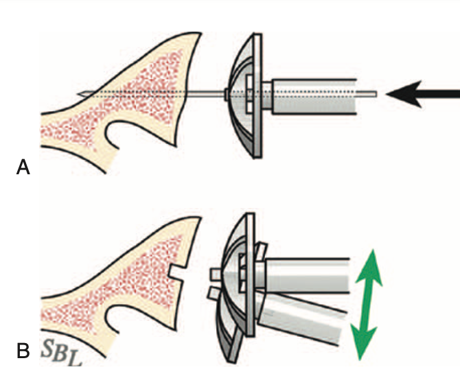
In contrast to the guide wire and cannulated reamer ("A" below), the wireless approach uses a non-cannulated nubbed reamer ("B" below) that can be angulated so that the bony glenoid face is completely reamed to a single concavity with minimal bone removal. 


While reaming, the surgeon can adjust the angle of the reamer so that blades contact and ream the entire glenoid bone surface (green arrows below).


The adequacy of the reaming can be verified by using a round backed trial to see if it tips when loaded (upper figure) or if it is stable on the reamed glenoid surface (bottom figure).


Wireless reaming can be used with any implant system offering a nubbed reamer that can be inserted into a shallow hole in the center of the glenoid.


As described above, the surgeon can adjust the angle of the reamer so that blades contact and prepare the entire glenoid bone surface (green arrows below).


And again, the completeness and adequacy of the reaming can be verified by using a round backed trial to see if it tips when loaded or if it is stable on the reamed glenoid surface.


Below are the preoperative and postoperative axillary truth views from a patient who had wireless reaming for B2 glenohumeral pathology.


The wireless preparation of the glenoid does not require a preoperative CT scan.

It also reduces the risk of over reaming with loss of the glenoid bone stock needed to support the glenoid component as shown in the upper two figures below. (see Version Correction via Eccentric Reaming Compromises Remaining Bone Quality in B2 Glenoids: A Computational Study)


Thanks as always to Steve Lippitt for his wonderful art - often used, seldom acknowledged.

You can support cutting edge shoulder research and education that are leading to better care for patients with shoulder problems, click on this link.

Follow on twitter: https://twitter.com/RickMatsen or https://twitter.com/shoulderarth
Follow on facebook: click on this link
Follow on facebook: https://www.facebook.com/frederick.matsen
Follow on LinkedIn: https://www.linkedin.com/in/rick-matsen-88b1a8133/

Here are some videos that are of shoulder interest
Shoulder arthritis - what you need to know (see this link).
How to x-ray the shoulder (see this link).
The ream and run procedure (see this link).
The total shoulder arthroplasty (see this link).
The cuff tear arthropathy arthroplasty (see this link).
The reverse total shoulder arthroplasty (see this link).
The smooth and move procedure for irreparable rotator cuff tears (see this link).
Shoulder rehabilitation exercises (see this link).

Saturday, November 29, 2014

Glenoid bone density in arthritic shoulders - effect of functional decentering


Glenoid subchondral bone density distribution in male total shoulder arthroplasty subjects with eccentric and concentric wear.

These authors state that "glenoid component loosening in total shoulder arthroplasty may be prevented by component placement on a congruent and adequate bony surface".

They measured bone density in three dimensions for the glenoids of 42 men (21 with eccentric and 21 with concentric wear patterns) with glenohumeral arthritis.

They divided the glenoid subarticular layers into three regions: calcified cartilage (≤1.5 mm), subchondral plate (2-4.5 mm) and cancellous bone (≥5 mm).

In concentric glenoids, the subchondral bone density distribution was homogeneous, with greater mineralization in the central zone, compared with the posterior, anterior, and superior zones. In the eccentric group, the subchondral bone density distribution was inhomogeneous. Mineralization was greatest in the posterior zone, 1,739.0 ± 172.6 HU (at 2.5 mm), followed by the inferior zone, 1,722.1 ± 186.6 HU (at 3 mm).

Comment: This study shows how thin the subchondral bone of the glenoid is. Thus attempts to 'normalize' glenoid version - by reaming the anterior aspect of the glenoid - may transgress the subchondral bone there, leaving the glenoid component supported only by less dense cancellous bone.

The increased density of the posterior bone in eccentric glenoids reflects the increased loading from a posteriorly uncentered head. As pointed about by a previous paper*, CT scans and MRIs taken with the arm at the side fail to reflect the degree of posterior displacement of the humeral contact point on the glenoid when the arm is in functional positions - they refer to this as 'functional malcentering'.
As an example, see the image below, taken with the arm at the side showing only a small amount of posterior displacement of the head on the glenoid, but severe posterior glenoid erosion. One can be certain that when the arm is elevated to a functional position, the humeral head would fall into the posterior concavity = functional malcentering.


For this reason, we prefer to judge the degree of posterior subluxation on axillary views taken with the arm in a functional position of elevation in the plane of the scapula.

*"Functional malcentering of the humeral head and asymmetric long-term stress on the glenoid: potential reasons for glenoid loosening in total shoulder arthroplasty.

We tested the hypothesis that functional malcentering of the humeral head during arm elevation exists in patients with glenohumeral osteoarthritis and influences long-term glenoid loading. Twenty-eight healthy volunteers and 10 patients with primary osteoarthritis, 10 with cuff-arthropathy, and 1 with dysplastic glenoid were examined. Open magnetic resonance imaging and 3-dimensional (3D) digital postprocessing techniques were applied in various arm positions. Osteoabsorptiometry was used to determine 3D subchondral mineralization patterns of the glenoid as an indicator of integral long-term stress distribution. At 30 degrees of abduction, 5 patients demonstrated malcentering of the humeral head posteriorly; all patients with cuff arthropathy had malcentering superiorly. At 90 degrees, most patients displayed significant (P < .001) malcentering in the superior and posterior direction. The shoulders showed maximal subchondral mineralization patterns in the direction of malcentering. Most patients with glenohumeral osteoarthritis displayed functional malcentering, which might be responsible for postoperative glenoid loosening in shoulder arthroplasty if not corrected intraoperatively."

Figure 3 from this article is particularly illuminating.

===


To see the topics covered in this Blog, click here

Use the "Search" box to the right to find other topics of interest to you.

You may be interested in some of our most visited web pages including:shoulder arthritis, total shoulder, ream and runreverse total shoulderCTA arthroplasty, and rotator cuff surgery as well as the 'ream and run essentials'

Saturday, April 12, 2014

Reaming the glenoid for total shoulder arthroplasty

A glenoid reaming study: how accurate are current reaming techniques?

The authors used plastic models of arthritic glenoids in an attempt to explore how the quality of the reamed surface was influenced by different reamers and by glenoid erosion patterns.

They found that for type A glenoids the center and direction of reaming were constant for all surgeons, whereas for type B glenoids the center and direction of reaming differed significantly between surgeons. The congruity of the reamed surface was better after flat reaming than after convex reaming. Whether the reamers were guided by a central K-wire or by a nipple had no significant effect on the reamed surface.

They concluded that reaming of a uniconcave glenoid is reproducible, but reaming of a biconcave glenoid was variable being influenced by the degree of erosion and deformity.

Comment: While it is recognized that the bad arthritic triad is a risk factor for glenoid component failure in total shoulder arthroplasty and while there is interest in means to 'correct' glenoid version in performing total shoulder arthroplasty we do not yet have evidence that 'normalizing' glenoid version (i.e. using asymmetrical reaming, glenoid grafting or augmented components) improves clinical outcomes of total shoulders. It is, however, recognized that aggressive reaming removes precious glenoid bone stock jeopardizing glenoid component fixation.


===
Consultation for those who live a distance away from Seattle.

Check out the new Shoulder Arthritis Book - click here.

Click here to see the new Rotator Cuff Book

To see the topics covered in this Blog, click here

Use the "Search" box to the right to find other topics of interest to you.

You may be interested in some of our most visited web pages including:shoulder arthritis, total shoulder, ream and runreverse total shoulderCTA arthroplasty, and rotator cuff surgery as well as the 'ream and run essentials'

See from which cities our patients come.

See the countries from which our readers come on this post.

To see other similar posts, click on the label of interest below.

Tuesday, January 1, 2013

Ream conservatively in shoulder arthroplasty

Glenoid morphology after reaming in computer-simulated total shoulder arthroplasty.

These authors open their article by stating "The most common cause of TSA failure is loosening of the glenoid component." They point out that posterior bone loss and retroversion are common features of glenohumeral osteoarthritis and that these characteristics complicate the secure and durable placement of a glenoid component. They indicate that preservation of glenoid bone stock, correction of glenoid version and maximization of glenoid component-glenoid bone contact may be competing priorities

This paper emphasizes the hazards of zealous attempts to normalize glenoid version by reaming. They used computer models from computed tomography scans of patients with advanced osteoarthritis. Computer-simulated reaming was performed to study the effect of  reaming depth, reamer placement, and version correction. They found that reamed surface area significantly increased with larger depths of reaming and smaller amounts of initial glenoid retroversion. Bone volume removed was related to reaming depth. They concluded that smaller amounts of initial glenoid retroversion allow for greater implant-bone surface contact.

Glenoid retroversion is common in patients needing shoulder arthroplasty. As we've pointed out in previous posts and as shown in the figure below, are also concerned about the amount of bone removed in trying to 'correct' a retroverted glenoid (upper two figures). For that reason we often choose to ream in retroversion to preserve glenoid bone stock (lower two figures), finding that preserving bone stock and achieving good component-bone contact produce a stable reconstruction.  Paradoxically, shoulders managed this way have greater glenohumeral stability than those in which version is 'corrected'.

--

Use the "Search" box to the right to find other topics of interest to you.

You may be interested in some of our most visited web pages including:shoulder arthritis, total shoulder, ream and runreverse total shoulderCTA arthroplasty,  and rotator cuff surgery.


Saturday, December 29, 2012

The eroded glenoid in total shoulders

Total shoulder arthroplasty does not correct the orientation of the eroded glenoid


This article brings up the important topic of management of the glenoid with posterior erosion - a very common finding in glenohumeral arthritis. Here's an example showing glenoid retroversion, posterior glenoid erosion and posterior humeral subluxation.


The authors sought to determine the extent to which the glenoid component position was governed by the preoperative erosion of the glenoid and whether excessive erosion of the glenoid was associated with perforation of the glenoid vault on insertion of a glenoid component. Using preoperative and postoperative CT scans the authors asssessed version, inclination, rotation, and offset of the glenoid relative to the scapula plane.

The surgical technique sought to position the glenoid perpendicular to the plane of the scapula. Asymmetrical reaming was used to change the orientation of the glenoid. The authors reference a method we described in 1994 for identifying the glenoid centerline to guide their reaming. They found that their preparation of the glenoid did not substantially change the version of the glenoid and that the keel of their glenoid component perforated the glenoid vault in 5 of 29 cases - especially in cases of severe posterior erosion in which the anterior cortex was perforated.

The authors do a nice job of pointing out the competing priorities in achieving a durable, stable, functional arthroplasty:
(1) normalizing glenoid version
(2) avoiding glenoid vault perforation
(3) preserving glenoid bone stock
(4) achieving glenohumeral stability


While it has been stated that glenoid penetration is associated with early loosening, we have not seen documentation of this statement.

Our goal in arthroplasty prioritizes the last two of these:  preserving glenoid bone stock and achieving glenohumeral stability as shown in this post regarding the ream and run and in this post regarding the total shoulder. 


The patient whose preoperative x-ray is shown above elected to have a ream and run. His post operative films so the humeral prosthesis centered in a glenoid that was only reamed enough to create a single concentric concavity.


---
Use the "Search" box to the right to find other topics of interest to you.

You may be interested in some of our most visited web pages including:shoulder arthritis, total shoulder, ream and runreverse total shoulderCTA arthroplasty,  and rotator cuff surgery.


Wednesday, September 28, 2011

How is glenoid reaming done?

This morning on rounds I got asked how is reaming done to convert an arthritic glenoid like this


to a smooth concentric glenoid surface like this


To help demonstrate, here's a video clip showing the actual reaming.


--

Use the "Search the Blog" box to the right to find other topics of interest to you.

You may be interested in some of our most visited web pages including:shoulder arthritis, total shoulder, ream and runreverse total shoulderCTA arthroplasty,  and rotator cuff surgery.

Tuesday, May 24, 2011

Ream and Run for Shoulder Arthritis - initial clinical results - research foundation 3

Based on the encouraging laboratory studies demonstrated in the previous posts we began offering the ream and run procedure to selected and informed patients with arthritis of the shoulder who wished to avoid the risk of glenoid component failure and the activity limitations recommended for total shoulder replacement. In this procedure, we expose the arthritic glenoid (shown below),

remove the damaged cartilage, and then ream the bone to a spherical concavity (shown below) before replacing the humeral side of the joint using the technique explained in our April 6 post.

We were truly excited to see that in many cases, after completely removing the cartilage from the surface of the glenoid, new soft tissue regenerated to cover the reamed bony surface as shown by the dark space between the metal ball and the bone of the glenoid shown on x-rays taken a year after surgery. In the x-rays shown below, this regenerative zone is marked with a red "R".


--

Use the "Search the Blog" box to the right to find other topics of interest to you.

You may be interested in some of our most visited web pages including: shoulder arthritis, total shoulder, ream and runreverse total shoulderCTA arthroplasty, and rotator cuff surgery.

Friday, May 13, 2011

Shoulder joint replacement: minimizing risk of loosening with surgical technique

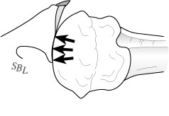
We have seen that the technique with which the polyethylene glenoid component is inserted is a major factor in its durability. One of the special challenges of the shoulder is that as the shoulder is moved, the ball does not stay centered in the socket, but rather translates across its surface. As shown in the figure below.
This translation causes the plastic socket to be loaded eccentrically. Eccentric loading can potentially cause the polyethylene component to wobble, warp and loosen by a mechanisms we have called "rocking horse loosening, diagrammed below.
Shoulder fellow Collins demonstrated that good carpentry in the preparation of the glenoid component can minimize wobble and warp when the shoulder is loaded eccentrically. The results of this study are shown below
The use of glenoid reaming to create the best fit is shown in the figure below.
When the surface of the glenoid bone is properly reamed, rocking horse loosening can be further resisted by a glenoid fixation system that has pegs in front of and behind its center to resist 'lift-off' as shown below on the right in contrast to the keel fixation as shown below on left.
The effectiveness of our approach has been clinically demonstrated by shoulder fellow Lazarus in his article on radiographic evaluation of keeled and pegged glenoid components. He showed that pegged components inserted by an experienced surgeons have a better track record.


Not infrequently in shoulder arthritis, the back of the bony glenoid is worn more than the front, creating a 'bi-concave' glenoid surface. 
When there is a major bi-concavity, we adjust the angle of the reamer to avoid excessive bone removal.


We find this vastly preferable to trying to fill the gap with 'putty carpentry' in which case the wedge of cement may displace leaving the glenoid component unsupported.

--

--

Use the "Search" box to the right to find other topics of interest to you.

You may be interested in some of our most visited web pages including:shoulder arthritis, total shoulder, ream and runreverse total shoulderCTA arthroplasty,  and rotator cuff surgery.


Wednesday, May 4, 2011

Total shoulder replacement: avoiding heat damage

One of the concerns about fixation of the glenoid component in total shoulder replacement is the observation that 'radiolucent lines (RLL's)' may develop between the bone on one hand and the component and cement on the other. These RLL's suggest that the connection of the component to bone is not as strong as it should be. Of greater concern is that these RLLs are often progressive, that is, the fixation of the component becomes less secure with time. This is well shown in the article by Walch et al in the SUNDAY, MAY 1, 2011 post. 


There has been little study of the factors that may contribute to the development and progression of RLL's, especially in cases where no lucent lines are seen immediately after surgery.

One factor that we have studied is the possibility of heat damage to the bone of the glenoid causing local death and resorption of the bone over time with progressive mechanical loosening.  In a study with shoulder fellows Churchill, Boorman and Fehringer, Glenoid cementing may generate sufficient heat to endanger the surrounding bonewe found that the amount of heat generated was related to the volume of cement used in fixing the glenoid component. This is because cement cures with an exothermic, or heat generating reaction. With larger amounts of cement, the temperature of bone can rise to a level that can kill the bone.

This observation led us to explore methods of glenoid fixation that used only minimal amounts of cement: precise 'carpentry' with the removal of a minimal amount of bone.

In second study by shoulder fellows Clinton and Lynch along with resident Olson, we found that reaming of the bone could cause thermal damage: Thermal effects of glenoid reaming during shoulder arthroplasty in vivo. The figure below was taken during surgery using a thermal camera. 

We found that irrigating the wound with cool saline solution at the time of reaming kept the temperature of the bone within safe limits.


--

Use the "Search the Blog" box to the right to find other topics of interest to you.

You may be interested in some of our most visited web pages including: shoulder arthritis, total shoulder, ream and runreverse total shoulderCTA arthroplasty, and rotator cuff surgery.