Showing posts with label steptech. Show all posts

Sunday, May 12, 2024

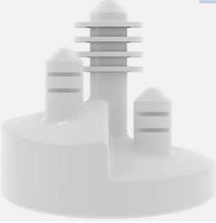
The stepped glenoid component


In their classic article, Stepped Augmented Glenoid Component in Anatomic Total Shoulder Arthroplasty for B2 and B3 Glenoid Pathology, the authors demonstrate that a stepped augmented glenoid component can restore premorbid glenoid anatomy in patients with asymmetric biconcave glenoid bone loss (Walch B2), with short-term clinical and radiographic results equivalent to those for patients without glenoid bone loss (Walch A1) treated with a non-augmented component. 


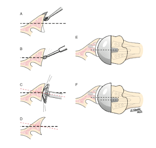
They found a greater risk of osteolysis around the central peg in patients with moderate-to-severe B3 glenoid pathology with this stepped augmented glenoid component. As demonstrated in the technique guide, use of this component involves reaming of the posterior glenoid to fit the step, which may diminish the bony support for the back of the component.

Should this component fail, the posterior bone stock available for conversion to a reverse total shoulder may be compromised. 

A recent paper, Total Shoulder Arthroplasty for Glenohumeral Arthritis Associated with Posterior Glenoid Bone Loss: Midterm Results of an All-Polyethylene, Posteriorly Augmented, Stepped Glenoid Component,  presented a 5 year followup of 35 shoulders receiving a stepped glenoid for the treatment of glenohumeral osteoarthritis with posterior glenoid bone loss. The average preoperative glenoid retroversion was 21.6˚. Although postoperative CT scans were obtained, the postoperative glenoid retroversion was not presented.
Two patients (6%) experienced prosthetic instability requiring revision.

The average Lazarus score (0 no radiolucency to 5 gross loosening) was 0.72. The average Yian score (0 no radiolucency to 18 radiolucent line around entire component) was 2.6. There was an increase in Lazarus score and decrease in Wirth score between 2- and 5-year follow-up. The severity of radiographic loosening correlated with patient-reported pain levels. 

The authors point out that these results are not inferior to those achieved with standard glenoid components in the treatment of glenohumeral arthritis with posterior bone loss.

Comment: Since posterior reaming removes posterior bone and the stepped component adds posterior polyethylene, it would be of interest to know the net change in glenoid retroversion in these patients.
Two figures from this series seem to suggest that substantial retroversion remains after insertion of the stepped component.


As pointed out in the two posts referenced below, future research is needed to determine the clinical value and means of "correcting" glenoid retroversion.

What happens when glenoid version and inclination are "corrected"?

Glenoid version: acceptors and correctors

You can support cutting edge shoulder research that is leading to better care for patients with shoulder problems, click on this link.

Follow on twitter/X: https://x.com/RickMatsen
Follow on facebook: https://www.facebook.com/shoulder.arthritis
Follow on LinkedIn: https://www.linkedin.com/in/rick-matsen-88b1a8133/

Here are some videos that are of shoulder interest
Shoulder arthritis - what you need to know (see this link).
How to x-ray the shoulder (see this link).
The ream and run procedure (see this link).
The total shoulder arthroplasty (see this link).
The cuff tear arthropathy arthroplasty (see this link).
The reverse total shoulder arthroplasty (see this link).
The smooth and move procedure for irreparable rotator cuff tears (see this link).
Shoulder rehabilitation exercises (see this link).

Thursday, December 20, 2018

Post operative glenoid component version - how much does it matter?

Clinical and Radiographic Outcomes of a Posteriorly Augmented Glenoid Component in Anatomic Total Shoulder Arthroplasty for Primary Osteoarthritis with Posterior Glenoid Bone Loss
These authors sought to evaluate the ability of a posteriorly stepped augmented glenoid component, used in 71 patients (mean 65 years of age) with primary glenohumeral osteoarthritis with B2 or B3 glenoid morphology, to correct preoperative retroversion and humeral head subluxation and to identify factors associated with radiographic radiolucency and patient-reported clinical outcomes.

At a median of 2.4 years (range, 1.9 to 5.7 years); the Penn Shoulder Score, range of motion, humeral head centering, and glenoid version were significantly improved. Patients with persistent posterior subluxation of the humeral head postoperatively had worse preoperative fatty infiltration of the teres minor and greater postoperative component retroversion. Patients with center-peg osteolysis had more preoperative joint-line medialization and posterior glenoid bone loss. Patients with more preoperative humeral head posterior subluxation had a lower PSS.

Postoperative humeral head centering on the glenoid component was measured as the anteroposterior humeral head glenoid alignment (HGA-AP) on an axillary radiograph as the amount of posterior displacement of the head center (black dot) with respect to the glenoid component centerline (solid green line) divided by the radius of curvature of the humeral head. In the example below the humeral head component is centered on the glenoid component centerline.

In the example below the humeral head center lies posterior to the glenoid component centerline


Comment: It was of interest to compare these results with those of another recent paper,
Does Postoperative Glenoid Retroversion Affect the 2-Year Clinical and Radiographic Outcomes for Total Shoulder Arthroplasty?

This study analyzed 71 patients having total shoulder arthroplasty with a conventional non-augmented glenoid component inserted with no specific effort to change the version of the glenoid. 

The authors compared the 21 shoulders in which the glenoid component was implanted in 15° or greater retroversion (mean ± SD, 20.7° ± 5.3°) with the 50 in which it was implanted in less than 15° retroversion (mean ± SD, 5.7° ± 6.9°). At the 2-year followup the mean (± SD) improvement in the SST (6.7 ± 3.6; from 2.6 ± 2.6 to 9.3 ± 2.9) for the retroverted group was not inferior to that for the nonretroverted group (5.8 ± 3.6; from 3.7 ± 2.5 to 9.4 ± 3.0). The percent of maximal possible improvement (%MPI) for the retroverted glenoids (70% ± 31%) was not inferior to that for the nonretroverted glenoids (67% ± 44%). 
No patient in either group reported symptoms of subluxation or dislocation. The radiographic results for the retroverted glenoid group were similar to those for the nonretroverted group with respect to central peg lucency (four of 21 [19%] versus six of 50 [12%]; p = 0.436. The mean percentages of posterior humeral head decentering were also similar (3.4% ± 5.5% versus 1.6% ± 6.0%; p = 0.223). The percentage of patients with retroverted glenoids undergoing revision (0 of 21 [0%]) was not inferior to the percentage of those with nonretroverted glenoids (three of 50; [6%]; p = 0.251).

The postoperative decentering was measured on an axillary view in essentially the same manner as in the other paper, except that the displacement is express as a percent of the head diameter rather than the radius. In the example below the humeral head is centered on the glenoid.

The following table attempts to compare the shoulders from each article with respect to postoperative glenoid component retroversion of less than 15 degrees and postoperative glenoid version of 15 degrees or more.


In both series the results appear similar for glenoid components inserted in more or less than 15 degrees of retroversion. There were no revisions for glenoid failure in either series.

It is recognized that long term studies will be needed to determine the relative effectiveness and longevity of different approaches to the retroverted glenoid.

=====
We have a new set of shoulder youtubes about the shoulder, check them out at this link.

Be sure to visit "Ream and Run - the state of the art"  regarding this radically conservative approach to shoulder arthritis at this link and this link

Use the "Search" box to the right to find other topics of interest to you.


You may be interested in some of our most visited web pages   arthritis, total shoulder, ream and runreverse total shoulderCTA arthroplasty, and rotator cuff surgery as well as the 'ream and run essentials'

Wednesday, May 24, 2017

Managing arthritic posterior instability


Many approaches have been suggested for managing the posteriorly decentered humeral head that has come to rest in a pathological posterior concavity. These include posterior bone grafting (a technically difficult procedure with associated problems of hardware failure and graft resorption), reaming the anterior ‘high side” (a procedure that sacrifices bone stock without improving stability), posteriorly augmented plastic glenoid components (devices than increase the force and pressure on the posterior polyethylene with the associated risks of cold flow and increased ‘rocking horse’ lever arm) , and reverse total shoulder (with its associated risks and limitations).

Here are the preoperative x-rays on a heavy set, active patient.  The AP view suggests 'standard' osteoarthritis.


However the axillary 'truth' view shows severe posterior decentering into a pathologic glenoid concavity resulting from severe posterior erosion.

Our approach to this pathoanatomy is to convert the biconcavity into a single concavity by conservative reaming without attempting to change glenoid version, to insert a standard (non-augmented) all polyethylene glenoid, and then to manage any tendency for excessive posterior translation using an anteriorly eccentric humeral head and a rotator interval plication.

The postoperative AP view is shown below


Along with the axillary view that shows the anteriorly eccentric humeral head component to be centered in the glenoid component, which has been inserted in retroversion. Note the slight anterior penetration of the central peg.

At surgery the shoulder was stable to posteriorly directed forces applied to the humeral head. Postoperatively, the patient was able to participate fully in the standard post total shoulder rehabilitation program, including assisted flexion on the evening of surgery.

Of note in this approach is that the head is stabilized in large part by the posterior soft tissues (blue arrows in the figure below),
rather than by loading the posterior aspect of an augmented glenoid component.


Time will tell the best approach for managing this complex pathology. Stay tuned!
===
Consultation for those who live a distance away from Seattle.

Click here to see the new Shoulder Arthritis Book

Click here to see the new Rotator Cuff Book

Use the "Search" box to the right to find other topics of interest to you.

You may be interested in some of our most visited web pages including:shoulder arthritis, total shoulder, ream and runreverse total shoulderCTA arthroplasty, and rotator cuff surgery as well as the 'ream and run essentials'

See from which cities our patients come.

See the countries from which our readers come on this post.

Wednesday, February 22, 2017

Posteriorly augmented glenoid components - an update

Radiographic results of augmented all polyethylene glenoids in the presence of posterior glenoid bone loss during total shoulder arthroplasty

These authors evaluated the clinical and radiographic results of an augmented all-polyethylene glenoid used in 21 patients for the treatment of glenoid osteoarthritis with an average posterior glenoid bone loss of 4.7 mm.

Inclusion criteria: any patient undergoing total shoulder arthroplasty for a diagnosis of glenohumeral osteoarthritis with a Walch B2 or C type glenoid morphology, glenoid retroversion of a minimum of 12°, presence of humeral head subluxation greater than 10%, posterior glenoid bone loss, intact rotator cuff and implantation of an augmented glenoid with minimum of 2-year follow- up.

The augmented glenoid component 


was inserted after posterior preparation by removing sclerotic bone with a high-speed bur and then placing a corresponding sized rasp guide. An oscillating rasp was used to finalize preparation of the posterior glenoid surface.


Significant improvements were demonstrated for American Shoulder and Elbow Surgeons Shoulder Assessment (52.3), Simple Shoulder Test (8.1), forward flexion (50°), external rotation (32°), and pain.

Preoperative retroversion averaged 20.8° (range, 12°-37°), humeral scapular alignment averaged 30% (range, 15%-50%), and humeral glenoid alignment averaged 8% (range, 0%-23.0%.). 

Postoperative retroversion averaged 9° (range, 0°-32°), humeral scapular alignment averaged 6.98% (range, 0%-23%), and humeral glenoid alignment averaged − 0.24% (range, − 8% to 3.5%). 

Central peg ingrowth was demonstrated in all patients, and complete component seating was achieved in 19 patients. No complications were encountered, and no clinical or radiographic failures were identified.

Comment: This is a well done study. As the authors point out and as pointed out in this post, Failure of a total shoulder glenoid component occurs after two years, longer term followup of these implants will be of great interest.

This study can be compared to other recent studies with two year followup on posteriorly augmented glenoid components as presented below.

First

These authors report on 14 shoulders having shoulder arthroplasty with an augmented glenoid component to treat posterior glenoid bone deficiency associated with advanced osteoarthritis.


According to a modified Neer result rating system, 36% of patients had an excellent result, 50% a satisfactory result, and 14% an unsatisfactory result.

At the most recent radiographs, four of the 12 shoulders that could be evaluated radiographically had more than mild glenohumeral subluxation. Three shoulders had moderate posterior subluxation, one shoulder had severe anterior subluxation. All of the shoulders with postoperative subluxation had some degree of subluxation present prior to surgery. 

The shoulder with severe anterior subluxation postoperatively had moderate posterior subluxation prior to surgery. This patient had Parkinsonism and sustained anterior instability resulting in dislocation, treated with closed reduction, followed by immobilization and physical therapy. 

There was no periprosthetic lucency in four shoulders, periprosthetic lucency was grade 1 in seven shoulders and grade 5 in one shoulder, and the glenoid component had shifted in position. This shoulder also had moderate posterior subluxation, and this glenoid component was considered radiographically loose.

The authors concluded, "Our results suggest patients undergoing total shoulder arthroplasty with an asymmetric glenoid component for osteoarthritis achieve satisfactory mid-term pain relief and improvement in function; however, instability is not always corrected. The advantage of this component seems marginal, and its use has been discontinued."


Second
Preliminary Results of a Posterior Augmented Glenoid Compared to an all Polyethylene Standard Glenoid in Anatomic Total Shoulder Arthroplasty

These authors report on 24 patients having total shoulder arthroplasty using a posteriorly augmented glenoid for arthritis with posterior glenoid wear. The degree of posterior wear and retroversion before surgery are not presented.


At two years after surgery, 60% of the shoulders had a periglenoid radiolucent line with an average radiographic line score of 1.10.

One glenoid was radiographically loose. 

Two shoulders demonstrated superior subluxation. 

Three were anteriorly subluxated.

Comment: These results speak to the challenges inherent in the use of posteriorly augmented components.

One of the rarely discussed concerns is the effects of using thick posterior polyethylene to manage the   posteriorly directed loads applied when the arm is elevated to the functional position of forward elevation, which is known to create the risk of functional decentering.

This is best explained by noting that when the arm is at the side with a posteriorly augmented glenoid, the net humeral joint reaction force (red arrow) is centered.


However, when the arm is elevated to a functional position, the net humeral joint reaction force (red arrow) is directed posteriorly against the posteriorly augmented polyethylene, subjecting it to the risk of cold flow.

 Furthermore, the point of application of the net humeral joint reaction force of the elevated arm creates an increased glenoid loosening moment (blue line), when the posterior polyethylene is thick.


It is possible that these mechanisms contributed to the development of lucent lines and instability with posteriorly augmented glenoid components in the series presented.

Another concern with removal of the posterior sclerotic glenoid bone for the insertion of an augmented component (as shown below)


 is that should the augmented component fail, the amount of posterior bone loss may compromise revision surgery.


Our approach to the retroverted glenoid is simple - see this link..

(1) We do not rely on preoperative CT scans because they cannot image the shoulder in the functional  position of forward elevation. Instead we prefer the simple standardized axillary view taken with the arm elevated 90 degrees in the plane of the scapula as shown below (this shoulder demonstrates the bad arthritic triad).


(2) We do not use preoperative planning software or patient specific drill guides, but rather ream the glenoid conservatively without trying to 'normalize' glenoid version as shown in this link.

(3) Finally, in a total shoulder arthroplasty for a retroverted glenoid, we place a standard glenoid component on the conservatively reamed glenoid, using an anteriorly eccentric humeral humeral head component if necessary to achieve centering of the articulation. 

This type of reconstruction is shown below on an axillary view taken with the arm in the functional position of forward elevation. Note the centered humeral head and the lack of glenoid lucent lines after two years of implantation.


This approach preserves the maximal amount of glenoid bone in contrast to what is required to fit the bone to a more complex back side geometry as explained in this link.

Sunday, June 12, 2016

When should we use a stepped posteriorly augmented glenoid component?

Total shoulder arthroplasty for glenohumeral arthritis associated with posterior glenoid bone loss: results of an all-polyethylene, posteriorly augmented glenoid component

Between May 2011 and January 2013, 22 shoulders in 19 patients (15 men and 4 women) underwent primary TSA by a single surgeon. In all cases, an all-polyethylene, posteriorly augmented, stepped glenoid component was implanted. 


In these cases preoperative glenoid retroversion measured 15 degrees or greater (14 of these cases had retroversion of 25 degrees or less).


At a mean follow-up of 36 months, 12 shoulders had osseous integration between the central-peg flanges, 6 had bone adjacent to the central-peg flanges but without identifiable osseous integration, and 1 showed osteolysis. 

While the overall clinical results showed improvement, two patients sustained a total of 3 episodes of humeral dislocation, one anterior and two posterior.


The authors point out that " implantation of a stepped glenoid component requires removal of some posterior bone. In glenoids in which there is both retroversion and glenoid medialization, there may be insufficient subchondral bone, volume, and/or density after preparation to support the posterior component." This is shown by the figure below.


Comment: In the osteoarthritic shoulder with posterior wear, the bone and cartilage have failed to hold up under the loads applied by the humeral head. With a posteriorly augmented glenoid component, the hope is that the thickened posterior polyethylene will be able to hold up under these same loads. 

This report shows that achieving prosthetic balance with this prosthesis can be difficult, even in the hands of an experienced surgeon.

The indications for the use of the stepped prosthesis remain to be determined. Many of the shoulders in this series had relatively low amounts of preoperative retroversion that are in the range usually managed with conventional components and soft tissue balancing.

Should this implant fail, the bone defect would be greater than that present before the arthroplasty as diagrammed below.


Sunday, November 1, 2015

Shoulder arthroplasty with anterior glenoid bone deficiency - augmented glenoid or posteriorly eccentric humeral head?

Total shoulder arthroplasty with an augmented component for anterior glenoid bone deficiency.

These authors present 5 patients having total shoulder arthroplasty using an anteriorly augmented glenoid component. The preoperative diagnoses were anterior glenoid erosion in 2 patients, and 1 patient each with malunited glenoid fracture, nonunited glenoid fracture, and post-traumatic arthritis. The mean age at the time of surgery was 67.4 years (range, 53-75 years).

At an average of 33.2 months (range, 21.9-43.2 months) after surgery no patient had demonstrated radiographic or clinical signs of glenoid component loosening or instability. 

Comment: Anterior glenoid erosion predisposes the shoulder to anterior instability. As shown in this figure from the paper, it can usually be identified on a standardized axillary x-ray.
These authors have reported the use of an augmented glenoid component in the management of five such shoulders with durable results.

An alternative approach is to use a posteriorly eccentric humeral head component with a standard glenoid component accepting the glenoid anteversion as shown in the case below. This approach preserves the maximal amount of glenoid bone stock.


The posteriorly eccentric humeral head component has also improved useful in the management of failed arthroplasty with anterior glenoid bone deficiency, again accepting the anteversion of the glenoid.


The posteriorly eccentric humeral head can also manage post traumatic deformity in which the head is posteriorly malunited.


=


Check out the new Shoulder Arthritis Book - click here.


Use the "Search" box to the right to find other topics of interest to you.

You may be interested in some of our most visited web pages including:shoulder arthritis, total shoulder, ream and runreverse total shoulderCTA arthroplasty, and rotator cuff surgery as well as the 'ream and run essentials'


Thursday, August 20, 2015

Augmented glenoid component - requires removal of posterior glenoid bone

There is a lot to be learned about evaluation and management of the arthritic glenoid as shown in a recent article. In this article the x-rays below illustrate some of these. The figure below shows the implantation of a 7 mm posteriorly augmented glenoid component  'recommended' by a three dimensional imaging and templating system. Note that this implantation requires removal of about half of the dense bone in the posterior glenoid.


This bone removal is also shown in the figures below copyrighted by Steve Lippitt comparing the bone loss with the StepTech to that of the conservative glenoid reaming approach that we embrace (using anteriorly augmented humeral head components and rotator interval plication if necessary to manage posterior instability).


It is apparent that if a stepped posterior glenoid component fails, the bone loss will greatly complicate any additional reconstruction.

Here are the eccentric heads used in conduction with conservative reaming.

===


Check out the new Shoulder Arthritis Book - click here.


Use the "Search" box to the right to find other topics of interest to you.

You may be interested in some of our most visited web pages including:shoulder arthritis, total shoulder, ream and runreverse total shoulderCTA arthroplasty, and rotator cuff surgery as well as the 'ream and run essentials'