Showing posts with label posterior decentering. Show all posts

Sunday, April 20, 2025

Severe B2 glenoid in an active 51 year old man

 A 51 year old general contractor, competitive bow hunter and swimmer presented with persistent and limiting left shoulder pain and grinding that has been refractory to arthroscopic "debridement" and intraarticular steroid injections.  On examination he had limited motion with 80 degrees of glenohumeral flexion, 0 degrees of external rotation, and internal rotation to the gluteal area. His x-rays at presentation show osteoarthritis with inferior and posterior decentering and severe B2 pathoanatomy.


After discusscion of the alternatives of non-operative management, an anatomic total shoulder and reverse total shoulder, he elected to proceed with a ream and run arthroplasty to avoid the risks and limitations of a plastic glenoid component.
CT scanning and the use of planning software were avoided. The procedure was performed under general anesthesia without a nerve block. A subscapularis peel was performed, preserving the long head tendon of the biceps. A standard free-hand anatomic neck cut was made in 30 degrees of retroversion and at a 135 degree angle with the humeral shaft.
His humeral head showed the "Friar Tuck" pattern of central cartilage loss.


Conservative glenoid reaming was performed without attempt to modify glenoid version. Patient-specific instrumentation and augmented reality were not used. Intraoperative trialing indicated that a 54 20 anteriorly eccentric humeral head provided the optimal balance of mobility and stability.  Neither a plastic glenoid component or bone cement was used. The postoperative x-rays are shown below.


Five months after surgery he reported " Shoulder doing good saw great improvements this month. Strength is improving and have resumed construction work with some limitations. Haven’t started shooting a bow yet but soon I think.  Thanks for a great shoulder" and provided the video shown below of his active motion.


House finch in cherry blossoms


You can support cutting edge shoulder research that is leading to better care for patients with shoulder problems, click on this link

Follow on twitter/X: https://x.com/RickMatsen
Follow on facebook: https://www.facebook.com/shoulder.arthritis
Follow on LinkedIn: https://www.linkedin.com/in/rick-matsen-88b1a8133/

Here are some videos that are of shoulder interest
Shoulder arthritis - what you need to know (see this link).
How to x-ray the shoulder (see this link).
The ream and run procedure (see this link).
The total shoulder arthroplasty (see this link).
The cuff tear arthropathy arthroplasty (see this link).
The reverse total shoulder arthroplasty (see this link).
The smooth and move procedure for irreparable rotator cuff tears (see this link)
Shoulder rehabilitation exercises (see this link).


Wednesday, July 3, 2024

B0 glenoid with functional decentering in a 44 year old man - what to do?

A 44 year old man had a previously asymptomatic left shoulder until he attempted to separate to young boys who were fighting at school. Since then he has had pain in his left shoulder, especially when he attempts to push with the arm in forward elevation. His ranges of motion are full and symmetrical to the opposite side. His pain is aggravated with cross body adduction and when he pushes forward and upward against resistance. Instability tests were all unremarkable.

An MRI taken with the arm at the side shows the humeral head centered in the glenoid. His posterior labrum appears somewhat hypertrophic (as seen in glenoid dysplasia) and appears detached from his glenoid.


His axillary "truth" view taken in the position of function and in the position most painful for him shows functional posterior decentering of the humeral head on a glenoid lacking a bony concavity posteriorly.


This case may meet the definition of a Walch B0 glenoid: pre-osteoarthritic posterior subluxation of the humeral head. The authors of that article state: "It appears that Walch B0 glenoid is a pathologic condition (initially dynamic, eventually evolving into a static condition) that may lead to posterior erosion of the glenoid, taking place once there is asymmetric increased posterior glenohumeral contact forces and possibly associated with increased glenoid retroversion."

Further discussion of this pathoanatomy can be found at
The B0 glenoid: why does the back of the glenoid wear out?

In the relevant and interesting ASES Podcast - Episode 107 - Glenohumeral Osteoarthritis Etiology Dr. Peter Chalmers and Dr. Brian Waterman conduct a roundtable interview on the etiology of glenohumeral osteoarthritis with Drs. Ben Zmistowski and Jean-David Werthel.

For our patient the question becomes "what can be done to prevent this shoulder from becoming arthritic? Will a posterior labral repair address the functional posterior decentering? Is there a bony procedure that would be more effective?

We are wondering. Share your thoughts at shoulderarthritis@uw.edu

You can support cutting edge shoulder research that is leading to better care for patients with shoulder problems, click on this link

Follow on twitter/X: https://x.com/RickMatsen
Follow on facebook: https://www.facebook.com/shoulder.arthritis
Follow on LinkedIn: https://www.linkedin.com/in/rick-matsen-88b1a8133/

Here are some videos that are of shoulder interest
Shoulder arthritis - what you need to know (see this link).
How to x-ray the shoulder (see this link).
The ream and run procedure (see this link).
The total shoulder arthroplasty (see this link).
The cuff tear arthropathy arthroplasty (see this link).
The reverse total shoulder arthroplasty (see this link).
The smooth and move procedure for irreparable rotator cuff tears (see this link).

Shoulder rehabilitation exercises (see this link).


Friday, June 21, 2024

The relationship of glenoid version and glenohumeral centering to clinical outcome in a series of 210 patients having anatomic arthroplasty followed for a mean of eight years.


Some shoulder surgeons devote substantial resources to correcting preoperative glenohumeral pathoantomy, while others are inclined to accepting features such as glenoid retroversion (see Glenoid version: acceptors and correctors). 

Recognizing that the surgeon is the method, it is interest to view the outcomes of different  approaches to anatomic total shoulder arthroplasty (ATSA).

The authors of Anatomic total shoulder arthroplasty for posteriorly eccentric and concentric osteoarthritis: a comparison at minimum 5-year follow-up present the average 8 year outcomes for a single-center series of 210 patients with refractory primary osteoarthritis treated with ATSA without attempt to correct glenoid version.

All cases were performed by one of three fellowship-trained shoulder surgeons. Preoperative 3D planning was not used for any of these cases. 

The shoulder was approached through the deltopectoral interval with a subscapularis peel. In cases with posteriorly eccentric wear, the humeral and glenoid sided capsular release during exposure was limited to the mid- sagittal plane (i.e. “6 o’clock”) to preserve posterior capsular tension. Glenoid reaming was limited to that necessary to create a single concavity, preserving bone stock without attempting to correct retroversion. 


Sufficient reaming was indicated by the absence of tipping when a pegless, round-backed trial component was loaded eccentrically. 


The glenoid component in the majority of cases utilized a fluted central peg for bone ingrowth with cemented peripheral pegs (Depuy-Synthes Anchor Peg; n = 204). Cementation technique involved meticulous drying with a pressurized carbon-dioxide spray


Particular attention was given to avoid cement on the backside of the implant.


A standard anatomic humeral arthroplasty was usually performed with conventional length stem (Depuy Global AP or Enovis Turon; n=204).

In cases in which excessive intraoperative posterior translation was identified with trial components in place, an anteriorly eccentric humeral head without or with rotator interval plication was considered to provide stability.


Preoperative and postoperative standardized axillary views were used to determine Walch classification, glenoid component seating, humeroglenoid alignment (HGA-AP) and version. 


The outcome measures included the Simple Shoulder Test, radiolucencies around the glenoid component, and revisions.

98 (47%) of the shoulders had posteriorly decentered humeral heads while 108 (51%) had centered humeral heads. 


77 shoulders had Walch type A glenoids and 122 had Walch type B glenoids. 


35 shoulders had preoperative glenoid retroversion >15 degrees


The mean preoperative SST score of 3.4 improved to a mean of 9.4 at 8 years after surgery. Two patients (1%) underwent re-operations during the study period. 

There was minimal change in glenoid version: the average postoperative retoversion was 7.0 degrees in comparison the preoperative average of 8.8 degrees.

Neither the final SST, change in SST or percentage of maximal improvement were correlated with pre- and postoperative humeral head centering, Walch classification or glenoid version. 

In patients with Walch B1 and B2 glenoids (n=110), there were no differences in outcome measures between patients with postoperative retroversion of more and less than 15 degrees.

While 15 of 51 patients (29%) with minimum 5-year radiographs had glenoid radioluciences, these radiographic findings were not associated with inferior clinical outcomes. 

On multivariable analysis glenoid component radiolucencies were most strongly associated with incomplete component seating (bottom two images below).


Comment: This study indicates that clinically significant and durable outcomes with low revision rates can be accomplished with a straightforward surgical technique in which glenoid bone preservation is prioritized.

You can support cutting edge shoulder research that is leading to better care for patients with shoulder problems, click on this link

Follow on twitter/X: https://x.com/RickMatsen
Follow on facebook: https://www.facebook.com/shoulder.arthritis
Follow on LinkedIn: https://www.linkedin.com/in/rick-matsen-88b1a8133/

Contact: shoulderarthritis@uw.edu

Here are some videos that are of shoulder interest
Shoulder arthritis - what you need to know (see this link).
How to x-ray the shoulder (see this link).
The ream and run procedure (see this link).
The total shoulder arthroplasty (see this link).
The cuff tear arthropathy arthroplasty (see this link).
The reverse total shoulder arthroplasty (see this link).
The smooth and move procedure for irreparable rotator cuff tears (see this link).

Shoulder rehabilitation exercises (see this link). 

Saturday, August 12, 2023

Subluxation in the arthritic shoulder - what is it and what should be done about it?

Subluxation of a joint is defined by the World Health Organization as an "incomplete or partial dislocation" 

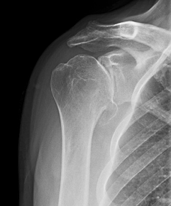
Posterior subluxation of the humeral head on the glenoid is shown below in an axillary view taken with the arm in a position of functional elevation. Because it loads the back of the shoulder, this "truth" view may be better at detecting posterior decentering than a CT taken with the arm at the side. See Answering the critical question: "To what degree is the humeral head functionally centered on the glenoid?" in 6 easy steps.


See Functional malcentering of the humeral head and asymmetric long-term stress on the glenoid: Potential reasons for glenoid loosening in total shoulder arthroplasty ("we found that only 25% of the patients demonstrated a fixed posterior malcentering in a relaxed situation, but 85% showed a functional de-centering during elevation of the arm").

The topic of arthritic shoulder subluxation is obviously of great interest in that the number of articles relating to it is increasing each year.


The amount of posterior subluxation of the humeral head on the glenoid can be determined on an axillary projection as described by Walch et al in Primary glenohumeral osteoarthritis: clinical and radiographic classification. The Aequalis Group


Another approach uses the scapular body - rather than the glenoid articular surface - as the reference. In this method a line is drawn from the tip of the medial border of the scapula to the center of the glenoid fossa; the degree of subluxation is defined as the amount of humeral head lying posterior to this line. As shown in the figure below modified from Approach to glenoid bone loss and deformity in B3 and C glenoids: Primary anatomic shoulder arthroplasty, this method will show a substantial amount of "subluxation" in B3 and C glenoids.


However, applying the original Walch method to this example finds that the humeral head is centered in the glenoid (i.e. not subluxated).


When subluxation is defined as the relationship of the humeral head to the scapular plane, it is strongly correlated with glenoid version, as can be seen from the data presented in Association Between Rotator Cuff Muscle Size and Glenoid Deformity in Primary Glenohumeral Osteoarthritis  (correlation coefficient 0.919)


However the relationship of the humeral head to the plane of the scapula does not reveal the degree of centering or decentering of the humeral head in the glenoid. This helps us understand that the difference between an A2 glenoid and a B3 glenoid lies not in the degree to which the humeral head is centered in the glenoid, but rather in the degree of retroversion


Similarly, the humeral head is often centered on the retroverted type C glenoid.

Version can be altered using eccentric reaming, posterior bone graft or augmented glenoid components - each of which may add complexity, cost, and complications.

Thus, in the treatment of symptomatic B3 and C glenoids, the surgeon needs to decide if altering glenoid version is worthwhile. See Glenoid version: acceptors and correctors

The authors of Early to midterm outcomes of anatomic shoulder arthroplasty performed on dysplastic glenoids studied 29 patients with Type C glenoids with minimum 2 year follow-up after anatomic total shoulders with standard components compared to a matched cohort of 58 A1 glenoids. No effort was made to correct glenoid version through reaming or posterior bone graft. They found similar outcomes scores, patient satisfaction, complication rates, and revision rates.

Treatment of posterior decentering is a different matter and can usually be accomplished without changing glenoid version as demonstrated in Management of intraoperative posterior decentering in shoulder arthroplasty using anteriorly eccentric humeral head components

A more extensive review of subluxation and its clinical importance can be found in  Subluxation in the Arthritic Shoulder The authors critically reviewed the use and misuse of the term "subluxation" in the characterization of arthritic shoulders. Their bullet points are listed below:

» The term “subluxation” means partial separation of the joint surfaces. In the arthritic shoulder, “arthritic glenohumeral subluxation” refers to displacement of the humeral head on the surface of the glenoid.


» The degree of arthritic glenohumeral subluxation can be measured using radiography with standardized axillary views or computed tomography (CT).


» Shoulders with a type-B1 or B2 glenoid may show more posterior subluxation on an axillary radiograph that is made with the arm in an elevated position than on a CT scan that is made with the arm at the side.


» The degree of arthritic glenohumeral subluxation is not closely related to glenoid retroversion.


» The position of the humeral head with respect to the plane of the scapula is related to glenoid retroversion and is not a measure of glenohumeral subluxation.


» Studies measuring glenohumeral subluxation before and after arthroplasty should clarify its importance to the clinical outcomes of shoulder reconstruction.


Applying the standard definition, "subluxation" refers to the displacement of the humeral head relative to the glenoid articular surface. In the diagram below, posterior displacement of the center of a circle that is fit to the humeral head articular surface (decentering) is shown with respect to the perpendicular bisector (red line) of a line segment connecting the anterior and posterior edges of the glenoid. The amount of displacement (the length of the line segment with double arrows) can be expressed as a percentage of the diameter of the circle.


The figure below shows the posterior displacement of the center of a circle fit to the humeral head articular surface with respect to the scapular body reference (long blue line). The amount of displacement (the length of the line segment with double arrows) can be expressed as a percentage of the diameter of the circle. Displacement of the humeral head relative to the plane of the scapular body is not the same as subluxation in that it does not indicate the degree of displacement of the joint surfaces. 


As shown in the diagram below, the degree of posterior subluxation ( decentering) of the humeral head realative to the glenoid (red symbols) for the different glenoid types is not related to the amount of glenoid retroversion. On the other hand the degree of posterior displacement of the humeral head relative to the scapular body (blue symbols) for the different glenoid types is essentially linearly related to the amount of glenoid retroversion. 


The amount of posterior subluxation of the humeral head relative to the glenoid is affected by the position of the arm when the image is made. In the graph below, note that the amount of posterior decentering for shoulders with B1 and B2 glenoids is greater for axillary views taken with the arm in a position of functional elevation (blue symbols, see x-ray at the top of this post) in comparison to that for CT scans taken with the arm positioned at the side (red symbols). Note also that the A1, A2 and B3 glenoids remain centered in both arm positions.


Comment: The measurement of subluxation of the humeral head in relation to the glenoid is an important element in understanding and managing the arthritic shoulder. The different glenoid types show characteristic patterns of glenohumeral subluxation that are not closely related to the degree of glenoid retroversion. The relationship of the humeral head to the body of the scapula is mostly related to glenoid retroversion and is not a measure of glenohumeral subluxation.


Consistency and appropriate use of the term subluxation” will enhance our understanding of arthritic glenohumeral pathoanatomy and its management.


Use of the same imaging technique and arm position before and after surgery enables the surgeon to evaluate the effectiveness of arthroplasty in re-centering the humeral head on the glenoid


Thanks to Mihir Sheth, UW shoulder fellow, for his help in preparing this post.

You can support cutting edge shoulder research that is leading to better care for patients with shoulder problems, click on this link.

Follow on twitter (X): https://twitter.com/shoulderarth
Follow on facebook: click on this link
Follow on facebook: https://www.facebook.com/frederick.matsen
Follow on LinkedIn: https://www.linkedin.com/in/rick-matsen-88b1a8133/

Here are some videos that are of shoulder interest
Shoulder arthritis - what you need to know (see this link).
How to x-ray the shoulder (see this link).
The ream and run procedure (see this link).
The total shoulder arthroplasty (see this link).
The cuff tear arthropathy arthroplasty (see this link).
The reverse total shoulder arthroplasty (see this link).
The smooth and move procedure for irreparable rotator cuff tears (see this link).
Shoulder rehabilitation exercises (see this link).

Monday, February 28, 2022

Answering the critical question: "To what degree is the humeral head functionally centered on the glenoid?" in 6 easy steps.

Understanding the arthritic shoulder and planning its management depends in large part on determining the anteroposterior position of the humeral head on the glenoid in a position of function.

Documenting the effectiveness of surgical management of the arthritic shoulder depends in large part on determining the postoperative anteroposterior position of the humeral head on the glenoid using the same method that was used preoperatively.

As explained in this video, these goals can most practically be accomplished by obtaining the axillary "truth" view taken with the arm forwardly elevated in a position of function. A proper axillary "truth" view will show the "eye" of the spinoglenoid notch (see arrow).


The axillary "truth" view can reveal the wide range of posterior decentering encountered in clinical practice as shown here (note the "eye" on each of these views):


Quantitating the amount of posterior decentering is easily measured on a properly taken axillary "truth" view using 6 straightforward steps:
1. A line segment (AC) is drawn from the anterior (A) to the posterior (C) edges of the glenoid. 
2. A perpendicular bisector to this line is drawn from the midpoint (B) of AC.
3. X is the center of a circle fit to the humeral articular surface 
4. A diameter of the circle (DF) is drawn through X and parallel to AC 
5. E is the intersection of DF with the perpendicular bisector drawn from B. 
6. The percentage of posterior decentering is (EF/DF – 0.5) × 100%. 

This particular shoulder demonstrates a preoperative posterior decentering of 24%.


Unfortunately, many axillary views are not "truth" views, see such an example below. Functional decentering cannot be measured on such a view.


Unfortunately, decentering in a position of function cannot be measured on a CT scan obtained with the arm at the side (see below):


The amount of decentering preoperatively in a position of function can be compared before and after surgery using the axillary "truth" view.


Examples of the utility of the axillary "truth" view can be seen in "Management of intraoperative posterior decentering in shoulder arthroplasty using anteriorly eccentric humeral head components" and in "Total shoulder arthroplasty with an anterior-offset humeral head in patients with a B2 glenoid"


You can support cutting edge shoulder research that is leading to better care for patients with shoulder problems, click on this link.


Follow on twitter: https://twitter.com/shoulderarth

Follow on facebook: click on this link

Follow on facebook: https://www.facebook.com/frederick.matsen

Follow on LinkedIn: https://www.linkedin.com/in/rick-matsen-88b1a8133/


Here are some videos that are of shoulder interest
Shoulder arthritis - what you need to know (see this link).
How to x-ray the shoulder (see this link).
The ream and run procedure (see this link).
The total shoulder arthroplasty (see this link).
The cuff tear arthropathy arthroplasty (see this link).
The reverse total shoulder arthroplasty (see this link).
The smooth and move procedure for irreparable rotator cuff tears (see this link).
Shoulder rehabilitation exercises (see this link).

Saturday, October 30, 2021

Management of severe B2 glenoid in an active young man


An active surfer/athlete presented with pain and stiffness of both shoulders and the x-rays shown below. Each of the axillary "truth" views shows posterior decentering of the humeral head on a biconcave glenoid.


After considering the options of an anatomic total shoulder, an anatomic total shoulder with a posteriorly augmented glenoid component, and a reverse total shoulder, the patient elected to proceed with a ream and run procedure on each shoulder, two years apart.

The four year post operative films for the right shoulder are shown below. Note the use of an anteriorly eccentric humeral head component to control posterior translation.

The two year postoperative films of the left shoulder are shown below. Note again the use of an anteriorly eccentric humeral head component. 


He is back to surfing, tennis, lifting weights and swimming 1,000 yards, activities he was unable to perform before his shoulder arthroplasties.

The active motion of both shoulders is shown below.


As expected, these shoulders had difficulty in regaining functional motion after their prolonged stiffness. In such cases, we use outpatient manipulation under anesthesia and complete muscle relaxation to supplement the patient's rehabilitation effort. In this case several manipulations were peformed on each shoulder. 


We asked him to comment on the use of manipulation in his case. He responded, "Regarding the MUA experience, with both shoulders the MUA benefits seemed to wear off within a couple of weeks until the ones I had done at the two year mark. I could tell each time within even a couple of days that I was tightening up again. But for some reason two years seems to be long enough that my body stops laying down so much scar tissue."


He adds, "one other thing I wanted to mention that has been extremely helpful. For the last few weeks once a day I spend about ten minutes with a baseball (lacrosse ball without seams would likely be better) and lay on my back and use body weight on the ball to work the sore part of the joint. It has made a big difference in soreness that would occur normally from lifting weights, playing tennis, swimming, which are all things I do on a fairly regular basis (each activity at least once a week)"


Comment: This is an exceptionally motivated young man, who has been completely dedicated to his rehabilitation program. His experience with manipulation as long as two years after surgery has been instructive. 


Follow on twitter: https://twitter.com/shoulderarth

Follow on facebook: https://www.facebook.com/frederick.matsen

Follow on LinkedIn: https://www.linkedin.com/in/rick-matsen-88b1a8133/


How you can support research in shoulder surgery Click on this link.

Here are some videos that are of shoulder interest
Shoulder arthritis - what you need to know (see this link)
Shoulder arthritis - x-ray appearance (see this link)
The smooth and move for irreparable cuff tears (see this link)
The total shoulder arthroplasty (see this link).
The ream and run technique is shown in this link.
The cuff tear arthropathy arthroplasty (see this link).
The reverse total shoulder arthroplasty (see this link).

Shoulder rehabilitation exercises (see this link).

This is a non-commercial site, the purpose of which is education, consistent with "Fair Use" as defined in Title 17 of the U.S. Code.          
Note that author has no financial relationships with any orthopaedic companies.