Showing posts with label stress shielding. Show all posts

Sunday, January 5, 2025

Stemless humeral component - is stress shielding an issue?

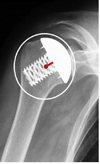
Every type and design of humeral component changes the way in which load is transferred from the articular surface to the bone. Thus, adaptive changes (read: "stress shielding) are inevitable.

The authors of Stress shielding following stemless anatomic total shoulder arthroplasty set out to assess the radiographic proximal humeral bone adaptations seen following stemless anatomic total shoulder arthroplasty. They prospectively followed 152 stemless total shoulder arthroplasties utilizing a single implant design 
performed by 21 shoulder arthroplasty surgeons.


At 2 years postoperatively, "stress shielding" was noted in 61 (41%) shoulders. A total of 11 (7%) shoulders demonstrated major stress shielding with 6 occurring along the medial calcar. At the final follow-up, no humeral implants were radiographically loose or migrated. There was no statistically significant difference in clinical and functional outcomes between shoulders with and without stress shielding. Thus the clinical importance of these adaptive changes was not demonstrated.

In reviewing some of the figures from this article, "stress shielding" may not be the principal cause of bone resorption (yellow arrow). Consider this comparison of the 3 month (left) and the 2 year (right) anterior posterior views of the same shoulder. The 2 year film was interpreted as "demonstrating severe stress shielding along the medial calcar".  Note, however that the cement originally beneath the glenoid component (green arrow) is no longer seen on the 2 year film and that the two year film appears to show shift of the glenoid component's position and lucencies around the pegs (red arrow). These findings bring up the possibility of particulate debris as a contributing factor to the humeral bone resorption at the yellow arrow.


Another figure (below) shows sequential axillary radiographs (L to R: 6 weeks, 6 months, 12 months, and 24 months postoperatively) and was interpreted as showing moderate stress shielding along the medial calcar and greater tuberosity regions. However, it appears that that the proximal humeral bone may have been cystic (yellow arrow) leading to what may be humeral component loosening (red arrows).


Thus it is difficult to know whether the changes seen in the humeral bone are a result of "stress shielding" or are a result of problems with humeral and glenoid component fixation, such as those shown below in other figures from this paper.


While the authors described that "Glenoid radiolucent lines were noted in 28 (18%) patients. These were Lazarus grade 1 in 6, grade 2 in 5, grade 3 in 13, and grade 4 in 2", the relationship of these findings to humeral adaptive changes was not reported.

These authors concluded "the occurrence and severity of bony adaptations had no effect on short-term patient clinical outcomes."

For more detail on this topic see Stemless humeral components: stress shielding

You can support cutting edge shoulder research that is leading to better care for patients with shoulder problems, click on this link

Follow on twitter/X: https://x.com/RickMatsen
Follow on facebook: https://www.facebook.com/shoulder.arthritis
Follow on LinkedIn: https://www.linkedin.com/in/rick-matsen-88b1a8133/

Here are some videos that are of shoulder interest
Shoulder arthritis - what you need to know (see this link).
How to x-ray the shoulder (see this link).
The ream and run procedure (see this link).
The total shoulder arthroplasty (see this link).
The cuff tear arthropathy arthroplasty (see this link).
The reverse total shoulder arthroplasty (see this link).
The smooth and move procedure for irreparable rotator cuff tears (see this link)
Shoulder rehabilitation exercises (see this link).


Saturday, December 28, 2024

Reverse total shoulder: what is the ideal humeral stem?

In contrast to the situation in an anatomic shoulder arthroplasty (below left), the center of rotation (center of yellow circle) in a reverse total shoulder (below right) is not closely aligned with the humeral medullary canal (red line). This results in a rotational moment arm (blue line) and a bending force each of which can contribute to loosening of the humeral stem in the humeral canal.


These loosening factors need be resisted by secure and durable fixation of the prosthesis to bone.

Recently there has been interest in short stemmed reverse humeral components.

However the short stem places the implant at risk for shift in component position as shown below.


Some have tried to manage this tendency for loosening by using tight fitting components, but these can increase the risk of stress shielding as shown below.


And can be difficult to revise in case of malposition, infection or fracture.


I found this recent article co-authored by Patrick Denard of relevance: Volumetric Humeral Canal Fill Ratio Effects Primary Stability and Cortical Bone Loading in Short and Standard Stem Reverse Shoulder Arthroplasty: A Biomechanical and Computational Study

The authors used cadaveric specimens to evaluate the effect of three-dimensional volumetric humeral canal fill ratios of reverse shoulder arthroplasty short and standard stems on the biomechanical stability and bone deformations in the proximal humerus. 

They compared standard stems inserted with low and high filling ratios and short stems inserted with low and high filling ratios


The specimens were cyclically loaded while optical recording allowed for spatial implant tracking and the quantification of cortical bone deformations in the proximal humerus. 

Both short and standard-length stem RSA humeral components implanted with a low canal filling ratio maintain dynamic bone loading in the medial calcar of the humerus similar to the native bone tested. This dynamic bone loading is desirable in the prevention of bone losss from stress shielding. Thus high canal fill ratios ( > 0.72) may correlate with the severity of bone resorption in the clinical setting.

However, short stems implanted with a low filling ratio (< 0.72have an increased risk for implant tilt and subsidence compared to those inserted with high filling ratios or standard length stems. Short stems inserted with higher filling ratios and standard length stems implanted with low or high filling ratios demonstrated higher primary stability.

 
A conclusion from the foregoing is that short stems are at risk for stress shielding if they are inserted with high filling ratios, and at risk for loosening (tilt and subsidence) if they are inserted with low filling ratios. 

So, one can reduce the risks of stress shielding and component loosening through the use of a standard length smooth stem inserted with a low filling ratio as seen in the x-ray below with a stable humeral component and no bone resorption two years after reverse total shoulder arthroplasty. Secure fixation is achieved through impaction autografting.

You can support cutting edge shoulder research that is leading to better care for patients with shoulder problems, click on this link

Follow on twitter/X: https://x.com/RickMatsen

Here are some videos that are of shoulder interest
Shoulder arthritis - what you need to know (see this link).
How to x-ray the shoulder (see this link).
The ream and run procedure (see this link).
The total shoulder arthroplasty (see this link).
The cuff tear arthropathy arthroplasty (see this link).
The reverse total shoulder arthroplasty (see this link).
The smooth and move procedure for irreparable rotator cuff tears (see this link)
Shoulder rehabilitation exercises (see this link). 


Friday, December 8, 2023

Stemless humeral components: stress shielding

A 68 year old woman had bilateral total shoulder arthroplasties performed by another surgeon four years prior to our consultation with her. She noted a painful clicking/clunking of the left shoulder on motion, which we're working up with a CT scan. Her x-rays are shown below. It is not clear why different systems were used for the two shoulders, but of interest is the difference in stress shielding following essentially simultaneous procedures (a "controlled study").

At four years after surgery, the right stemless component (below left) shows localized absence of bone beneath the inferior aspect of the humeral head (green arrow).

At four years after surgery, the left short stemmed component (below right) shows reduced bone density proximal to the diaphysis into which the component has been tightly inserted (black arrows). The red arrows show the localized osteopenia of the lateral humerus while the yellow arrows show the absence of bone medially.


As pointed out in a prior post, with stemmed implants loads applied to the humeral head are distributed by the stem to the humeral diaphysis. However, with the stemless humeral arthroplasty, loads applied to the prosthetic humeral head are distributed to the humeral metaphysis (blue rectangle), bypassing the humeral cortex unless there is contact between the head and humeral neck cut (red arrows). The degree of cortical loading is a function of implant design and surgical technique.


In this post we'll take a closer look at what's been published about stress shielding and adaptive changes after stemless humeral arthroplasty. There's a lot here so you may want to skip to the Comment at the bottom to see a summary.

Computer model studies
Comparison of proximal humeral bone stresses between stemless, short stem, and standard stem length: a finite element analysis used finite element analysis to investigate the effect of humeral component length on stresses in the proximal humerus. The authors found that cortical bone stresses in the most proximal slice for the standard and short stem models were significantly reduced compared with the intact and stemless models. These reductions persisted in the second cortical slice for the standard stem compared with the intact, stemless, and short models. They concluded that reducing stem length produced humeral stresses that more closely matched the intact stress distribution in proximal cortical bone. Opposing trends presented in the proximal trabecular bone, probably because of differences in load transfer when shorter stems are used.


The effect of stemless humeral component fixation feature design on bone stress and strain response: a finite element analysis analyzed the effect of several different humeral component fixation design features 
The implant–bone contact area was greatest for peripheral, followed by central and boundary-crossing designs. All implants elicited similar bone stress variations, which were greatest 0 to 5 mm beneath the resection and laterally. The simulated potential cortical response was also similar for all implants, with the greatest simulated resorbing potential 0 to 15 mm beneath the resection, and very little expected remodeling. Differences between implants were most prominent within the simulated potential trabecular response, with the central implants having the least bone volume percentage expected to resorb.
Bone adaptation impact of stemless shoulder implants: a computational analysis used a computer model to explore the influence of 5 stemless designs on bone adaptation of the humerus (Eclipse, Global Icon, SMR, Simpliciti, and Sidus stemless systems). For the designs not possessing a collar that covers the entire resected surface of the humerus, conditions of contact and no contact were simulated between the humeral head components and the bone surface.


The bone adaptation process was evaluated by comparing differences in bone density between the implanted models and the intact model of the humerus.

The Eclipse-based design was associated with the largest bone mass loss, whereas the SMR based design caused the least. When contact was simulated between
 the resected bone surface and the humeral head components of the SMR-, Simpliciti-, and Sidus-based designs and, bone resorption increased.
The authors concluded that when considering the bone adaptation process, the results suggest that the SMR-based implant presents the best performance and that contact between the humeral head component and the resected bone surface should be avoided.
Another comparison of simulations of remodeling with resurfacing and stemless implants can be found at Bone remodelling of the humerus after a resurfacing and a stemless shoulder arthroplasty


Clinical studies

Stress shielding of the humerus in press-fit anatomic shoulder arthroplasty: review and recommendations for evaluation emphasized the importance of standardizing the technique of taking the x-rays, the views taken, and include measurements obtained immediately after surgery and at final followup. Bone changes should be documented in 6 zones.


Notably, the clinical papers reviewed below show a wide spread in assessment methods and in the reported rates of stress shielding in the humerus after stemless arthroplasty.

Clinical and radiographic outcomes of the Simpliciti canal-sparing shoulder arthroplasty system: a prospective two-year multicenter study. evaluated 147 patients with glenohumeral arthritis treated with the Simpliciti total shoulder system at two year followup. 


There was no reported evidence of migration, subsidence, osteolysis, or loosening of the humeral components.

Another study of the Simpliciti was Early fixation of the humeral component in stemless total shoulder arthroplasty which used radiostereometric analysis radiographs obtained in 24 patients at one day, six weeks, six months, one year, and two years postoperatively. At two years, median translation along the x-, y-, and z-axis was -0.12 mm, -0.17 mm,, and 0.09 mm. Median rotation around the x-, y-, and z-axis was 0.12°, -0.98°, and 0.09°. Overall, 20 prostheses stabilized within 12 months postoperatively. Four cases had continuous shift in position.

The authors of Stress shielding following stemless anatomic total shoulder arthroplasty found that with the Sidus stemless component, 41% of shoulders demonstrated stress shielding at 2 years postoperatively. 7% demonstrated severe stress shielding with 6 occurring along the medial calcar (red arrow). 


The authors of Screw fixation in stemless shoulder arthroplasty for the treatment of primary osteoarthritis leads to less osteolysis when compared to impaction fixation compared their results with the Eclipse Shoulder Prosthesis 


to their results with the Sidus Stem-free Shoulder System 


they found no osteolysis of the medial calcar at a mean of 42 months in 18 patients having the Eclipse; however with the Sidus, osteolysis of the medial calcar was present in seven of the 21 patients at a mean of 36 months after surgery (see above). 


Results for the Eclipse implant were reported in Midterm results of stemless shoulder arthroplasty: a prospective study for 78 patients at a minimum follow-up of 5 years. Bone mineral density was reduced in 34.9% of the older population.


Nine-year outcome after anatomic stemless shoulder prosthesis: clinical and radiologic results presented results for 49 patients receiving the Eclipse shoulder prosthesis at mean of 9 years. Localized lowering of bone density was seen in 29% of the patients.


Radiological changes do not influence clinical mid-term outcome in stemlesshumeral head replacements with hollow screw fixation: a prospective radiological and clinical evaluation used the Eclipse shoulder prosthesis 73 patients, 37% of which had localized bone loss.

Survival of stemless humeral head replacement in anatomic shoulder arthroplasty: a prospective study reported outcomes for 75 patients having the Eclipse shoulder prosthesis with a mean followup of 126 months. Stress shielding around the humeral component was not detected. 


Mid-term results with an anatomic stemless shoulder prosthesis in patients with primary osteoarthritis evaluated 38 patients at an average of 37 months after insertion of a TESS stemless shoulder prosthesis. No signs of stress shielding were noted.


Results of a new stemless shoulder prosthesis: Radiologic proof of maintained fixation and stability after a minimum of three years’ follow-up reported 36 months followup in 63 patients having the TESS stemless shoulder prosthesis; no stress shielding was reported.

The authors of Short-term radiographic analysis of a stemless humeral component for anatomic total shoulder arthroplasty reported the two year radiographic outcomes for 54 patients receiving the Equinoxe Stemless. Stress shielding was observed in 4 patients (7%) with the medial calcar being the most common location of stress shielding. Three of the 4 patients had evidence of partial resorption while 1 patient (25%) had evidence of complete resorption.

Proximal humeral bone loss in stemless shoulder arthroplasty: potential factors influencing bone loss and a new classification system. found that women patients were more likely to experience bone loss after stemless arthroplasty with the Affinis Short Stemless Anatomic Shoulder Prosthesis. Shown below are anteroposterior radiographs demonstrate bone loss grades at the proximal humerus. Grade 0: no bone loss, grade I: bone loss with rounding off of the medial calcar, grade II: bone loss with the corner of the metallic fin at the medial calcar exposed, and grade III: bone loss up to the central stem at the medial calcar. Arrows indicate areas of bone loss.


At a median of 85 months after surgery the bone loss was as shown in the table below.

SPECT/CT demonstrates the osseointegrative response of a stemless shoulder prosthesis. considered the metaphyseal bone metabolic activity in 28 patients after insertion of the Affinis Short.


The SPECT/CT data suggested that the primary osseointegration of the stemless shoulder prosthesis was almost completed 3 months after implantation. 


Reliability of stemless shoulder arthroplasty in rheumatoid arthritis:observation of early lysis around the humeral component compared outcomes for the Affinis Short in 35 patients with RA to 35 patients with osteoarthritis.The proportion of progressive proximal humeral bone loss after TSA was 33% in the RA group and 13% in the control group. Two examples of severe bone loss are shown below.


Impact of humeral stem length on calcar resorption in anatomic total shoulder arthroplasty was a  retrospective review of TSA patients treated with three different-length humeral implants (canal-sparing, short, and standard-length designs). Patients were matched 1:1:1 based on both gender and age (±4 years), resulting in 40 patients per cohort. The canal-sparing implant was the Enovis CS Edge. At 3 months, calcar resorption was observed in 20% of the canal sparing cohort, while the short and standard designs demonstrated resorption in 55% and 52.5%, respectively. At 12 months, calcar resorption was seen in 65% of the canal-sparing design, while both the short and standard designs had a 77.5% rate of resorption. The severity of calcar resorption for the canal-sparing cohort was significantly lower at all time points when compared to the short stem.


Comment: It seems clear that humeral stress shielding is not-infrequently noted after insertion of a stemless implant (as is also the case for implants with short and standard length stems). Risk factors for greater amounts of stress shielding may include inflammatory arthritis and female sex. Many factors may influence the rate and amount of identified stress shielding, including bone quality, implant design, surgical technique, implant position, x-ray position and technique, measurement technique, postoperative patient activity, and length of followup. There is suspicion on some parts that bone resorption around the humeral component may be increased in total shoulder arthroplasty because of polyethylene debris.  Medial calcar resorption seems to be the most common location of adaptive change. The clinical significance of stress shielding is as yet undetermined.

You can support cutting edge shoulder research and education that are leading to better care for patients with shoulder problems, click on this link.

Follow on twitter: https://twitter.com/RickMatsen or https://twitter.com/shoulderarth
Follow on facebook: click on this link
Follow on facebook: https://www.facebook.com/frederick.matsen
Follow on LinkedIn: https://www.linkedin.com/in/rick-matsen-88b1a8133/

Here are some videos that are of shoulder interest
Shoulder arthritis - what you need to know (see this link).
How to x-ray the shoulder (see this link).
The ream and run procedure (see this link).
The total shoulder arthroplasty (see this link).
The cuff tear arthropathy arthroplasty (see this link).
The reverse total shoulder arthroplasty (see this link).
The smooth and move procedure for irreparable rotator cuff tears (see this link).
Shoulder rehabilitation exercises (see this link).

Wednesday, December 22, 2021

Avoiding stress shielding around the humeral implant - are short stems the answer or the problem?

Short stem humeral implants have been introduced to the market based on the proposition that they are "bone preserving". One might ask:

Do they require less bone removal than a standard stemmed implant? 

Do they result in less stress shielding and adaptive bone loss than a standard stemmed implant?

Because short stems achieve fixation through a snug fit of the prosthesis in the proximal humerus, stress shielding is commonly observed (see 

Radiographic changes differ between two different short press-fit humeral stem designs in total shoulder arthroplasty and 

Postoperative radiographic findings of an uncemented convertible short stem for anatomic and reverse shoulder arthroplasty and 

Radiologic bone adaptations on a cementless short-stem shoulder prosthesis).

The challenge with short stems is to achieve fixation without stress shielding. See examples of stress shielding with short stemmed components below.


The authors of The effect of humeral implant thickness and canal fill on interface contact and bone stresses in the proximal humerus studied the effects of implant thickness and canal fill on load transfer, contact, and stress shielding in a finite element study comparing three generic short-stem implant models with different cross-sectional thickness (thinner ( 8 mm), medium (12 mm),  and thicker (16 mm).


 They determined (1) the amount of bone-to-implant contact, (2) changes in cortical and trabecular bone stresses from the intact state, and (3) changes in cortical and trabecular strain energy densities which can predict bone remodeling or stress shielding.

They found that the thinner implant with the lowest canal fill ratio produced significantly lower changes in stress from the intact state in both cortical and trabecular bone. In addition, the thinner implant resulted in a substantially lower volume of bone predicted to stress shield and resorb when compared with the medium and thicker stems.


They concluded that thinner implants and lower canal fill may be beneficial over thicker sizes, provided equal initial fixation can be achieved. The thinner implant has a greater degree of load sharing and increases the mechanical load placed on surrounding bone, reducing the risk of stress shielding and bone resorption.


The question becomes "how is fixation of a thin stem achieved?". Furthermore, if a thin stem is used, what is the advantage of a short stem? The amount of bone removed is the same for both as pointed out in Anatomic short-stem humeral component in total shoulder arthroplasty: a retrospective analysis.


With a standard length stem, impaction autografting can be used to achieve secure fixation of a thin stem on one hand and avoid problems with stress shielding on the other. 


The authors of Radiographic outcomes of impaction-grafted standard-length humeral components in total shoulder and ream-and-run arthroplasty: is stress shielding an issue? evaluated humeral stress shielding after shoulder arthroplasty performed with a smooth, standard-length humeral stem fixed with impaction autografting.


Prior to placement of the final component, cancellous autograft harvested from the humeral head was placed in the humeral canal and pressed into place using a humeral impactor with the same stem geometry as the implant. Autograft was progressively inserted until the impactor fit tightly within the humerus. The final uncoated, smooth, stemmed, fixed-angle humeral component with the desired head geometry was then driven into the prepared canal. 
At two years after surgery, the radiographic appearances were evaluated by an independent experienced shoulder surgeon from another institution not involved in the care of these patients.  The metaphysical and diaphysial filling ratios were measured as shown below.


The filling ratios were small, showing a substantial preservation of bone stock.

The overall radiographic results are shown below


These authors concluded that the independent assessment of the 2-year radiographic and clinical outcomes of a conventional smooth humeral stem inserted with impaction autografting demonstrated the clinical utility of this bone-preserving approach to humeral component fixation with minimal complications; good clinical outcomes; and low rates of bone loss, component subsidence, and shift in position.

Impaction allografting is a useful method for addressing failed prior short stem humeral component fixation - no additional bone is removed


Several other articles are of relevance. The authors of Cementless Curved Short Stem Shoulder Prostheses with a Proximal Porous Coating: Ingrowth Properties at 2-5 Years Radiological Follow-up with Clinical Correlation found a strong correlation between filling ratio and bone loss.


Symptomatic Aseptic Loosening of a Short Humeral Stem Following Anatomic Total Shoulder Arthroplasty

These authors point out that the rate of aseptic humeral component loosening with standard-length stems is low (0.3%) and that presence of humeral loosening gives rise to concern for periprosthetic shoulder infection.


They conducted a retrospective multi-institutional review of anatomic total shoulders performed with a  grit-blasted, rectangular short humeral stem without ingrowth coating.


Twenty-three (12.5%) patients presented with a painful shoulder and met the authors' criteria for humeral loosening at a mean follow-up of 1.5 years (range: 1.5 months – 3.4 years); half had gross humeral subsidence. Thirteen (7.1%) of these underwent revision shoulder arthroplasty where a loose stem was confirmed. 


The rate of symptomatic aseptic humeral loosening in this series was 10.9% (20/184) with 5.4% undergoing revision surgery. Patients with symptomatic aseptic humeral loosening were more likely to be male (90.5%) than those patients without symptoms (52.8%; p<0.001). Of the 105 male shoulders included in this cohort, 19 (18.0%; 19/105) developed symptoms and radiographic findings concerning for aseptic loosening. Ten (9.5%; 10/105) had a revision for aseptic loosening.


The authors concluded that the early humeral loosening rate for this stem design "far outpaces previously reported rates and this study likely under-reports the true incidence of clinically-significant loosening as it only contains limited short-term follow-up". 


Comment: Based on the above information, short stemmed humeral components do not provide added value in comparison to impaction autografted thinner standard length humeral stems.

=====

Follow on facebook: https://www.facebook.com/frederick.matsen

Follow on LinkedIn: https://www.linkedin.com/in/rick-matsen-88b1a8133/


How you can support research in shoulder surgery Click on this link.

Here are some videos that are of shoulder interest
Shoulder arthritis - what you need to know (see this link)
Shoulder arthritis - x-ray appearance (see this link)
The smooth and move for irreparable cuff tears (see this link)
The total shoulder arthroplasty (see this link).
The ream and run technique is shown in this link.
The cuff tear arthropathy arthroplasty (see this link).
The reverse total shoulder arthroplasty (see this link).