Showing posts with label bone graft. Show all posts

Wednesday, May 3, 2017

Reverse total shoulder - to graft or not to graft?

Glenoid bone grafting in primary reverse total shoulder arthroplasty

These authors sought to determine results, complications, and rates of failure of glenoid bone grafting in 41 shoulders having primary reverse shoulder arthroplasty.

These surgeons used bone grafting to achieve between 30% and 50% contact  between the implant and bone. In specific instances of superior bone loss, graft was used to promote inferior tilt of the implant. In the setting of posterior or anterior defects, graft was used to  restore glenoid version. Cancellous graft was used in the setting of  lesser defects. In larger glenoid deficiencies, the use of structural grafts was considered.

Thirty-four received corticocancellous grafts and 7 structural grafts. At a minimum follow-up of 2 years, they found that preoperative severe glenoid erosion and increasing body mass index were significantly associated with worse American Shoulder and Elbow Surgeons scores.

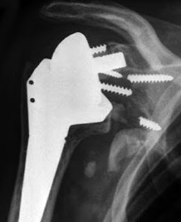
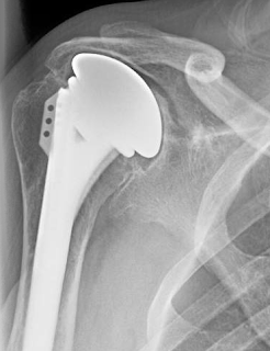
Two sets of x-rays were presented.


On radiographic evaluation, 7 patients had grade 1 or grade 2 glenoid lucency. Glenoid bone graft incorporation was observed in 31 patients. Twelve patients suffered from grade 1 or grade 2 scapular notching. All of the patients with structural grafts showed graft incorporation and no signs of glenoid lucency. None of the patients needed revision surgery. 

Comment: As the authors point out, major glenoid erosion is an indication of severe disease. Patients with severe disease are more likely to have poor outcomes.

The decision to graft and what type of graft to use depends on a number of factors, including the type of baseplate fixation of the particular implant, the experience of the surgeon, the pathoanatomy, and the quality of the residual bone.

In our practice, we use a small drill inserted into the central glenoid vault as a 'dip stick' to determine the adequacy of the extant bone. If a bone depth of approximately 3 cm is present, we conclude that there is sufficient bone to stabilize the implant and ream around a tap placed along the path of this drill.

The use of bone grafting in reverse total shoulder is also discussed in this link.

Our standard reverse technique is shown in this link.

A bit more about bone grafting is detailed in the articles and discussion below

Management of glenoid bone loss in primary and revision reverse total shoulder arthroplasty

These authors reviewed the literature on the management of glenoid defects in reverse shoulder arthroplasties and presented their approach.


They present a classification system:

 For contained defects, they have used impaction allografting beneath the base plate


For eccentric defects, they have used structural grafts


or allograft composites.


If there is concern about the stability of the construction, the authors consider a two stage procedure in which the  glenoid augmentation is performed without implanting the base plate combined with a humeral hemiarthroplasty. The reverse arthroplasty is done as a secondary procedure after confirming graft incorporation with CT scans.

The system used by the authors includes a central bone ongrowth peg. For cases of glenoid deficiency they use an extended length peg.


Comment: This is a useful review of the challenges posed by defects in glenoid bone when performing a reverse total shoulder.

It can be compared to anther recent publication:


These authors point out that large glenoid defects pose difficulties in shoulder arthroplasty. They reviewed 44 patients (20 men and 24 women; mean age, 69 years) having a bulk structural graft behind the baseplate of a reverse total shoulder to manage structure defects.  The grafts consisted of a humeral head autograft in 29, iliac crest autograft in 1, or femoral head allograft in 14.

These cases used a prosthesis with a bone ingrowth stem on the baseplate


inserted over the bone graft as shown below.


Postoperative scores for the bone graft cohort were significantly lower than those in a cohort without grafting. Complete or partial incorporation was shown radiographically in 81% of grafts. 

Six baseplates were considered loose. The major (clinically significant) complication rate was 13.6%. There were 2 graft failures that caused clinical loosening of the baseplate and required revision. Two infections occurred (1 autograft and 1 allograft). One was revised in a 2-stage procedure, and 1 elected to retain the antibiotic spacer. One patient with clinical humeral loosening required revision (autograft). One postoperative dislocation (autograft) occurred. The patient was treated with a closed reduction in the operating room and immobilized for 6 weeks. No further dislocations occurred. Other minor (not clinically significant) complications included 6 patients with scapular notching of grades 1 or 2, and 4 patients with radiolucent lines of 2 mm around the humeral stem but without evidence of gross loosening. This resulted in a total complication rate of 36%.


Comment: There is no question that bone deficiency complicates shoulder arthroplasty. The question is when to use a graft and when to work with the bone stock available. That is a decision that can be made only by the operating surgeon based on the glenoid, the prosthesis used and the surgical technique.

We prefer to avoid structural grafts whenever possible because of uncertainty regarding quality, healing, and resistance to resorption. As pointed out in this paper, bone graft failures can cause clinical loosening of the baseplate.

Our preferred method for reverse total shoulder arthroplasty uses a central screw on the baseplate that provides immediate purchase in glenoid cortical bone without having to depend on bone ingrowth into a central peg and that provides lateral offset of the glenosphere to make up for bone deficiency.


In cases of glenoid bone deficiency, we use a small drill as a 'dip stick' to probe the glenoid to find the appropriate area that offers the best bone stock.  The depth of solid bone is then verified with the tap: 30 mm is ideal.


The baseplate is then screwed into position, assuring that it locks securely in place when tightened.

Here is a recent case of failed shoulder hemiarthroplaty with severe glenoid deficiency.


Secure baseplate and glenosphere fixation was achieved by the method described.

This method may be considered instead of bone grafting when there is sufficient remaining bone of quality.


Friday, April 15, 2016

Reverse total shoulder and glenoid bone defects

Reverse total shoulder arthroplasty with structural bone grafting of large glenoid defects

These authors retrospectively reviewed 44 patients (20 men and 24 women; mean age, 69 years) who had undergone primary or revision reverse total shoulder arthroplasty (RTSA) using a bulk structural graft to augment the glenoid bone behind the baseplate.

The grafts consisted of a humeral head autograft in 29, iliac crest autograft in 1, or femoral head allograft in 14. The graft was shaped to fit the defect.  The graft was provisionally held with Kirschner wires inserted at an angle that did not impede placement of the baseplate. The new surface was reamed with the standard reamers to fit the back of the baseplate. The final fixation of the graft was achieved using screws through the baseplate, spanning the graft into the native glenoid.

Average follow-up was 40.6 ± 16 months. Improvements were seen in the functional outcome scores at the latest follow-up.

The grafting cohort was compared with an age- and sex-matched cohort of RTSA patients without glenoid grafting. Postoperative scores for the bone graft cohort were significantly lower than those in the cohort without grafting.

21 grafts (51.7%) fully incorporated, 12 (29.3%) partially incorporated, and 8 (19.5%) were not incorporated.


Six baseplates were considered loose. Complications included 2 infections, 1 dislocation, 1 humeral loosening, and 2 instances of clinical aseptic baseplate loosening. Six patients showed mild scapular notching.

Comment: These authors have taken on some difficult reconstructions. The loads applied to the baseplate fixation in a reverse total shoulder will challenge the healing of the graft as well as the security of the screw and peg fixation. 6 of the 41 baseplates were loose.

The authors point out that the results of reverses with grafts are not as good as those without grafts, but they do not address the important and difficult questions "when is the glenoid bone loss of sufficient severity to merit consideration of a bone graft?" and "when is the residual glenoid too compromised to support the fixation of a graft?"

===


Check out the new Shoulder Arthritis Book - click here.


Use the "Search" box to the right to find other topics of interest to you.

You may be interested in some of our most visited web pages including:shoulder arthritis, total shoulder, ream and runreverse total shoulderCTA arthroplasty, and rotator cuff surgery as well as the 'ream and run essentials'


Wednesday, October 28, 2015

Glenoid Bone-Grafting in Revision to a Reverse Total Shoulder Arthroplasty - another example of the 4 Ps

Glenoid Bone-Grafting in Revision to a Reverse Total Shoulder Arthroplasty

These authors have an extensive experience with using reverse total shoulder as a revision procedure, performing143 such cases between 2005 and 2010. 102 patients did not undergo grafting, but in 41 they used bone grafting because of glenoid bone deficiency. 40 of the shoulders had follow-up of more than two years (mean, 3.1 years). 16 had central glenoid defects, 13 had posterior defects, 10 had superior defects and 1 had an inferior defect. The arthroplasty systems used included the Comprehensive Reverse Shoulder (20 shoulders),  the Encore Reverse Shoulder Prosthesis (10 shoulders), the Delta III  (1 shoulder) Delta Xtend (7 shoulders) and the Aequalis Reversed Shoulder (2 shoulders).  Fifteen prostheses had a lateral center of rotation, and twenty-five had a medial center of rotation.

Seven of the 40 patients  required another revision surgery because of glenoid loosening (four patients), instability (two patients), or infection (one patient). Of the five structural grafts, one of them failed at 1.2 years secondary to glenoid loosening. The other three failures for glenoid loosening involved cancellous allograft only (two shoulders), or mixed cancellous autograft and allograft (one shoulder).

The two year revision-free survival (76%) and the glenoid radiographic loosening-free survival (89%) were both worse than that for patients who had not required glenoid bone grafting.

Increasing age, revision of a total anatomic or reverse shoulder arthroplasty in contrast to a hemiarthroplasty, and smoking were associated with the need for revision surgery.

Increased rates and grades of glenoid lucency were seen in patients with an increased BMI, a history of total shoulder arthroplasty, an implant with a lateral center of rotation, and in those who smoked.

Comment: The use of a reverse total shoulder in the revision of a failed arthroplasty is known to be among the most challenging applications of this type prosthesis (to whit there were 6 intraoperative humeral fractures in this series). This is a very interesting report in that it indicates the need to individualize the technique when a reverse total shoulder is used to manage a failed joint replacement. In this series five different types of prostheses with three different types of bone graft where used to mange four different types of bone defects after three different types of prior joint replacement (hemiarthroplasty, total shoulder arthroplasty or reverse total shoulder). Once again we see the effect of (1) patient factors (age, BMI, smoking status), (2) shoulder problem characteristics (type of prior arthroplasty, amount and location of the bone defect), and (3) the procedure (type of graft, type of prosthesis). We don't have data on the physician providing the treatment, but that may have had an effect on the survival rate as well.

A major takeaway is that compromised bone quantity and quality can contribute to the risk of glenoid component failure in revision reverse total shoulders: those shoulders needing bone graft, shoulders of smokers and those shoulders that had the removal of a previously placed glenoid component were at greater risk for failure of the reversed glenoid component.

Here's a thought-provoking x-ray, not from this article.


And here's a recent case from our practice - fortunately one without a glenoid bone defect. Note the use of a glenoid component with a long screw for obtaining secure fixation of the glenosphere to the glenoid bone.


=


Check out the new Shoulder Arthritis Book - click here.


Use the "Search" box to the right to find other topics of interest to you.

You may be interested in some of our most visited web pages including:shoulder arthritis, total shoulder, ream and runreverse total shoulderCTA arthroplasty, and rotator cuff surgery as well as the 'ream and run essentials'


Wednesday, July 1, 2015

Reverse total shoulder arthroplasty - bone grafting - can we tell if it reabsorbs?


Effectiveness of CT for the detection of glenoid bone graft resorption following reverse shoulder arthroplasty.

Some surgeons use glenoid bone grafting in cases of reverse shoulder arthroplasty (RSA) to manage glenoid bone deficiency or to increase the offset of the glenosphere from the native glenoid bone. Whenever bone graft is used there is a concern about the possibility of the graft reabsorbing, leaving the glenoid component unsupported. When grafts have been employed, computed tomography (CT) has been used to detect glenoid bonegraft resorption- typically identified by a gap between the bone graft and baseplate.

These authors used a cadaver model to ask if a simulated bone graft resorption gap is detectable by CT scan after a RSA with glenoid bone grafting. They performed RSA with glenoid bone grafting on four cadaver shoulders. Glenoid bone graft resorption gaps were simulated by fixing the implant at six different gap widths (0, 1, 2, 4, 6 and 8mm). CT scans were acquired for each gap and gap widths were measured on  DICOM images in the axial and coronal directions.

The sensitivity of CT imaging to positively identify bonegraft resorption was 38%, with an accuracy of 46%.  Observers tended to visualize no-gap for most conditions. Resorption gap width measurements were consistently underestimated. Metal artifact prevented identification of simulated bonegraft resorption gaps and observers most often determined that there was bonegraft-to-implant contact on CT, when in fact a gap was present.

They concluded that CT scanning was not reliable for assessing bone graft resorption following RSA.

Comment:  We prefer to avoid bone grafts in RSA because of the concern that motivated this article: the risk of failure of graft healing and resorption. If we wish to increase the lateral offset, we use a glenosphere with an extended neck. As the case below from this week illustrates, cases of glenoid deficiency often have glenoid sclerosis that can enable secure fixation of a glenoid base plate that has a strong central screw and peripheral locking screws.


===

Use the "Search" box to the right to find other topics of interest to you.


You may be interested in some of our most visited web pages including:shoulder arthritis, total shoulder, ream and run, reverse total shoulder, CTA arthroplasty, and rotator cuff surgery as well as the 'ream and run essentials'


See from which cities our patients come.


See the countries from which our readers come on this post.


Thursday, December 26, 2013

Does bone grafting solve the problem of glenoid bone deficiency?

Structural bone grafting for glenoid deficiency in primary total shoulder arthroplasty.

These authors reviewed 25 shoulders (representing about 1% of the total number of primary total arthroplasties they performed over the same time period) having a structural bone graft (humeral head autograft and screw fixation) as a glenoid bone augmentation prior to glenoid component placement. Mean clinical and radiographic followup were about 8 years.

On radiographic evaluation, 10 shoulders had glenoids at risk for component loosening. Two of these shoulders were symptomatic and underwent revision surgery to address glenoid component loosening.

Although most of the patients were clinically improved, this study points to the challenges of total shoulder arthroplasty in the management of a retroverted glenoid with posterior humeral instability. The large forces on the back of the glenoid threaten fixation of the graft and the component.

We have used the ream and run procedure for carefully selected patients with this type of glenohumeral pathoanatomy, avoiding the risk of glenoid component failure in the bad arthritic triad (BAT).


===

**Check out the new Shoulder Arthritis Book - click here.**

To see the topics covered in this Blog, click here


Use the "Search" box to the right to find other topics of interest to you.

You may be interested in some of our most visited web pages including:shoulder arthritis, total shoulder, ream and runreverse total shoulderCTA arthroplasty, and rotator cuff surgery as well as the 'ream and run essentials'


Friday, December 20, 2013

Revision shoulder arthroplasty with glenoid bone graft reconstruction

Outcomes after shoulder arthroplasty revision with glenoid reconstruction and bone grafting.

These authors report 17 patients who underwent reconstruction surgery for massive glenoid bone loss after a prior total shoulder using an autologous iliac crest bone graft and secondary revision arthroplasty.

Constant scores improved from 24 ± 17 and to 40 ± 13. CT scans revealed adequate glenoid bone stock restoration with no relevant graft resorption or loosening of the glenoid.

This is a complex two-stage procedure. In the first step the humeral component was revised and the glenoid component removed. A tricortical bone graft from the ipsilateral iliac crest, contoured and fixed to the native glenoid by cannulated 2.7-mm titanium screws. Three months later a second surgery was performed at which the screws were removed and a cemented keeled polyethylene glenoid component inserted.

Comment: These authors have demonstrated a possible approach to the failed glenoid component with bone loss: a two stage graft reconstruction.

Because of concern regarding the possibility of infection associated with glenoid component failure and the poor quality of residual bone at the time of revision, we usually remove the glenoid component, avoid grafting of any type and revise the humeral component to articulate with the residual glenoid bone as shown here, here and here. This is a single stage approach that avoids the risk of a second glenoid component failure.

==
===

Check out the new Shoulder Arthritis Book - click here.

Use the "Search" box to the right to find other topics of interest to you.

You may be interested in some of our most visited web pages including:shoulder arthritis, total shoulder, ream and runreverse total shoulderCTA arthroplasty, and rotator cuff surgery as well as the 'ream and run essentials'