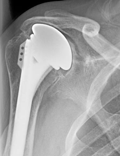
Showing posts with label glenoid deficiency. Show all posts

Wednesday, January 13, 2021

Reverse total shoulder in the bone-deficient glenoid - the alternative center line

Mid-term outcomes of reverse shoulder arthroplasty using the alternative center line for glenoid baseplate fixation: a case-controlled study

These authors point out that secure glenoid baseplate fixation is essential for a successful reverse total shoulder (RSA). In cases of glenoid bone loss, use of the anatomic glenoid center line may not provide sufficient bone support for fixation. Anteversion of the baseplate and central screw fixation along the alternative center line is a described method for achieving baseplate fixation in such cases. 


The authors comparde the outcomes of RSA using the anatomic or alternative center line using a retrospective case-controlled study. 66 patients treated with the anatomic center-line technique

for baseplate fixation were matched 3:1 based on sex, indication for surgery, and age with 22 patients treated with the alternative center-line technique. The mean age was 74.2 years (range, 58-89 years) and mean follow-up period of 53 months (range, 24-130 months). 


monoblock central-screw glenoid baseplate was used in all cases. One of the features of this design is that the preponderance of the fixation is provided by a large central compression screw.


 In cases in which the anatomic center line was used, the baseplate was inserted along the standard glenoid center line. Alternative center-line placement of the baseplate was used to achieve primary baseplate

fixation in cases in which it was determined preoperatively or intraoperatively that there was inadequate bone to support fixation of the center screw.


If <80% coverage of the baseplate could be obtained on host bone, structural grafting with either humeral head autograft or femoral head allograft was used to augment baseplate support as shown in the case below.


Attempts were made to achieve secondary fixation by resting the peripheral rim of the glenosphere on the host glenoid bone or bone graft to distribute the load observed through the baseplate fixation.

Often, a glenosphere with a lip extension was used to achieve this goal (glenosphere sizes of 36 mm – 4, 40 mm neutral, and 40 mm – 4). 


Postoperatively, all patients were managed with the same rehabilitation protocol consisting of wearing a shoulder immobilizer with a self-directed therapy protocol focused on only pendulum exercises for the first 6 weeks, followed by an activeassisted stretching program. Strengthening and lifting were delayed for 3 months.


At the final follow-up, they found no significant differences in patient reported measures, including the Simple Shoulder Test score, American Shoulder and Elbow Surgeons score, visual analog scale pain score, and Single Assessment Numeric Evaluation score


The overall improvements in these measures and all active motions and functional tasks of internal rotation were not different.


No radiographic evidence of glenoid loosening was found in either group. 


Two patients in each cohort (3% of the anatomic group and 9% of the alternative group) experienced an acromial fracture.


Low-grade scapular notching developed in 15.2% of the anatomic group and 18.2% of the alternative center line group.


Comment: This report demonstrates that - in experienced hands - placing the central screw in the thickest part of the glenoid neck can provide good fixation if coupled with bone grafting when adequate support by native bone cannot be achieved. 


We have found it useful to use a small diameter drill to confirm the orientation that will provide the maximum (ideally 3 cm) of screw fixation.


While the surgeon attempted to achieve secondary fixation by resting the peripheral rim of the glenosphere on the host glenoid bone, it is important that this contact not interfere with complete seating of the glenopshere on the baseplate.


As the authors point out, these results may not be generalizable to practitioners who have less experience or those who use other reverse shoulder devices, such as those without a central compression screw (see an example of such an implant below).


To see a YouTube of our technique for a reverse total shoulder arthroplasty, click on this link.

=====
To support our research to improve outcomes for patients with shoulder problems, click here.
To subscribe to this blog, enter your email in the box to your right that looks like the below


===
How you can support research in shoulder surgery Click on this link.

We have a new set of shoulder youtubes about the shoulder, check them out at this link.

Be sure to visit "Ream and Run - the state of the art" regarding this radically conservative approach to shoulder arthritis at this link and this link

Use the "Search" box to the right to find other topics of interest to you.

You may be interested in some of our most visited web pages  arthritis, total shoulder, ream and runreverse total shoulderCTA arthroplasty, and rotator cuff surgery as well as the 'ream and run essentials'

Wednesday, May 3, 2017

Reverse total shoulder - to graft or not to graft?

Glenoid bone grafting in primary reverse total shoulder arthroplasty

These authors sought to determine results, complications, and rates of failure of glenoid bone grafting in 41 shoulders having primary reverse shoulder arthroplasty.

These surgeons used bone grafting to achieve between 30% and 50% contact  between the implant and bone. In specific instances of superior bone loss, graft was used to promote inferior tilt of the implant. In the setting of posterior or anterior defects, graft was used to  restore glenoid version. Cancellous graft was used in the setting of  lesser defects. In larger glenoid deficiencies, the use of structural grafts was considered.

Thirty-four received corticocancellous grafts and 7 structural grafts. At a minimum follow-up of 2 years, they found that preoperative severe glenoid erosion and increasing body mass index were significantly associated with worse American Shoulder and Elbow Surgeons scores.

Two sets of x-rays were presented.


On radiographic evaluation, 7 patients had grade 1 or grade 2 glenoid lucency. Glenoid bone graft incorporation was observed in 31 patients. Twelve patients suffered from grade 1 or grade 2 scapular notching. All of the patients with structural grafts showed graft incorporation and no signs of glenoid lucency. None of the patients needed revision surgery. 

Comment: As the authors point out, major glenoid erosion is an indication of severe disease. Patients with severe disease are more likely to have poor outcomes.

The decision to graft and what type of graft to use depends on a number of factors, including the type of baseplate fixation of the particular implant, the experience of the surgeon, the pathoanatomy, and the quality of the residual bone.

In our practice, we use a small drill inserted into the central glenoid vault as a 'dip stick' to determine the adequacy of the extant bone. If a bone depth of approximately 3 cm is present, we conclude that there is sufficient bone to stabilize the implant and ream around a tap placed along the path of this drill.

The use of bone grafting in reverse total shoulder is also discussed in this link.

Our standard reverse technique is shown in this link.

A bit more about bone grafting is detailed in the articles and discussion below

Management of glenoid bone loss in primary and revision reverse total shoulder arthroplasty

These authors reviewed the literature on the management of glenoid defects in reverse shoulder arthroplasties and presented their approach.


They present a classification system:

 For contained defects, they have used impaction allografting beneath the base plate


For eccentric defects, they have used structural grafts


or allograft composites.


If there is concern about the stability of the construction, the authors consider a two stage procedure in which the  glenoid augmentation is performed without implanting the base plate combined with a humeral hemiarthroplasty. The reverse arthroplasty is done as a secondary procedure after confirming graft incorporation with CT scans.

The system used by the authors includes a central bone ongrowth peg. For cases of glenoid deficiency they use an extended length peg.


Comment: This is a useful review of the challenges posed by defects in glenoid bone when performing a reverse total shoulder.

It can be compared to anther recent publication:


These authors point out that large glenoid defects pose difficulties in shoulder arthroplasty. They reviewed 44 patients (20 men and 24 women; mean age, 69 years) having a bulk structural graft behind the baseplate of a reverse total shoulder to manage structure defects.  The grafts consisted of a humeral head autograft in 29, iliac crest autograft in 1, or femoral head allograft in 14.

These cases used a prosthesis with a bone ingrowth stem on the baseplate


inserted over the bone graft as shown below.


Postoperative scores for the bone graft cohort were significantly lower than those in a cohort without grafting. Complete or partial incorporation was shown radiographically in 81% of grafts. 

Six baseplates were considered loose. The major (clinically significant) complication rate was 13.6%. There were 2 graft failures that caused clinical loosening of the baseplate and required revision. Two infections occurred (1 autograft and 1 allograft). One was revised in a 2-stage procedure, and 1 elected to retain the antibiotic spacer. One patient with clinical humeral loosening required revision (autograft). One postoperative dislocation (autograft) occurred. The patient was treated with a closed reduction in the operating room and immobilized for 6 weeks. No further dislocations occurred. Other minor (not clinically significant) complications included 6 patients with scapular notching of grades 1 or 2, and 4 patients with radiolucent lines of 2 mm around the humeral stem but without evidence of gross loosening. This resulted in a total complication rate of 36%.


Comment: There is no question that bone deficiency complicates shoulder arthroplasty. The question is when to use a graft and when to work with the bone stock available. That is a decision that can be made only by the operating surgeon based on the glenoid, the prosthesis used and the surgical technique.

We prefer to avoid structural grafts whenever possible because of uncertainty regarding quality, healing, and resistance to resorption. As pointed out in this paper, bone graft failures can cause clinical loosening of the baseplate.

Our preferred method for reverse total shoulder arthroplasty uses a central screw on the baseplate that provides immediate purchase in glenoid cortical bone without having to depend on bone ingrowth into a central peg and that provides lateral offset of the glenosphere to make up for bone deficiency.


In cases of glenoid bone deficiency, we use a small drill as a 'dip stick' to probe the glenoid to find the appropriate area that offers the best bone stock.  The depth of solid bone is then verified with the tap: 30 mm is ideal.


The baseplate is then screwed into position, assuring that it locks securely in place when tightened.

Here is a recent case of failed shoulder hemiarthroplaty with severe glenoid deficiency.


Secure baseplate and glenosphere fixation was achieved by the method described.

This method may be considered instead of bone grafting when there is sufficient remaining bone of quality.


Saturday, July 2, 2016

Revision of a dislocated trabeculated metal prosthesis

A middle aged patient had an attempted reverse total shoulder using a trabeculated metal bone ingrowth prosthesis (below left). On insertion of the glenoid component, the anterior glenoid fractured causing the surgeon to abandon the reverse total shoulder and to convert the implant to a hemiarthroplasty (below right).

On presentation to us, the hemiarthroplasty was chronically dislocated into a major anterior glenoid defect.


The patient desired an attempt at conversion of this dislocated implant to a reverse.

After a difficult removal of the dislocated ingrowth humeral component, we used the technique described in a recent article (see this link), exploring with a drill to find the best bone into which  the central screw of the baseplate could be inserted.


We accepted the necessity of anteverting the glenoid component and achieving secure fixation by inserting the screw into the base of the scapular spine.


 And filling in the anterior glenoid defect with bone graft.


After a 36 neutral glenosphere was fixed in place, we cemented the humeral component to achieve control of height and version. The osteotomized proximal humerus was repaired to the humeral component and cancellous allograft added.


Comment: This case illustrates several points. First, trabeculated metal ingrowth prostheses make revision surgery very difficult. We have not found a use for them in our practice, preferring to fix the humeral component with impaction grafting (which facilitates revision should it become necessary). 

Second, care is needed in the insertion of the reverse glenoid component - difficulties in exposure may tempt the surgeon to place the baseplate in an excessively anterior position risking glenoid fracture as occurred here. Third, it is possible to salvage cases of severe glenoid insufficiency using secure fixation of the baseplate screw into the scapular spine. Fourth, because it is a sphere, version of the glenosphere is not critically important to stability as long as anterior and posterior abutment of the humerus against the glenoid is avoided.

===


Use the "Search" box to the right to find other topics of interest to you.

You may be interested in some of our most visited web pages including:shoulder arthritis, total shoulder, ream and runreverse total shoulderCTA arthroplasty, and rotator cuff surgery as well as the 'ream and run essentials'


Tuesday, April 19, 2016

Patient-specific guides in cadaveric glenoid defects - is there evidence of cost-effectiveness?

A patient-specific guide for optimizing custom-made glenoid implantation in cases of severe glenoid defects: an in vitro study.

These authors created glenoid defects in 10 cadaveric shoulders. Computed tomography images were used to plan reversed shoulder arthroplasty and design patient-specific glenoid components. A patient-specific positioning guide was designed for 5 specimens. The remaining 5 specimens were implanted without the guide. 


The patient-specific positioning guide significantly reduced the angular deviations from the planned glenoid implant positioning (P < .05) but not offset, version, inclination or roll. It improved the positioning of the screws and the postoperative total intraosseous screw length.

Comment: This article does not present the learning curve, incremental cost or incremental time associated with the use of patient-specific instrumentation in this cadaver study to weigh against the difference in the two groups as shown below.


===


Check out the new Shoulder Arthritis Book - click here.

Use the "Search" box to the right to find other topics of interest to you.

You may be interested in some of our most visited web pages including:shoulder arthritis, total shoulder, ream and runreverse total shoulderCTA arthroplasty, and rotator cuff surgery as well as the 'ream and run essentials'


Friday, March 4, 2016

Glenoid deficiency - use of a mini-glenoid component in seven patients

Total shoulder arthroplasty using an inlay mini-glenoid component for glenoid deficiency: a 2-year follow-up of 9 patients.

From 2010 to 2013, these authors performed 388 primary TSAs and 53 revision shoulder arthroplasties. In 9 an inset mini-glenoid component was used because of glenoid bone loss and severe retroversion.  Six were primary arthroplasties and 3 were revisions (1 hemiarthroplasty conversion; 1 prior TSA with a loose glenoid and bone loss; and 1 case of  multiple revisions with severe medial glenoid bone loss.  Four shoulders were classified as Walch type A2 glenoids, 2 were classified as Walch type C, and 3 were unable to be classified.

Each patient had a custom glenoid model made from computed tomography reconstructions before surgery. The glenoid models were used to determine the location for the planned mini-glenoid component. 


The custom glenoid components used in this study were premanufactured by various companies. Each miniature glenoid contained a single central peg that measured less than 1 cm in depth.


These authors report on 7 of these patients (9 shoulders) for which two year followup was available. The patients were clinically improved and there were no revisions at two years.  

Comment: Glenoid bone deficiency creates challenges in glenoid component fixation.


This paper presents an approach using custom mini inlay glenoid components. Data on radiolucent lines at the time of followup are not provided in this report. Problems with glenoid component failure are usually not evident in the first two years (the period of follow-up for this study).  When a glenoid component fails the shoulder is left with even greater glenoid bone deficiency than before surgery.

As shown below, the mini-glenoid component does not cover the surface of the glenoid bone and may not resolve the potential for contact between the lateral glenoid bone and the medial humerus. The cost of this approach is not provided in the paper.


Our current practice is to avoid glenoid component implantation in situations where bone quantity and quality is marginal. We await further evidence on the longer term value of the mini glenoid.

===


Check out the new Shoulder Arthritis Book - click here.


Use the "Search" box to the right to find other topics of interest to you.

You may be interested in some of our most visited web pages including:shoulder arthritis, total shoulder, ream and runreverse total shoulderCTA arthroplasty, and rotator cuff surgery as well as the 'ream and run essentials'