Showing posts with label trabecular metal. Show all posts

Sunday, February 18, 2024

Does the innovation of trabecular metal backed glenoid component improve 5 year total shoulder outcomes?

A Multi-Centre, Randomized Controlled Trial Comparing a Second-Generation Uncemented Trabecular Metal-backed versus Cemented Polyethylene Glenoid Component in Total Shoulder Arthroplasty: Five-year Results reports quality of life, clinical, patient-reported, and radiographic outcomes at five years from a randomized controlled trial comparing a second-generation uncemented trabecular metal-backed glenoid (TM, 46 patients, below right) versus a cemented non-ingrowth polyethylene glenoid (POLY, 47 patients, below left) in patients undergoing a total shoulder arthroplasty (TSA).


There were no preoperative differences between groups with respect to age, sex, or WOOS scores.
At followup, there were no statistical or clinically relevant differences in WOOS or patient-reported outcomes between the two groups.

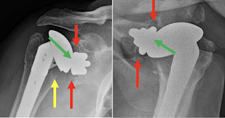
Metal debris was observed in 11 (23.9%) of the TM shoulders without apparent impact on clinical outcomes.


One TM patient experienced glenoid loosening in the setting of an infection. 

One POLY patient had a minor intraoperative periprosthetic fracture of the glenoid which resolved non-operatively. 

Otherwise there was no radiographic evidence of glenoid loosening in either group.

Comment: This randomized clinical trial showed a minimal rate of glenoid component failure at five years after anatomic total shoulder arthroplasty for both the uncemented trabecular metal-backed glenoid and the cemented non-ingrowth polyethylene glenoid in patients undergoing a total shoulder arthroplasty. They noted no differences in outcome between the two glenoid components.

The authors point out that the osseous integration seen with the TM glenoid has the negative consequence that occurs in the revision setting where prosthesis removal can be extremely challenging and lead to extensive bone loss.

You can support cutting edge shoulder research and education that are leading to better care for patients with shoulder problems, click on this link.

Follow on twitter: https://twitter.com/RickMatsen or https://twitter.com/shoulderarth
Follow on facebook: click on this link
Follow on facebook: https://www.facebook.com/frederick.matsen
Follow on LinkedIn: https://www.linkedin.com/in/rick-matsen-88b1a8133/

Here are some videos that are of shoulder interest
Shoulder arthritis - what you need to know (see this link).
How to x-ray the shoulder (see this link).
The ream and run procedure (see this link).
The total shoulder arthroplasty (see this link).
The cuff tear arthropathy arthroplasty (see this link).
The reverse total shoulder arthroplasty (see this link).
The smooth and move procedure for irreparable rotator cuff tears (see this link).
Shoulder rehabilitation exercises (see this link).


Sunday, November 21, 2021

Does the innovation of a trabecular metal glenoid component provide increased value in total shoulder arthroplasty?

Evaluation of Clinical and Radiographic Outcomes After Total Shoulder Arthroplasty with Inset Trabecular Metal-Backed Glenoid

These authors assessed clinical and radiographic outcomes at midterm follow-up of anatomic total shoulder arthroplasty using a Zimmer Biomet trabecular metal glenoid component placed with full backside support using an inset technique. The glenoid components were used in a press-fit manner, which is "off label" in the U.S..


Of 39 patients who having 41 total shoulder arthroplasty procedures with the Zimmer glenoid component, 35 patients (37 shoulders) were available for a minimum 2 year (average 7.2 years, range, 2-11) clinical follow-up. 


At final follow-up, the average shoulder elevation was 153 ± 22° and external rotation was 53 ± 12°. Average ASES scores were 86.8 ± 19.0, and VAS scores were 1.3 ± 2.4. Nine shoulders (27%) had metallic debris. Metal debris was not associated with inferior clinical outcomes.


However, the 39% of shoulders with radiolucencies around the glenoid had inferior clinical outcomes (due to the small number of cases, some of these differences are not statistically significant).


 No patient had a reoperation.

Comment: This study of minimum 2 year results shows that good outcomes can be obtained with a trabecular metal glenoid component in type A glenoid pathology; this type of pathoanatomy is the most straightforward of all the glenoid types. The study does not provide data on the use of this implant for the more problematic glenoid types: B1, B2, B3, C, and D.


Even for the type A glenoids, the study does not provide evidence that the trabecular metal component is superior to the commonly used all-polyeythene component. For example, this study found a 39% rate of radiolucency, which is not lower than that reported for all-polyethylene glenoids (see this link).


In one of the examples shown in the paper, there is a small amount of metal debris, but apparently secure component fixation with a substantial radiographic joint space (green arrows), centering of the humeral head in the glenoid and no evidence of osteolysis or stress shielding.


In the other illustrated case, the humeral head is superiorly decentered on the glenoid, no radiographic joint space is seen (green arrows) suggesting polyethylene wear, osteolysis around the glenoid (red arrows), and humeral osteolysis (yellow arrow). It is possible that the osteolysis resulted from polyethylene wear debris. Polyethylene wear has been recognized as an issue with metal backed components (see this link).
A final issue with trabecular metal glenoid components is the difficulty of their removal, as may become indicated for infection or for conversion to a reverse total shoulder in the event of rotator cuff failure. Extraction of the trabecular metal leaves a substantial glenoid defect as well as posing a risk for fracture.

Here are the x-rays of a recent case that presented to us for evaluation - note the loosening of the glenoid component and the osteolysis.


Follow on twitter: https://twitter.com/shoulderarth

Follow on facebook: https://www.facebook.com/frederick.matsen

Follow on LinkedIn: https://www.linkedin.com/in/rick-matsen-88b1a8133/


How you can support research in shoulder surgery Click on this link.

Here are some videos that are of shoulder interest
Shoulder arthritis - what you need to know (see this link)
Shoulder arthritis - x-ray appearance (see this link)
The smooth and move for irreparable cuff tears (see this link)
The total shoulder arthroplasty (see this link).
The ream and run technique is shown in this link.
The cuff tear arthropathy arthroplasty (see this link).
The reverse total shoulder arthroplasty (see this link).

Shoulder rehabilitation exercises (see this link).

This is a non-commercial site, the purpose of which is education, consistent with "Fair Use" as defined in Title 17 of the U.S. Code.          

Note that author has no financial relationships with any orthopaedic companies.


Thursday, October 1, 2020

Trabecular metal-backed glenoid components in total shoulder arthroplasty: what is their value?

 Modern trabecular metal-backed glenoid components in total shoulder arthroplasty: What is the evidence?

These authors queried the US National Library of Medicine (PubMed/MEDLINE), the Cochrane Database of Systematic Reviews and EMBASE for publications from January 1980 to October 2019 utilizing keywords pertinent to total shoulder arthroplasty, trabecular metal, and clinical outcomes.


They found seven articles to include in their analysis (322 operated shoulders, mean follow-up range: 2–4 years).


The survival rate of modern trabecular metal-backed glenoid components was 96% (309 out of 322 cases) at 43 months mean follow-up, while the rate of aseptic loosening was 0.3% (1 out of 322 cases).  


Fracture of the glenoid component occurred in six cases (overall rate 1.9% of the patients’ cohort), five of which required revision surgery. Among those, there were two cases of intra-operative periprosthetic fractures.


Six cases (1.9% of the patients of this review) of instability were found postoperatively, but only one of them required surgery.


Twenty-five cases (7.8% of the patient of this review) of complications not requiring reoperation were noted. Heterotopic ossification was the most common among them (9 out of 322 cases; 2.8%).


There were eight patients with osteopenia behind the glenoid plate and pegs, suggestive of stress shielding of the glenoid bone stock.


There were 35 cases (10.9%) with glenoid component radiolucency (one of them required revision), and 37 cases (11.5%) of metal debris formation, with four of them undergoing revision.


These authors concluded that there was "low quality evidence to show that the use of modern trabecular metal-backed glenoid components in total shoulder arthroplasty may be safe and effective at short-term follow-up. However, this analysis showed alarmingly high rates of both radiolucency of the glenoid component and metal debris formation which raise concern for potential failure of this glenoid component in the long term. Therefore, we feel that modern trabecular metal-backed glenoid components should be still used with caution as part of a structured surveillance or research program until we know if there is a detriment to the prosthesis in the medium to long term."


Here is a related post on an article entitled, Outcomes of Trabecular Metal–backed glenoid components in anatomic total shoulder arthroplasty 


These authors state that "the current design of the Trabecular Metal–backed glenoid component (Zimmer) was released in 2009. Although over 10,000 of these glenoid components have been implanted worldwide, evidence on either the intermediate- or long-term survival of Trabecular Metal–backed glenoid components in anatomic TSA is very limited."

After a Class 2 recall in 2005 because of concern for fracture at the junction of the base and the trabecular metal keel (see this link), this component was cleared by the FDA (see this link) based on its being substantially equivalent to the predicate recalled devices.


The authors investigated implant survival and two year (average 41 month) clinical outcomes for 36 of 47 patients who received a Trabecular Metal–backed glenoid component (see this link) during primary anatomic total shoulder arthroplasty performed by an individual surgeon.

Three patients showed signs of osteolysis, 4 had radiographic evidence of metal debris, and 1 patient had a catastrophic failure after a fall. Of the 47 TSAs, 5 (11%) were revised to a reverse TSA for subscapularis failure and pain. The authors concluded that Trabecular Metal–backed glenoids had a 25% rate of radiographic metal debris and osteolysis at a minimum 2-year follow-up in this series with one catastrophic failure.


This publication on failures of metal backed glenoids is consistent with a prior post, reproduced below

Early Debris Formation with a Porous Tantalum Glenoid Component: Radiographic Analysis with 2-Year Minimum Follow-up.

These authors point out that a first-generation porous tantalum glenoid component previously demonstrated failure, usually preceded by the appearance of intra-articular metallic debris. An example of component dissociation with this first-generation component is shown here.


After redesign, the component was reintroduced in 2009.


The authors reviewed 68 patients receiving a Trabecular Metal porous tantalum glenoid component (73 components; 5 patients underwent staged bilateral procedures) inserted without polymethylmethacrylate cement (representing off-label usage in the U.S.).

Sixty-six (90%) of the 73 components were evaluated at a minimum of 2 years of follow-up (mean radiographic follow-up of 50.8 months; range, 24 to 68 months). Of these, 92.4% demonstrated minimal or no glenoid radiolucency. Overall, the prevalence of metallic tantalum debris formation was 44% (29 of 66). Sequential radiograph review demonstrated that the incidence of metallic debris formation increased for each year of follow-up, with radiographs from 2, 3, 4, and ≥5 years of follow-up demonstrating a metallic debris incidence of 23%, 36%, 49%, and 52%, respectively.


The severity of metallic debris formation also increased with follow-up duration.


Here's an example of Grade 1, debris noted at the bone-metal interface;

of Grade 2, debris visible in soft tissues intra-articularly;

of Grade 3, visible but incomplete cracking or fracturing of the metal component;


They concluded that the development of metallic debris, increasing in both overall incidence and degree of severity over time, raises concern for potential failure of this glenoid component.

Comment: Metal backed glenoid components continue to manifest problems not present with all-polyethylene components. They demonstrate an increased rate of revision because of loosening, front side and back side polyethylene wear, component dissociation, fracture, instability, and cuff failure (possibility related to the increased thickness of the components) - see this link and the figure below.


 If an arthroplasty with bone ingrowth components requires revision because of infection, cuff failure or instability, removal of the components can result in substantial problems with bone integrity. Such bone damage may compromise secure fixation of a reverse total shoulder glenoid component.

This article presents another issue with metal-backed glenoid components, that of metallic debris, that appears to increase in rate and severity with time after implantation. The mechanism for this debris formation is unclear, but it could be that micromotion of the component pulls the porous trabeculated metal apart.

It can be seen from the list of Young's moduli below (in GPa), that the elastic modulus of polyethylene is closest to that of cortical and cancellous bone:
Cancellous Bone 0.4
Ultra high molecular weight polyethylene 0.5
PMMA bone cement 2
Cortical Bone 8
Titanium 112
Tantalum 186
Cobalt chrome 200.

The Young's modulus of a porous material can be modified by changing the degree of porosity. This is demonstrated in a recent article regarding porous tanalum (see this link). Here is the abstract:


"Relatively high cost of manufacturing and inability to produce modular all tantalum implants has limited its widespread acceptance, in spite of its excellent in vitro and in vivo biocompatibility. In this article, we report how to process Ta to create net shape porous structures with varying porosity using Laser Engineered Net Shaping (LENS™) for the first time. Porous Ta samples with relative densities between 45 to 73% have been successfully fabricated and characterized for their mechanical properties. In vitro cell materials interactions, using human osteoblast cell line hFOB, have been accessed on these porous Ta structures and compared with porous Ti control samples. The results show that the Young’s modulus of porous Ta can be tailored between 1.5 to 20 GPa by changing the pore volume fraction between 27 and 55%. In vitro biocompatibility in terms of MTT assay and immunochemistry study showed excellent cellular adherence, growth and differentitation with abundant extracellular matrix formation on porous Ta structures compared to porous Ti control. These results indicate that porous Ta structures can promote enhanced/early biological fixation. The enhanced in vitro cell-materials interactions on porous Ta surface are attributed to chemistry and its high wettability and surface energy relative to porous Ti. Our results show that these laser processed porous Ta structures can find numerous applications, particularly among older patients, for metallic implants because of their excellent bioactivity."

Nevertheless, based on the evidence available, metal backed glenoids may not offer to the patient advantages over an all polyethylene component as discussed below.

Radiographic and clinical outcomes of total shoulder arthroplasty with an all-polyethylene pegged bone ingrowth glenoid component: prospective short- to medium-term follow-up.

These authors reviewed 80 total shoulders using an all polyethylene glenoid component.  The glenoid was reamed minimally to preserve subchondral bone, which was given priority above correcting retroversion.

1 of 80 shoulders was revised for aseptic glenoid loosening.  81.6% had a radiolucency grade of 0 or 1. Nearly 90% had a glenoid seating grade of A or B. Grade 2 or 3 bone around the central peg was seen in 88.2%. 

No statistical association existed between Walch glenoid types and radiolucency grades, bone grades around the central peg, perfect radiolucency grade, seating grade, and grade 3 bone around the central peg. 

===


Our approach to total shoulder arthroplasty can be viewed by clicking here.


To subscribe to this blog, enter your email in the box to your right that looks like the below


===
How you can support research in shoulder surgery Click on this link.

We have a new set of shoulder youtubes about the shoulder, check them out at this link.

Be sure to visit "Ream and Run - the state of the art" regarding this radically conservative approach to shoulder arthritis at this link and this link

Use the "Search" box to the right to find other topics of interest to you.

You may be interested in some of our most visited web pages  arthritis, total shoulder, ream and runreverse total shoulderCTA arthroplasty, and rotator cuff surgery as well as the 'ream and run essentials'

Thursday, August 6, 2020

Trabecular metal glenoid components

Cemented versus Uncemented Fixation of Second Generation Trabecular Metal Glenoid

Components: Minimum 5-year Outcomes


In 2003, the first generation Trabecular MetalTM (Zimmer, Warsaw, IN, USA) metal backed polyethylene glenoid component was introduced. The first generation Trabecular MetalTM-backed glenoid components exhibited a high failure rate due to fracture at the component keel/tantalum disc  interface and was taken off the market in 2005. The second-generation TM glenoid was introduced in 2009.


Apparently surgeons implant this glenoid component using either cement or using a press-fit without cement. Cementation is performed with cement applied on the component’s back side, covering only the peripheral polyethylene surface.


This study compared outcomes for two surgeons, one of whom routinely cemented the glenoid component and utilized a lesser tuberosity; the second surgeon routinely implanted the glenoid component without cement and utilized a subscapularis peel.


Minimum 5 year followup was available for 55 shoulders; 27 in the cemented group (21 with full radiographic follow-up) and 28 in the uncemented group (22 with full radiographic follow-up). 


No patients required revision surgery. 


Groups had similar preoperative ROM, but uncemented patients had greater follow-up forward flexion (p=0.03), external rotation (p<0.01) and lateral elevation (p=0.03) when compared to cemented patients. 


ASES scores (89.8 cemented vs. 94.1 uncemented, p=0.21) were also similar. 


The uncemented group demonstrated significantly higher rates of radiolucent lines and higher frequency of mild metal debris. Mid-term radiographs showed a metal debris rate of 24% in the cemented group and 27% in the uncemented group.


No implants required revision. 

Below are the radiographs showing a shoulder with a cemented TM glenoid at 5.7 year followup. There is intra-articular metal debris, radiolucency, and superior translation of the humeral head on the glenoid component. 
Comment: While the significance of metal debris has not been rigorously defined, it may indicate migration of the component as reflected in the table above. 

It is noted that the insertion of this component requires removal of a substantial amount of bone, which may complicate revision (see prior post at this link). 

In total shoulder arthroplasty, cement on the backside of the component is unnecessary if the bone is properly prepared and the glenoid component properly seated. 

=====
To see our technique for total shoulder arthroplasty, click on this link.

To see our new series of youtube videos on important shoulder surgeries and how they are done, click here.

Use the "Search" box to the right to find other topics of interest to you.


You may be interested in some of our most visited web pages  arthritis, total shoulder, ream and runreverse total shoulderCTA arthroplasty, and rotator cuff surgery as well as the 'ream and run essentials'


Monday, July 4, 2016

Anteriorly unstable trabeculated metal prosthesis with rocking horse glenoid loosening

Here's another example of an anteriorly unstable trabeculated metal prosthesis referred to us for revision to a reverse because of refractory anterior instability, failed subscapularis reconstruction, and rocking horse loosening of the glnoied. See our most recent post (link here).


This was revised to a reverse. Because, as is often the case in revising an ingrowth humeral component the tuberosity cracked on humeral component removal, some proximal cement was needed to assure rotational stability,


By contrast, the typical reverse with intact tuberosities can usually be stabilized with impaction grafting alone.


===


Use the "Search" box to the right to find other topics of interest to you.

You may be interested in some of our most visited web pages including:shoulder arthritis, total shoulder, ream and runreverse total shoulderCTA arthroplasty, and rotator cuff surgery as well as the 'ream and run essentials'


Saturday, July 2, 2016

Revision of a dislocated trabeculated metal prosthesis

A middle aged patient had an attempted reverse total shoulder using a trabeculated metal bone ingrowth prosthesis (below left). On insertion of the glenoid component, the anterior glenoid fractured causing the surgeon to abandon the reverse total shoulder and to convert the implant to a hemiarthroplasty (below right).

On presentation to us, the hemiarthroplasty was chronically dislocated into a major anterior glenoid defect.


The patient desired an attempt at conversion of this dislocated implant to a reverse.

After a difficult removal of the dislocated ingrowth humeral component, we used the technique described in a recent article (see this link), exploring with a drill to find the best bone into which  the central screw of the baseplate could be inserted.


We accepted the necessity of anteverting the glenoid component and achieving secure fixation by inserting the screw into the base of the scapular spine.


 And filling in the anterior glenoid defect with bone graft.


After a 36 neutral glenosphere was fixed in place, we cemented the humeral component to achieve control of height and version. The osteotomized proximal humerus was repaired to the humeral component and cancellous allograft added.


Comment: This case illustrates several points. First, trabeculated metal ingrowth prostheses make revision surgery very difficult. We have not found a use for them in our practice, preferring to fix the humeral component with impaction grafting (which facilitates revision should it become necessary). 

Second, care is needed in the insertion of the reverse glenoid component - difficulties in exposure may tempt the surgeon to place the baseplate in an excessively anterior position risking glenoid fracture as occurred here. Third, it is possible to salvage cases of severe glenoid insufficiency using secure fixation of the baseplate screw into the scapular spine. Fourth, because it is a sphere, version of the glenosphere is not critically important to stability as long as anterior and posterior abutment of the humerus against the glenoid is avoided.

===


Use the "Search" box to the right to find other topics of interest to you.

You may be interested in some of our most visited web pages including:shoulder arthritis, total shoulder, ream and runreverse total shoulderCTA arthroplasty, and rotator cuff surgery as well as the 'ream and run essentials'