Showing posts with label glenoid loosening. Show all posts

Saturday, February 17, 2024

Risk factors for glenoid loosening and cuff tears after anatomic shoulder arthroplasty.

The authors of Risk Factors for Rotator Cuff Tears and Aseptic Glenoid Loosening After Anatomic Total Shoulder Arthroplasty reviewed the factors associated with increased risk of cuff tears and glenoid loosening at two years after anatomic total shoulder arthroplasty in 2,699 patients from an international multicenter study who received the Equinoxe platform implants. 

Postoperative cuff tears were reported in 2.5%; 1.3% had revision surgery because of a cuff tear. Patients were more likely to experience postoperative cuff tears if they had prior surgery on the affected side or were treated with a small glenoid component.

Aseptic glenoid loosening was reported in 4.0%; 3.4% had revision surgery because of glenoid loosening. Patients were more likely to experience glenoid loosening if they were ≤62 years of age at the time of surgery, treated with a small glenoid component, or treated with smooth pegged or keeled glenoid components (as opposed to hybrid glenoid components) 


Comment: a few thoughts about these risk factors

Patient factors:

(1) Patient age ≤62 years - this once more brings up the issue of "how do we choose the treatment of osteoarthritis in younger patients?".

(2) Prior surgery - the nature of the prior surgery is not reported. It seems likely that some of these prior surgeries may have been rotator cuff related, which could account for the observed increase in post-arthroplasty cuff tears.

Surgeon controlled factors:

(1) Small glenoid component - it is not clear whether the increased risk associated with the use of a small glenoid component is related to the size of the native glenoid, to less bony support of the component,  to lesser experience of the surgeon or to other factors.

(2) Non-ingrowth pegged or keeled glenoid components - many surgeons currently prefer glenoid components that provide the opportunity for bony ingrowth instead of smooth pegs or keels.

We've still got a lot to learn.

You can support cutting edge shoulder research and education that are leading to better care for patients with shoulder problems, click on this link.

Follow on twitter: https://twitter.com/RickMatsen or https://twitter.com/shoulderarth
Follow on facebook: click on this link
Follow on facebook: https://www.facebook.com/frederick.matsen
Follow on LinkedIn: https://www.linkedin.com/in/rick-matsen-88b1a8133/

Here are some videos that are of shoulder interest
Shoulder arthritis - what you need to know (see this link).
How to x-ray the shoulder (see this link).
The ream and run procedure (see this link).
The total shoulder arthroplasty (see this link).
The cuff tear arthropathy arthroplasty (see this link).
The reverse total shoulder arthroplasty (see this link).
The smooth and move procedure for irreparable rotator cuff tears (see this link).
Shoulder rehabilitation exercises (see this link).

Tuesday, August 20, 2019

Total shoulder arthroplasty - the risk of loosening of a keeled glenoid component

Risk factors for loosening of cemented glenoid components in anatomical shoulder arthroplasty

These authors examined the risk factors for glenoid component loosening in 471 shoulder arthroplasties with a mean followup of 8.1 years. The authors used a cemented keeled glenoid component iWrightMedicalGroup).


A flat-back glenoid component was used in the first 186 cases and a convex-backed component in the following 285 cases. Preparation of the glenoid was different among surgeons. Some surgeons preferred only a slight reaming and protection of the subchondral bone layer, and some preferred to take down the bone layer.

A total of 137 glenoid components (29.1%) were radiographically judged to be at risk for loosening.


Univariate analysis revealed that 
1. Excessive reaming on the native glenoid with removal of subchondral bone was associated with a 3.7-fold higher risk for glenoid component loosening (p< 0.001).

2. A glenohumeral mismatch <6mm was associated with higher radiographic loosening rates (p< 0.03), and the risk increased by 19% with every millimeter of less mismatch.

3. The use of a flat-back glenoid component was associated with a 3.1-fold higher risk for radiographic loosening compared to convex-back glenoids (p< 0.001).

4. B2 glenoids were associated with a higher risk for radiographic loosening compared to A1 (2.3-fold), A2 (3.6-fold), and B1 (2.7-fold) glenoids (p< 0.001).

Comment: This is an important long-term followup study. It lends further support to (1) preservation of the subchondral bone by minimizing glenoid reaming, (2) avoiding the overconstraint associated with conforming glenohumeral anatomy (i.e. a small degree of mismatch), and (3) avoiding a flat backed glenoid component.

It would be of interest to see the results of a multivariate analysis to see if the apparent risk associated with a B2 glenoid was actually due to a tendency for surgeons to excessively ream the anterior glenoid bone in an attempt to "correct" the glenoid pathoanatomy (upper figure below). Our approach is to minimize the reaming of the glenoid by accepting glenoid retroversion (lower figure below).


We found that insertion of an all polyethylene pegged glenoid component in >15 degrees of retroversion was not associated with inferior clinical results at two years (see Does Postoperative Glenoid Retroversion Affect the 2-Year Clinical and Radiographic Outcomes for Total Shoulder Arthroplasty?)

===
We have a new set of shoulder youtubes about the shoulder, check them out at this link.

Be sure to visit "Ream and Run - the state of the art"  regarding this radically conservative approach to shoulder arthritis at this link and this link

Use the "Search" box to the right to find other topics of interest to you.


You may be interested in some of our most visited web pages   arthritis, total shoulder, ream and runreverse total shoulderCTA arthroplasty, and rotator cuff surgery as well as the 'ream and run essentials'


Sunday, October 28, 2018

Total shoulder arthroplasty - what matters?

Radiographic comparison of finned, cementless central pegged glenoid component and conventional cemented pegged glenoid component in total shoulder arthroplasty: a prospective randomized study

These authors prospectively compare radiographic lucency between a finned, cementless central pegged glenoid component


and a conventional cemented pegged glenoid component 


on immediate postoperative and minimum 2-year follow-up radiographs.

Fifty-four patients undergoing total shoulder arthroplasty were prospectively randomized to receive one of the two glenoid components. 

Patients who had undergone revision surgery or had died before evaluation were excluded. 

Fifty patients met inclusion criteria; 42 were available for followup with the original glenoid implant in place. The mean follow-up duration was 35 months (24-64 months).

At a mean of 35 months after surgery, there were no significant differences in glenoid radiolucency between the two components.  

Comment:  This is an important randomized control trial with short term radiographic followup without clinical outcome data.

It is recognized that glenoid component failure is often delayed, usually appearing 10 years after surgery. A recent study of 333 patients, Results of a convex-back cemented keeled glenoid component in primary osteoarthritis: multicenter study with a follow-up greater than 5 years, found radiologic loosening of 0.3% at 5 years and 48.5% at 10 years.

It is hoped that these authors will be able to follow this cohort for another eight years to assess the radiographic and clinical status of the shoulders. To that point it is of interest that some of the shoulders in this series showed early glenoid loosening

while others showed superior displacement of the humeral component relative to the glenoid - a situation known to risk subsequent rocking horse loosening of the glenoid component.


It is of interest that the great majority of these cases were done for glenoid type A pathology:
79% type A 21% type B
This distribution of pathoanatomy is different from that in most reports. For example in the above referenced study of 333 shoulders, there were
54% type A, 42% type B, 4% other

In type A pathoanatomy, shown below, the humerus is centered in the glenoid.
 Many believe that type B glenoids (shown below) are at higher risk for glenoid component failure. 

It seems likely that these authors treated many of their type B glenoids with another type of arthroplasty.

Seven complications (13%) occurred in the 54 patients initially enrolled in the study:
one intraoperative proximal humerus metaphyseal fracture
one postoperative traumatic rupture of the subscapularis tendon
one intraoperative nondisplaced greater tuberosity fracture 
two glenohumeral dislocations (1 anterior and 1 posterior) 
one postoperative infection 
one small anterior glenoid fracture that occurred during glenoid reaming


=====
We have a new set of shoulder youtubes about the shoulder, check them out at this link.

Be sure to visit "Ream and Run - the state of the art"  regarding this radically conservative approach to shoulder arthritis at this link and this link

Use the "Search" box to the right to find other topics of interest to you.


You may be interested in some of our most visited web pages   arthritis, total shoulder, ream and runreverse total shoulderCTA arthroplasty, and rotator cuff surgery as well as the 'ream and run essentials'

Monday, September 24, 2018

Total shoulder - failure at 10 years

A 60 year old active patient presented to an outside medical center with pain, stiffness and x-rays showing degenerative joint disease.


A total shoulder replacement was performed. Note the white area of cement inserted underneath the face of the glenoid implant (arrow)
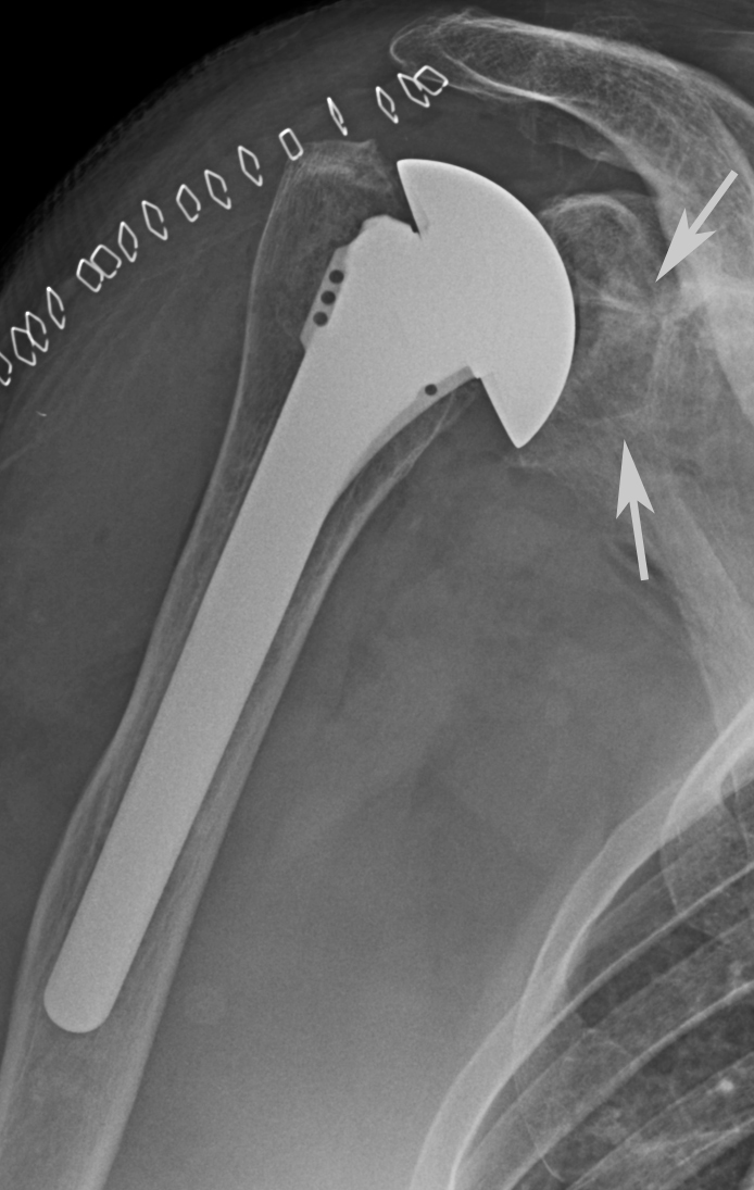
After surgery she developed progressive pain and loss of function. Eight years later x-rays of the shoulder showed loosening of the glenoid component (upper arrow) and loosening of the humeral component with a shift in position of the humeral component (lower arrow). Of note is the degradation of the cement originally placed beneath the glenoid component. It is possible that cement debris contributed to the failure of her joint replacement.


Use the "Search" box to the right to find other topics of interest to you.


You may be interested in some of our most visited web pages   arthritis, total shoulder, ream and runreverse total shoulderCTA arthroplasty, and rotator cuff surgery as well as the 'ream and run essentials'

Friday, June 1, 2018

Glenoid components can shift position after total shoulder arthroplasty

Sequential 3-dimensional computed tomography analysis of implant position following total shoulder arthroplasty

These authors sought to evaluate glenoid component position over time using 3-dimensional computed tomography (CT) analysis with minimum 2-year follow-up in 20 patients having primary TSA. 

Fourteen patients had a standard anchor peg glenoid component and 6 had a posteriorly augmented glenoid component (Global STEPTECH).

Each patient had a CT scan of the shoulder before surgery, within 2 weeks of surgery, and at a minimum of 2-years after surgery. 7 of the 20 glenoids showed evidence of component shift and/or grade 1 central peg osteolysis on the third scan were considered at risk of loosening: 6 had component shift (3 with increased inclination alone, 1 had increased retroversion alone, and 2 had both increased inclination and retroversion). 

Significantly more patients with glenoid component shift had grade 1 central peg osteolysis compared with those without shift (83% vs 7%, P = .002). One clinical failure occurred, with the patient undergoing revision to reverse TSA for rotator cuff deficiency. 

As shown in the table below, more severe types of glenoid pathology did not have a greater chance of being at risk
of the 11 Walch type A glenoids, 4 (36%) were at risk
of the 5 Walch B glenoids, 1 (20%) was at risk
of the 2 Walch C glenoids, none (0%) were at risk
of the 2 "other" glenoid types, both (100%) were at risk.

Of the 14 standard glenoid components 4 (29%) were at risk
Of the 6 StepTech posteriorly augmented glenoid components 3 (50%) were at risk.


This article demonstrates an intense (3 CT scans per shoulder) effort to evaluate the radiographic changes in the glenoid component after total shoulder arthroplasty. Interestingly, the results in 20 shoulders do not suggest that more severe forms of glenoid pathology are associated with a greater chance of the component being "at risk" and do not suggest that augmented glenoid components have less chance of the glenoid being at risk

While the paper suggests that sequential 3D CT imaging has the potential to be useful and clinically applicable for evaluating TSA component position over time, the cost, radiation exposure, and Human Subjects considerations would seem to limit this utility.

=====
Use the "Search" box to the right to find other topics of interest to you.


You may be interested in some of our most visited web pages including:shoulder arthritis, total shoulder, ream and runreverse total shoulderCTA arthroplasty, and rotator cuff surgery as well as the 'ream and run essentials'

Sunday, January 14, 2018

The critical shoulder angle after total shoulder, what does it reflect?

These authors define the 'critical shoulder angle' ( CSA ) at followup after a total shoulder as the angle between a line drawn from the inferior to superior glenoid rim and a line drawn from the inferior glenoid component edge to the inferolateral aspect of the acromion on a true anteroposterior (AP) radiograph.


They suggest that a larger critical shoulder angle (CSA) may cause superior glenoid component loading and more rapid component loosening.

They studied the relationship between the CSA and glenoid component loosening in midterm follow-up in 61 primary total shoulders for osteoarthritis with an average followup of 5.0 years without surgical revision. Standard true anteroposterior radiographs postoperatively and at longest follow-up were graded. An “at-risk” glenoid was defined as grade 3 or higher lucency. 

The average CSA was 32° ± 5°, median midterm lucency grade was 2 (range, 0-5), and median progression of lucency grade was 1 (range, −1 to 4). 

At midterm follow-up, 20% of TSAs were grade 3 or higher mean glenoid lucency, with an average CSA of 36°. 

There was a statistically significant correlation between CSA and both glenoid lucency grade (odds ratio, 1.20 per degree CSA) and progression of lucency grade (odds ratio, 1.24). 

An increase in CSA of 10° was associated with a 6.2-fold increased odds of having an at-risk glenoid.


Comment: This paper brings up some interesting points. 
First 20% of the total shoulders had glenoid components that were 'at risk' for failure. 
Second, the study did not include the preoperative or the immediate postoperative CSA measurements, so the prognostic value of CSA in these patients is unknown. 
Third, the authors suggest that CSA is a 'modifiable' risk factor for glenoid loosening, but it is not clear if they are suggesting that the CSA be modified by shortening the acromion or by placing the glenoid component in a more inferiorly directed position. 
Fourth, it is not clear if the post total shoulder CSA should be determined by a line drawing from the inferior to the superior rim of the glenoid component or a line drawn from the inferior to the superior rim of the residual bony glenoid. 
Finally, it is not clear whether the association of CSA and glenoid loosening is because the increase in CSA is causing the loosening or because the loosening of the glenoid (typified by superior tilting) is increasing the CSA as suggested by the two radiographs below from this article. 
The one on the left shows an initial CSA of 28 and grade 1 lucency, while the one on the right shows a same shoulder followup CSA of 32 or 40 (depending on the glenoid reference) and grade 4 lucency. 
This may, in fact, be an example of rocking horse loosening with rather massive osteolysis.


====

Monday, September 4, 2017

Rocking horse loosening in a superiorly unstable total shoulder

Recently we found this interesting Grashey view that nicely demonstrates superior rocking horse glenoid component loosening associated with superior instability after a total shoulder.
While no clinical information was available, the image is interesting in that it demonstrates the possible consequence when the humeral head is not centered in the prosthetic glenoid. This film also shows a substantial amount of backside cement, which we've found to predispose to glenoid component loosening.


===
The reader may also be interested in these posts:


Information about shoulder exercises can be found at this link.

Use the "Search" box to the right to find other topics of interest to you.


Monday, April 17, 2017

After a total shoulder, when is glenoid loosening aseptic?

A man in his mid sixties had a total shoulder for arthritis. Five years afterwards he noted a feeling of instability in his shoulder on active motion. Eight years after the arthroplasty he presented to us for a revision. On exam his shoulder was painful on active and passive motion. A distinct "clunk" could be felt when he flexed his arm. His AP views show a large humeral stem with the head sitting a bit high and perhaps a suspicion of lucency around the glenoid.


His axillary "truth" view showed obvious radiolucent lines around the cement of the keeled glenoid.


At his revision the humeral component was removed; the glenoid was grossly loose. The residual glenoid was smoothed. No glenoid bone grafting was performed. A new humeral component with an anteriorly eccentric humber head was inserted with impaction allografting. The residual glenoid defect is seen on the post operative x-ray.


His culture results, particularly from the glenoid, were positive for Propionibacterium (Cutibacterium) as shown below

Humeral head - 0.1
Humeral stem - no growth
Capsule - no growth
Collar memberane - 1.1
Glenoid cement #1 - 1
Glenoid cement #2 - 1
Glenoid component - 2

He was managed with the red protocol.

Ten months after surgery his shoulder was comfortable and stable, his active range of motion was progressing with PT. His x-rays showed apparent filling-in of the glenoid defect.


Comment: This case suggests the possibility of a Propionibacterium infection localized primarily to the glenoid.  In our practice of revision arthroplasty we usually avoid grafting the glenoid defect. It is of interest that in this case the glenoid surface appears to have reconstituted without grafting.


Our current management of apparently aseptic shoulder arthroplasty failure can be seen in this link

===


Information about shoulder exercises can be found at this link.

Use the "Search" box to the right to find other topics of interest to you.


Tuesday, January 24, 2017

Total shoulder arthroplasty failure

Optimizing follow-up after anatomic total shoulder arthroplasty

These authors sought to establish a data-based follow-up schedule minimizing unnecessary patient and health care system costs without sacrificing the quality of patient care. They reviewed 2786 consecutive anatomic total shoulder replacements to identify the common modes of failure and times to failure.

208 shoulders (7.5%) required reoperation. Early failure mechanisms included instability, rotator cuff tears, and infection, with 63% of these reoperations occurring within 2 years.  The most common failure mechanism was instability, resulting in 89 reoperations (3.2%). Later failures included mechanical failures (including component loosening) and periprosthetic fractures, with no identifiable peak occurrence. After 2 years, TSA failed at an average rate 1.1% per year.

Comment: In this large study total shoulder arthroplasties continued to fail at an almost linear rate as long as 20 years after surgery. In examining the chart below, the primary cause of failure in the first year was instability (green dots) (left arrow). It would be of interest to know more about the 89 cases with instability, in that this is not usually a common indication for revision.  Component failure (teal dashes) was not a prominent cause of failure until after five years, but then accounted for an progressively increasing percentage of the failures after that)(right arrow). 


"It's not over until it's over." As emphasized in this article, Glenoid Component Failure in Total Shoulder Arthroplasty is the long term issue in the survival of total shoulder arthroplasty.

The importance of long term followup is illustrated by this case.

The patient presented with this x-ray and a Simple Shoulder Test Score of 1/12

She had a total shoulder as shown below.


Two years after surgery, her SST was 12/12 with the x-ray shown below.


Thirteen years after surgery, her SST dropped to 2/12 and she presented with glenoid component failure as shown below.

We have found that asking patients to complete the Simple Shoulder Test annually provides an inexpensive, sensitive, convenient and practical method for the necessary long term followup after shoulder arthroplasty as demonstrated in this article:

Patient functional self-assessment in late glenoid component failure at three to eleven years after total shoulder arthroplasty The routine use of SST as a patient reported outcome may enable the early identification of component failure without having the patient return to the office.

Here's another recent case example
x-ray at 2.5 years after shoulder arthroplasty showing glenoid loosening and osteolysis


x-ray at 3 years after arthroplasty showing increased osteolysis and progressive loosening. SST at the time of presentation to us was 5/12.


===


To see the topics covered in this Blog, click here

Use the "Search" box to the right to find other topics of interest to you.

You may be interested in some of our most visited web pages including:shoulder arthritis, total shoulder, ream and runreverse total shoulderCTA arthroplasty, and rotator cuff surgery as well as the 'ream and run essentials'


Monday, October 24, 2016

Too tight stem -> too high head -> rocking horse glenoid loosening

A stem that is tight in the diaphysis can result in a too high humeral component that can, in turn, lead to rocking horse loosening of the glenoid component as shown by the x-rays of the right shoulder below (middle aged man who presented to us with a painful stiff shoulder).


We revised this shoulder by removal of the loose glenoid component, smoothing the bony glenoid surface without bone grafting or glenoid component reimplantation and by replacing the humeral component with a smaller stem inserted at the desired height and fixed with impaction allografting.


A three months after surgery he has a comfortable, functional shoulder with radiographic evidence of glenoid healing and a centered humeral head in both the AP
and axillary views.


Comment: Impaction grafting of a small stem allows proper positioning of the stem. The management of a loose glenoid does not require bone grafting to fill the defect or reimplantation of a new glenoid component.

===

Saturday, October 22, 2016

Humeral and glenoid component loosening - aseptic or culture positive? What can we learn?

Here are the films of the right shoulder of a man in his late 60s with glenohumeral arthritis


 and a posteriorly biconcave glenoid.

He had a total shoulder with mono block chrome-cobalt humeral component and a Hylamer glenoid.


He did very well after surgery, returning to gym workouts and a wide range of outdoor physical activities. His x-ray at three monte after surgery is shown below.

One year after surgery, he was still doing well. His X-ray at that time is shown below.

Seven years after surgery, his shoulder was becoming somewhat painful, but not so much that he wanted anything done. His x-rays at that time show glenoid component wear and humeral subsidence.


Thirteen years after his surgery, his symptoms were worsening as were the x-ray findings. He elected to give it a bit more time.


 Three months later the symptoms and x-ray changes (see below)


led to a revision surgery. Preoperative blood tests were normal.

At surgery the joint fluid was cloudy, but gram stains showed no neutrophils.

The humeral and glenoid components were loose and there was a thick humeral membrane. Frozen sections showed "Humeral membrane, right shoulder, excision: - Synovial tissue with extensive fibrosis and marked foreign body giant cell reaction with associated polarizable foreign material. - No neutrophils identified." 


The retrieved glenoid component showed substantial wear.


 Based on these findings, we considered the diagnosis of detritic synovitis (see this link).


He had a single stage revision to a hemiarthroplasty using Vancoymycin allograft for humeral fixation.


As a precaution we placed the patient on oral antibiotics (Augmentin) until the culture results were finalized.

In a week after surgery, the culture results came back as shown below


Note that the preoperative culture of his unprepared skin was strongly positive for Propionibacterium.
The humeral and glenoid explants and the membrane deep to the glenoid component were also strongly positive for Propionibacterium, while the humeral membrane specimens and joint fluid were unimpressive.
These findings emphasize that Propionibacterium is not uniformly distributed throughout the shoulder. Had the cultures been limited to the humeral membrane and joint fluid, the presence of Propionibacterium may have been overlooked.

With these culture results he was converted to the red protocol (see this link) of IV Ceftriaxone and Rifampin. 

Clinically, he is doing well with 140 degrees of assisted elevation and minimal shoulder discomfort.

Comment: This case is an excellent example of the complexities of the evaluation and management of a shoulder with delayed loosening of the total shoulder components. It is not possible to know if the Propionibacterium recovered from this shoulder were introduced from the patient's skin at the index surgery 13 years prior to the revision or if they arrived in the shoulder subsequently.

We have so much left to learn.

===