These authors point out that although eccentric reaming, augmented glenoid implants, bone grafting, and reverse arthroplasty have been used to manage bone loss and retroversion, there is no consensus on treatment.
Posteriorly augmented glenoid components (full-wedged, half-wedged, and stepped polyethylene posteriorly) are currently available for use in the United States.
Most clinical studies on augmented implants are case series, typically reported by the designers or consultants associated with the specific implant.
Some reports show increased radiolucency and failure rates when posteriorly augmented glenoids are used.
With studies with low levels of evidence, small numbers of patients, and short-term followup periods, the theoretically superior results of stepped implants and wedged implants in comparison with traditional methods using conventional glenoid components have not been shown.
Comment: From this review, we cannot determine the indications for or the incremental clinical value to patients of augmented glenoid components.
Furthermore, as shown in the article below, it is not clear how much "correction" of glenoid retroversion is necessary in practice:
While glenoid retroversion and posterior humeral head decentering are common preoperative features of severely arthritic glenohumeral joints, the relationship of postoperative glenoid component retroversion to the clinical results of total shoulder arthroplasty (TSA) is unclear. Studies have indicated concern for inferior outcomes when glenoid components are inserted in 15° or more retroversion.
In a population of patients undergoing TSA in whom no specific efforts were made to change the version of the glenoid, these authors asked whether at 2 years after surgery patients having glenoid components implanted in 15° or greater retroversion had (1) less improvement in the Simple Shoulder Test (SST) score and lower SST scores; (2) higher percentages of central peg lucency, higher Lazarus radiolucency grades, higher mean percentages of posterior decentering, and more frequent central peg perforation; or (3) a greater percentage having revision for glenoid component failure compared with patients with glenoid components implanted in less than 15° retroversion. They examined the records of 201 TSAs performed using a standard all-polyethylene pegged glenoid component

inserted after conservative glenoid reaming without specific attempt to modify preoperative glenoid version.

Of these, 171 (85%) patients had SST scores preoperatively and between 18 and 36 months after surgery. Ninety-three of these patients had preoperative radiographs in the database and immediate postoperative radiographs and postoperative radiographs taken in a range of 18 to 30 months after surgery. Twenty-two patients had radiographs that were inadequate for measurement at the preoperative, immediate postoperative, or latest followup time so that they could not be included. In comparison to those included in the analysis, the excluded patients did not have substantially different mean age, sex distribution, time of followup, distribution of diagnoses, American Society of Anesthesiologists class, alcohol use, smoking history, BMI, history of prior surgery or preoperative glenoid version. They analyzed the two year outcomes in the remaining 71 TSAs, comparing the 21 in the retroverted group (the glenoid component was implanted in 15° or greater retroversion (mean ± SD, 20.7° ± 5.3°)) with the 50 in the non-retroverted group ( the glenoid component was implanted in less than 15° retroversion (mean ± SD, 5.7° ± 6.9°)).
The mean (± SD) improvement in the SST (6.7 ± 3.6; from 2.6 ± 2.6 to 9.3 ± 2.9) for the retroverted group was not inferior to that for the nonretroverted group (5.8 ± 3.6; from 3.7 ± 2.5 to 9.4 ± 3.0). The percent of maximal possible improvement (%MPI) for the retroverted glenoids (70% ± 31%) was not inferior to that for the nonretroverted glenoids (67% ± 44%). The 2-year SST scores for the retroverted (9.3 ± 2.9) and the nonretroverted glenoid groups (9.4 ± 3.0) were similar (mean difference, 0.2; 95% CI, - 1.1 to 1.4; p = 0.697). No patient in either group reported symptoms of subluxation or dislocation. The radiographic results for the retroverted glenoid group were similar to those for the nonretroverted group with respect to central peg lucency (four of 21 [19%] versus six of 50 [12%]; p = 0.436; odds ratio, 1.7; 95% CI, 0.4-6.9), average Lazarus radiolucency scores (0.5 versus 0.7, Mann-Whitney U p value = 0.873; Wilcoxon rank sum test W = 512, p value = 0.836), and the mean percentage of posterior humeral head decentering (3.4% ± 5.5% versus 1.6% ± 6.0%; p = 0.223). The percentage of patients with retroverted glenoids undergoing revision (0 of 21 [0%]) was not inferior to the percentage of those with nonretroverted glenoids (three of 50; [6%]; p = 0.251).
The authors concluded that in this series of TSAs, postoperative glenoid retroversion was not associated with inferior clinical results at 2 years after surgery.
Here are two other articles on this interesting topic:
Total shoulder arthroplasty in patients with a B2 glenoid addressed with corrective reaming
These authors conducted a retrospective series of consecutive patients who underwent total shoulder arthroplasty with a Walch B2 glenoid addressed using partially corrective glenoid reaming and a standard (non-augmented) cemented all-polyethylene pegged glenoid prosthesis. The mean preoperative retroversion measured 18° (range, –1° to 36°), superior inclination was 8° (range, –11° to 27°), and posterior subluxation was 67% (range, 39%-91%).There were no exclusion criteria based on radiographic severity of glenoid deformity.

Glenoid deformities were addressed using a high side, corrective ream with the goal of achieving an estimated minimum of 80% glenoid support and a final retroversion angle within 10° to 15° of neutral. In more severe deformities, the implant was placed slightly more retroverted in favor of excessive reaming, which would compromise glenoid fixation.
Functional outcome scores were available for 59 of 92 eligible subjects (64%) at a mean of 50 months after surgery. The Simple Shoulder Test improved from 4.5 to 9.1; the visual analog scale improved from 7.4 to 1.4, and the American Shoulder and Elbow Shoulder Standardized Assessment improved from 35.4 to 84.3.
Radiographs were evaluated at a mean of 31 months: 38 had no glenoid radiolucent lines, 13 glenoids had grade 1, 2 had grade 2, and 5 had grade 3 lucencies.
22.7% of shoulders with > 20° of preoperative retroversion demonstrated progression of radiolucency of at least 1 grade in the Lazarus classification. 27.8
27.8% Of the shoulders with < 20° of preoperative retroversion developed progression of at least 1 grade of radiolucency.
No shoulders had radiographic evidence of glenoid loosening or subsidence. No patients had subjective complaints of shoulder instability.
Total shoulder arthroplasty for glenohumeral arthritis associated with posterior glenoid bone loss: results of an all-polyethylene, posteriorly augmented glenoid component
Between May 2011 and January 2013, 22 shoulders in 19 patients (15 men and 4 women) underwent primary TSA by a single surgeon. In all cases, an all-polyethylene, posteriorly augmented, stepped glenoid component was implanted.

At a mean follow-up of 36 months, 12 shoulders had osseous integration between the central-peg flanges, 6 had bone adjacent to the central-peg flanges but without identifiable osseous integration, and 1 showed osteolysis.

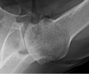
The authors point out that " implantation of a stepped glenoid component requires removal of some posterior bone. In glenoids in which there is both retroversion and glenoid medialization, there may be insufficient subchondral bone, volume, and/or density after preparation to support the posterior component." This is shown by the figure below.

In the osteoarthritic shoulder with posterior wear, the bone and cartilage have failed to hold up under the loads applied by the humeral head. With a posteriorly augmented glenoid component, the hope is that the thickened posterior polyethylene will be able to hold up under these same loads.

Total shoulder arthroplasty for glenohumeral arthritis associated with posterior glenoid bone loss: results of an all-polyethylene, posteriorly augmented glenoid component
Between May 2011 and January 2013, 22 shoulders in 19 patients (15 men and 4 women) underwent primary TSA by a single surgeon. In all cases, an all-polyethylene, posteriorly augmented, stepped glenoid component was implanted.

In these cases preoperative glenoid retroversion measured 15 degrees or greater (14 of these cases had retroversion of 25 degrees or less).

At a mean follow-up of 36 months, 12 shoulders had osseous integration between the central-peg flanges, 6 had bone adjacent to the central-peg flanges but without identifiable osseous integration, and 1 showed osteolysis.
While the overall clinical results showed improvement, two patients sustained a total of 3 episodes of humeral dislocation, one anterior and two posterior.

This report shows that achieving prosthetic balance with this prosthesis can be difficult, even in the hands of an experienced surgeon.
The indications for the use of the stepped prosthesis remain to be determined. Many of the shoulders in this series had relatively low amounts of preoperative retroversion that are in the range usually managed with conventional components and soft tissue balancing.
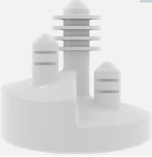
Should this implant fail, the bone defect would be greater than that present before the arthroplasty as diagrammed below.

See also "Posterior Glenoid Wear in Total Shoulder Arthroplasty: Eccentric Anterior Reaming Is Superior to Posterior Augment" at this link.
Long term followup of well-characterized patients treated with the different methods for managing glenoid retroversion will be required to define the relative risks, benefits, effectiveness and durability of each of them.
===
We have a new set of shoulder youtubes about the shoulder, check them out at this link.
We have a new set of shoulder youtubes about the shoulder, check them out at this link.
Be sure to visit "Ream and Run - the state of the art" regarding this radically conservative approach to shoulder arthritis at this link and this link
Use the "Search" box to the right to find other topics of interest to you.
You may be interested in some of our most visited web pages arthritis, total shoulder, ream and run, reverse total shoulder, CTA arthroplasty, and rotator cuff surgery as well as the 'ream and run essentials'